90 minutes endo resto
04/11/2024
Francesca Cerutti
Warning: Undefined variable $post in /var/www/vhosts/styleitaliano-endodontics.org/endodontics.styleitaliano.org/wp-content/plugins/oxygen/component-framework/components/classes/code-block.class.php(133) : eval()'d code on line 2
Warning: Attempt to read property "ID" on null in /var/www/vhosts/styleitaliano-endodontics.org/endodontics.styleitaliano.org/wp-content/plugins/oxygen/component-framework/components/classes/code-block.class.php(133) : eval()'d code on line 2
Endodontic treatment is not only about shaping, cleaning and filling the root canal system. The Literature agrees on the fact that the coronal restoration is a mandatory element to ensure that the endodontic treatment remains successful over time, but the main problem is that, often, the coronal restoration is delayed due to lack of time, due to the fact that the patient id referred and someone else will restore the tooth, due to the organization of the clinic, etc.
What if the person who did the root canal treatment also restored the tooth? Who knows better if there is the need for a fiber post and which fiber post is best than the person who did the root canal treatment?
When the conditions are favorable, restoring the tooth immediately can be convenient and safer, because we know that with temporary restorations there is the risk to have leakage, loss of the restoration, fracture of the tooth, etc.
Here is the report of a case managed in a single session.
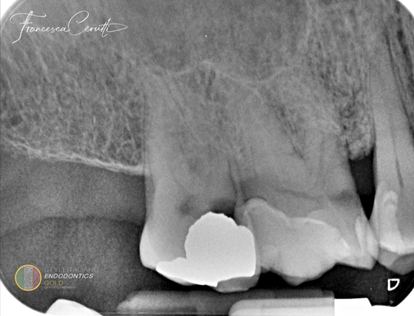
Fig. 1
A 65 years old patient came to my attention complaining about intense pain on the upper right hemiarch.
At the clinical examination, tooth 1.7 was hyper responsive to cold and presented an old amalgam restoration.
The periapical X-ray showed the presence of a secondary decay on tooth 1.7.
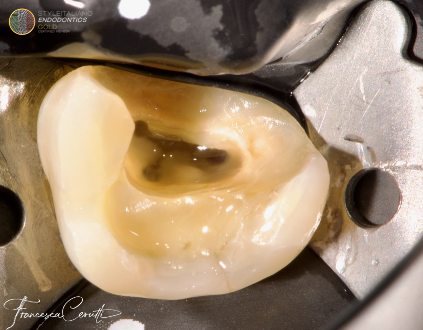
Fig. 2
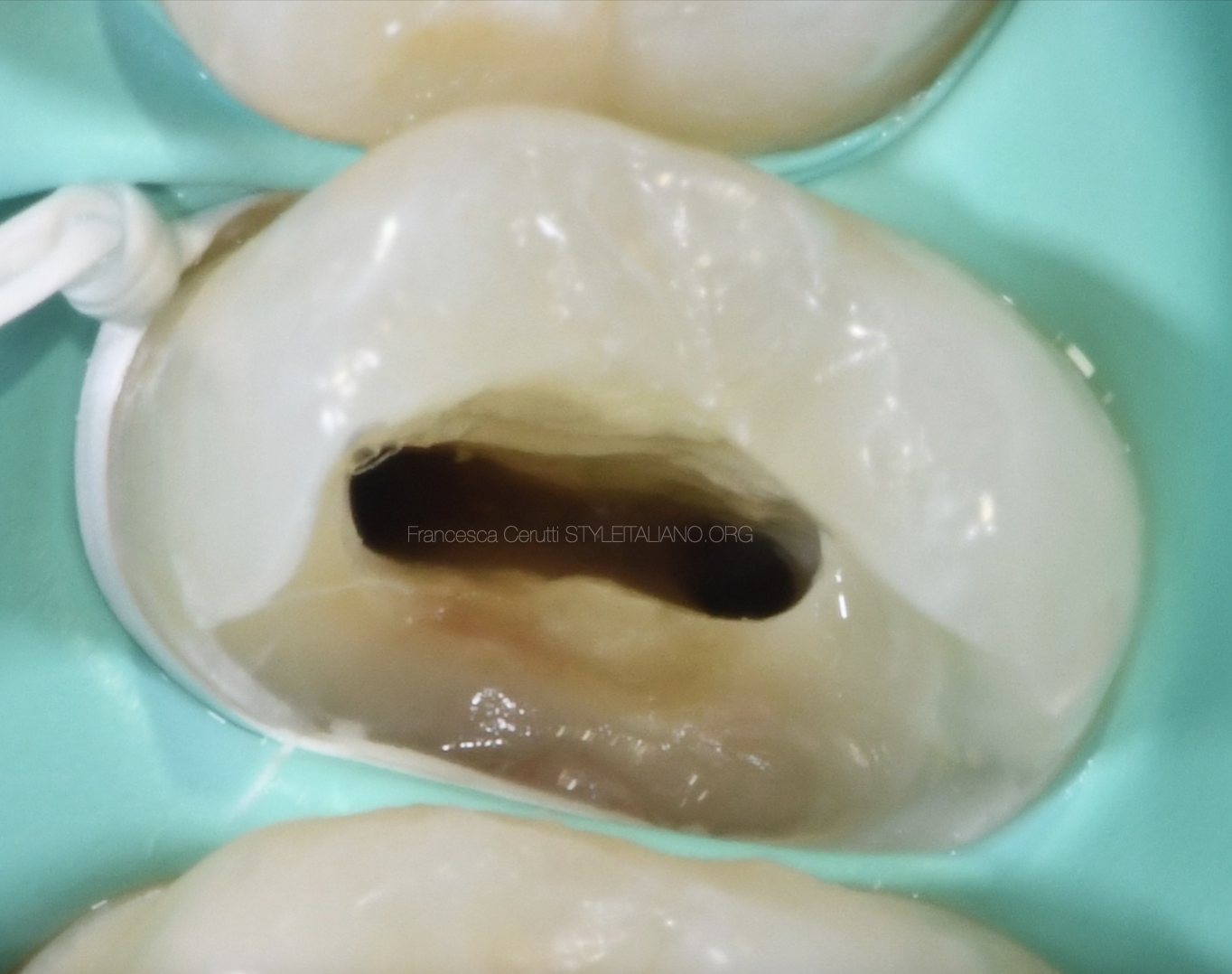
After the anesthesia, I isolated the tooth with rubber dam. Then I removed the existing restoration and the decay and started designing the access cavity
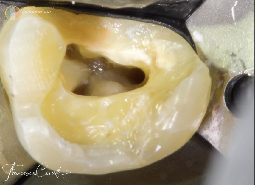
Fig. 3
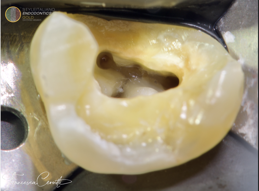
The color change on the pulp chamber floor clearly suggests the presence of four root canal openings.
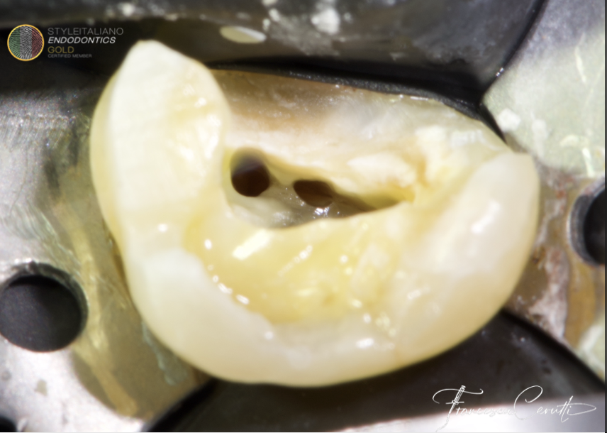
Fig. 4
I shaped the MB1, DB and P canal with martensitic files at 500 RPM and 4.0 Torque, then I started shaping the MB2.
The inclination of the MB2 opening suggests the presence of a confluence between MB1 and MB2.
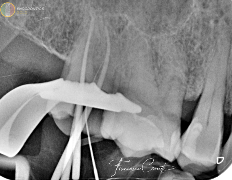
Fig. 5
The intra operative X-ray with gutta percha points in the MB1, DB and P and a k-file in the MB2 is useful to determine the fit of the master cones and to confirm the confluence of MB1 and MB2.
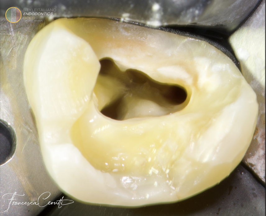
Fig. 6
The root canals shaped, cleaned and ready to be filled.
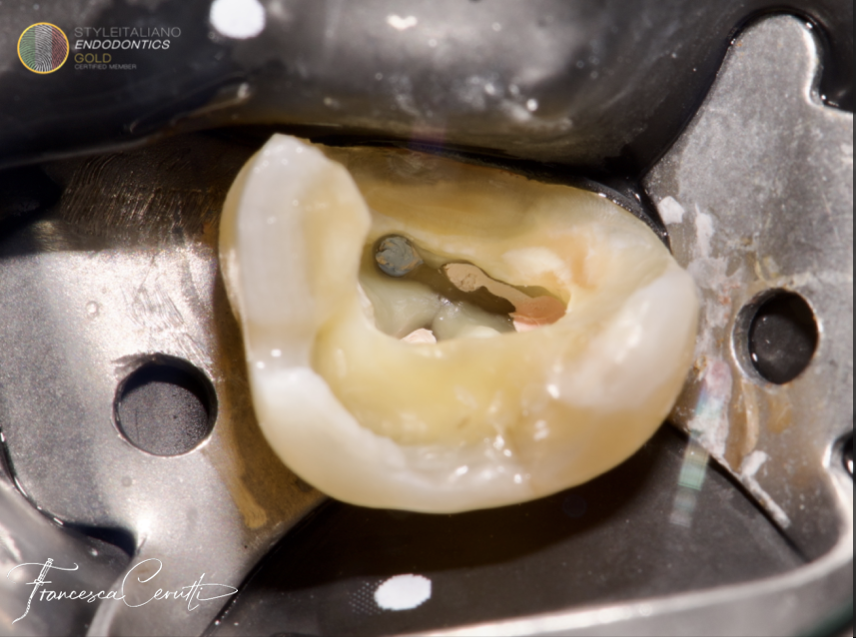
Fig. 7
After root canal filling with single cone and hydraulic sealer
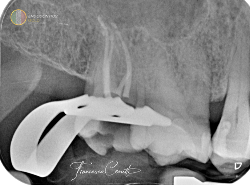
Fig. 8
X-ray to check the goodness root canal filling. If something had gone wrong, I would have fixed it immediately, before the setting of the sealer.
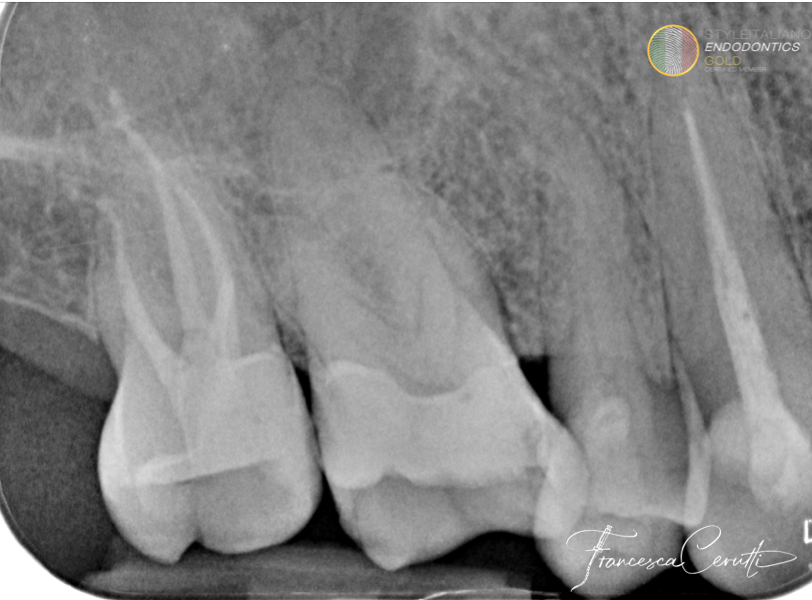
Fig. 9
Post operative X-ray
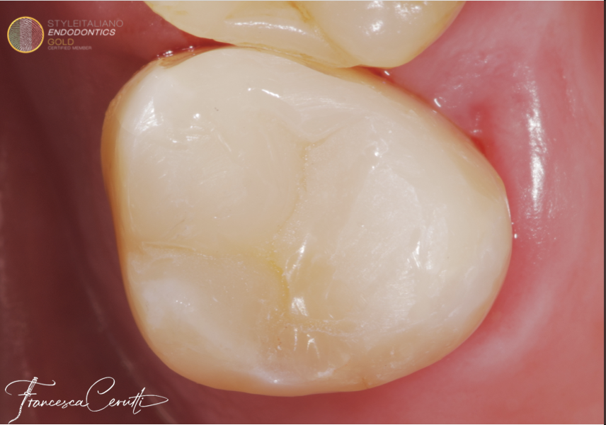
Fig. 10
Post operative picture
Conclusions
Being able to restore the tooth immediately, with a direct restoration or a build up followed by an indirect restoration, is a convenient and effective way to organize the clinical workflow for ETT.
Bibliography
Mergoni G, Ganim M, Lodi G, Figini L, Gagliani M, Manfredi M. Single versus multiple visits for endodontic treatment of permanent teeth. Cochrane Database Syst Rev. 2022;12(12):Cd005296.
Craveiro MA, Fontana CE, de Martin AS, Bueno CE. Influence of coronal restoration and root canal filling quality on periapical status: clinical and radiographic evaluation. J Endod. 2015;41(6):836-40.
Yee K, Bhagavatula P, Stover S, Eichmiller F, Hashimoto L, MacDonald S, et al. Survival Rates of Teeth with Primary Endodontic Treatment after Core/Post and Crown Placement. J Endod. 2017.
Migliau G, Besharat LK, Sofan AA, Sofan EA, Romeo U. Endo-restorative treatment of a severly discolored upper incisor: resolution of the "aesthetic" problem through Componeer veneering System. Annali di stomatologia. 2015;6(3-4):113-8.
Dos Reis-Prado AH, Abreu LG, Tavares WLF, Peixoto IFDC, Viana ACD, de Oliveira EMC, Bastos JV, Ribeiro-Sobrinho AP, Benetti F. Comparison between immediate and delayed post space preparations: a systematic review and meta-analysis. Clin Oral Investig. 2021 Feb;25(2):417-440. doi: 10.1007/s00784-020-03690-x. Epub 2021 Jan 8. PMID: 33417064.


