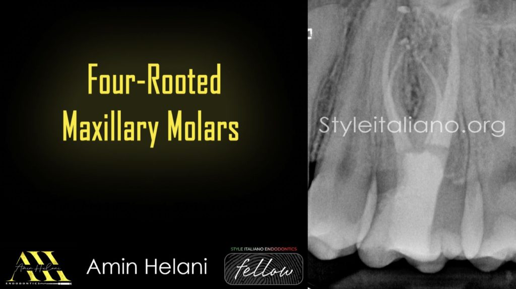
Four-Rooted Maxillary Molars
02/08/2024
Fellow
Warning: Undefined variable $post in /home/styleendo/htdocs/styleitaliano-endodontics.org/wp-content/plugins/oxygen/component-framework/components/classes/code-block.class.php(133) : eval()'d code on line 2
Warning: Attempt to read property "ID" on null in /home/styleendo/htdocs/styleitaliano-endodontics.org/wp-content/plugins/oxygen/component-framework/components/classes/code-block.class.php(133) : eval()'d code on line 2
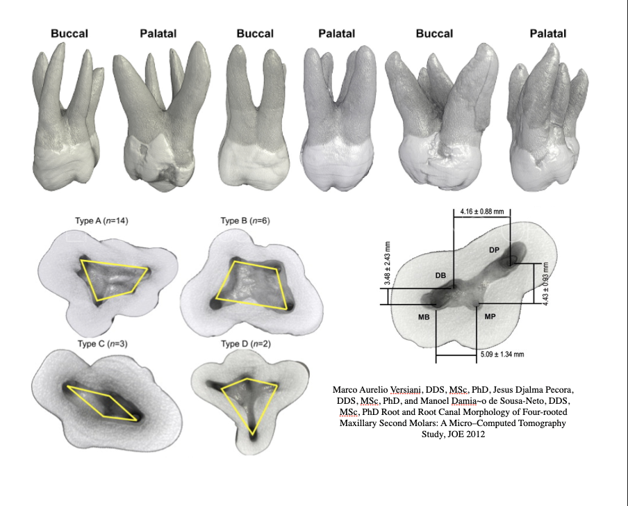
The maxillary first molar typically exhibits complex root canal anatomy, commonly presenting with three roots—two buccal and one palatal. However, anatomical variations, such as the presence of a fourth root, can occur and significantly impact endodontic treatment.
Recognizing these variations is crucial for successful root canal therapy.
This article discusses the endodontic management of four-rooted maxillary first molars, highlighting the importance of carefully reading the periapical Radiographs and advanced imaging techniques like cone-beam computed tomography (CBCT) in accurate diagnosis and treatment planning.
This anomaly poses challenges in terms of effective treatment planning. Radiographic examinations play a crucial role in identifying these variations preoperatively, facilitating appropriate endodontic interventions. Understanding the prevalence and morphology of four-rooted maxillary first molars is essential for dental practitioners to deliver precise and successful treatments.
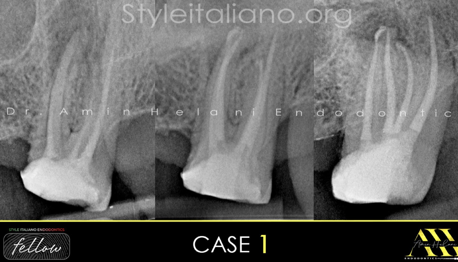
Fig. 1
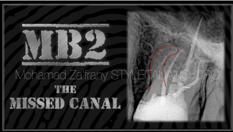
Case 1: A 56-year-old female patient reported to the clinic with a chief complaint of mild pain in left maxillary second molar since 2 days, and gave history of root canal treatment done 15 years ago.
Pre-Op Radiograph revealed radiolucency around the apex of mb root.
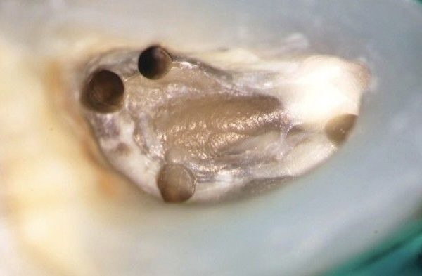
After the access opening, the anatomy of the pulp chamber floor (floor map) revealed an unusual canal orifice on the palatal side,
Indicating the presence of a second separated palatal root.
Shaping: VDW R25 Blue
Irrigation: NaOCL 5%, EDTA 17% with Ultrasonic Activation
Obturation: Single Cone with BC Sealer.
Video of the procedure.
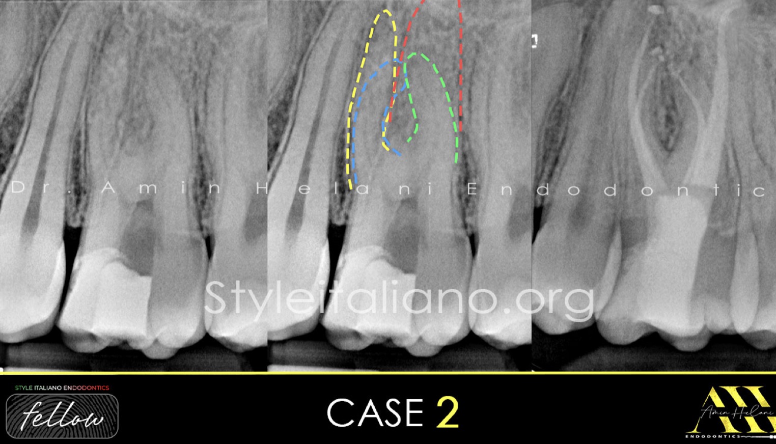
Fig. 2
Case 2: A 35-year-old woman complaining of sever pain especially during mastication.
Clinical examination revealed tenderness to percussion and palpation of peri-radicular zone of the left maxillary first molar.
Pre-Op Radiograph revealed the presence of 4 separated roots.
Shaping: VDW R25 Blue
Irrigation: NaOCL 5%, EDTA 17% with Ultrasonic Activation
Obturation: Single Cone with BC Sealer and WVC.
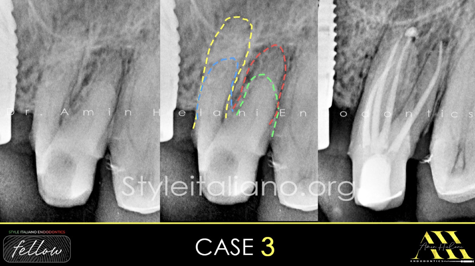
Fig. 3
Case 3: A 60-year-old male patient was referred to our clinic by a colleague who was unable to locate all the canals while preparing for new crowns. The patient has no symptoms.
Pre-Op Radiograph revealed the presence of 4 separated roots.
Shaping: VDW R25 Blue
Irrigation: NaOCL 5%, EDTA 17% with Ultrasonic Activation
Obturation: Single Cone with BC Sealer.
Fig. 4
• Dr. Amin Helani graduated in 2014 from the University of Aleppo in Syria.
• In 2021, he received a specialist degree in Endodontics from the German Society of Endodontics.
• He works as a Microscopic Endodontist for advanced root canal treatment in Braunschweig, Germany.
• He has been offering lectures, webinars, and hands-on courses to both general dentists and endodontists.
• He is a member of the German Society for Endodontology and Traumatology.
• He is a fellow member of Style Italiano Endodontics.
Conclusions
In conclusion, four-rooted maxillary first molars are a rare but significant anatomical variation that dental professionals should be aware of. Proper recognition of this variant through a radiographic evaluation is crucial for successful endodontic procedures .Utilizing advanced diagnostic tools like high quality pre-op X-ray and CBCT improves the accuracy of diagnosis and treatment outcomes, ensuring the longevity and functionality of the treated tooth.
Bibliography
References:
- Libfeld H, Rotstein I. Incidence of four-rooted maxillary second molars: literature review and radiographic survey of 1,200 teeth. J Endod. 1989;15:129–131.
- Ozcan E, Aktan AM, Arı H. A case report: unusual anatomy of maxillary second molar with 3 mesiobuccal canals. Oral Surg Oral Med Oral Pathol Oral Radiol Endod. 2009;107
- Zmener O, Peirano A. Endodontic therapy in a maxillary second molar with three buccal roots. J Endod. 1998;24:376–377.
- Kumar MR, Areekan MS, Simon EP, Raveendran C, Krishna A, Jayaraj D. Morphologic variations of roots and root canals in maxillary second molar: Case reports. IP Indian J Conserv Endod. 2020;5:92–96.