Bridging the Gap: How Today’s Endodontics Overcome Past Limitations
29/05/2024
Fellow
Warning: Undefined variable $post in /var/www/vhosts/styleitaliano-endodontics.org/endodontics.styleitaliano.org/wp-content/plugins/oxygen/component-framework/components/classes/code-block.class.php(133) : eval()'d code on line 2
Warning: Attempt to read property "ID" on null in /var/www/vhosts/styleitaliano-endodontics.org/endodontics.styleitaliano.org/wp-content/plugins/oxygen/component-framework/components/classes/code-block.class.php(133) : eval()'d code on line 2
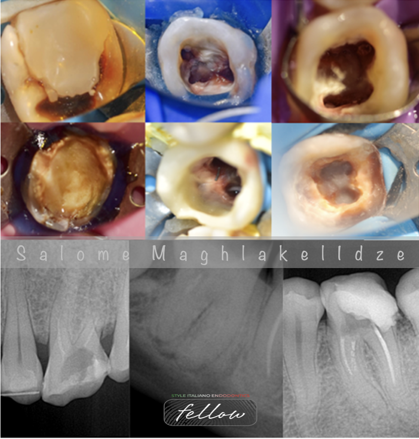
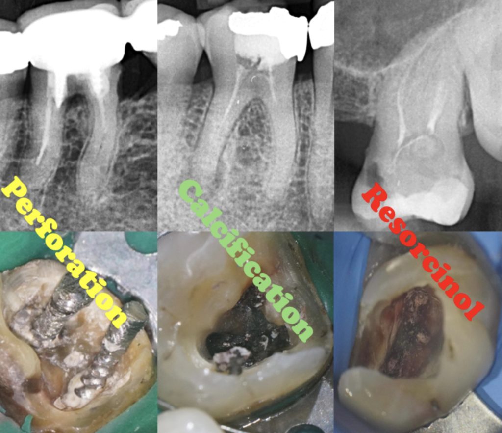
As modern dentistry continually strives for excellence, we frequently face the challenge of dealing with complications from older endodontic treatments, which often present unexpected difficulties. Calcifications, perforations, and stubborn obturation materials that are difficult to remove pose significant challenges in treatment. To effectively manage these cases, careful and thorough planning is essential. This involves a comprehensive understanding of the underlying problems so that they can be addressed efficiently and effectively, ensuring the best possible outcomes for our treatments.
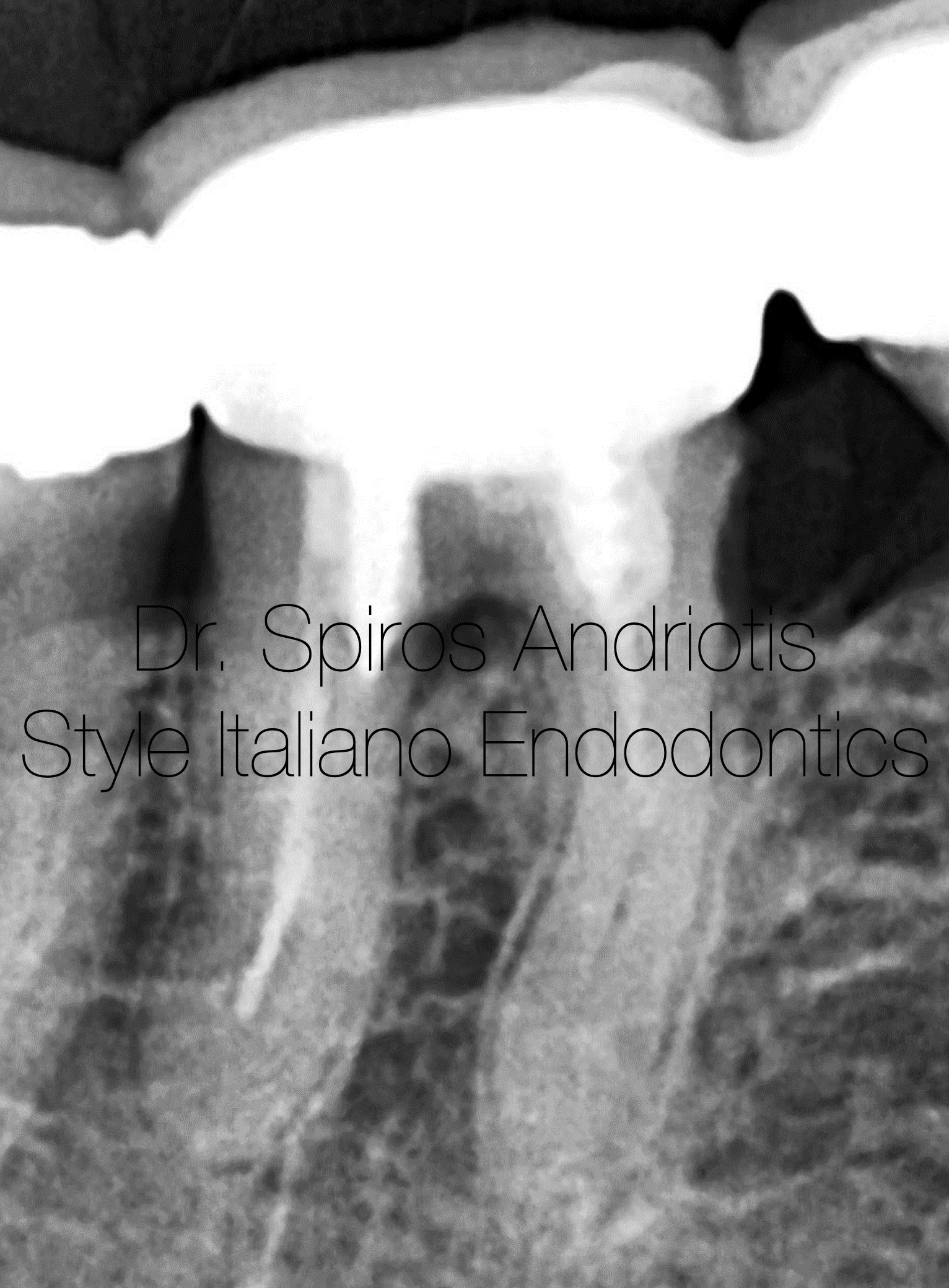
Fig. 1
Case #1:
Female patient, 60 years of age, free medical history.
LR6 with mild symptoms of discomfort.
Previous endodontic treatment, screw posts, metal/ceramic prosthesis. The treatment was carried out in Switzerland prior 30 years.
Diagnosis: Symptomatic apical periodontitis.
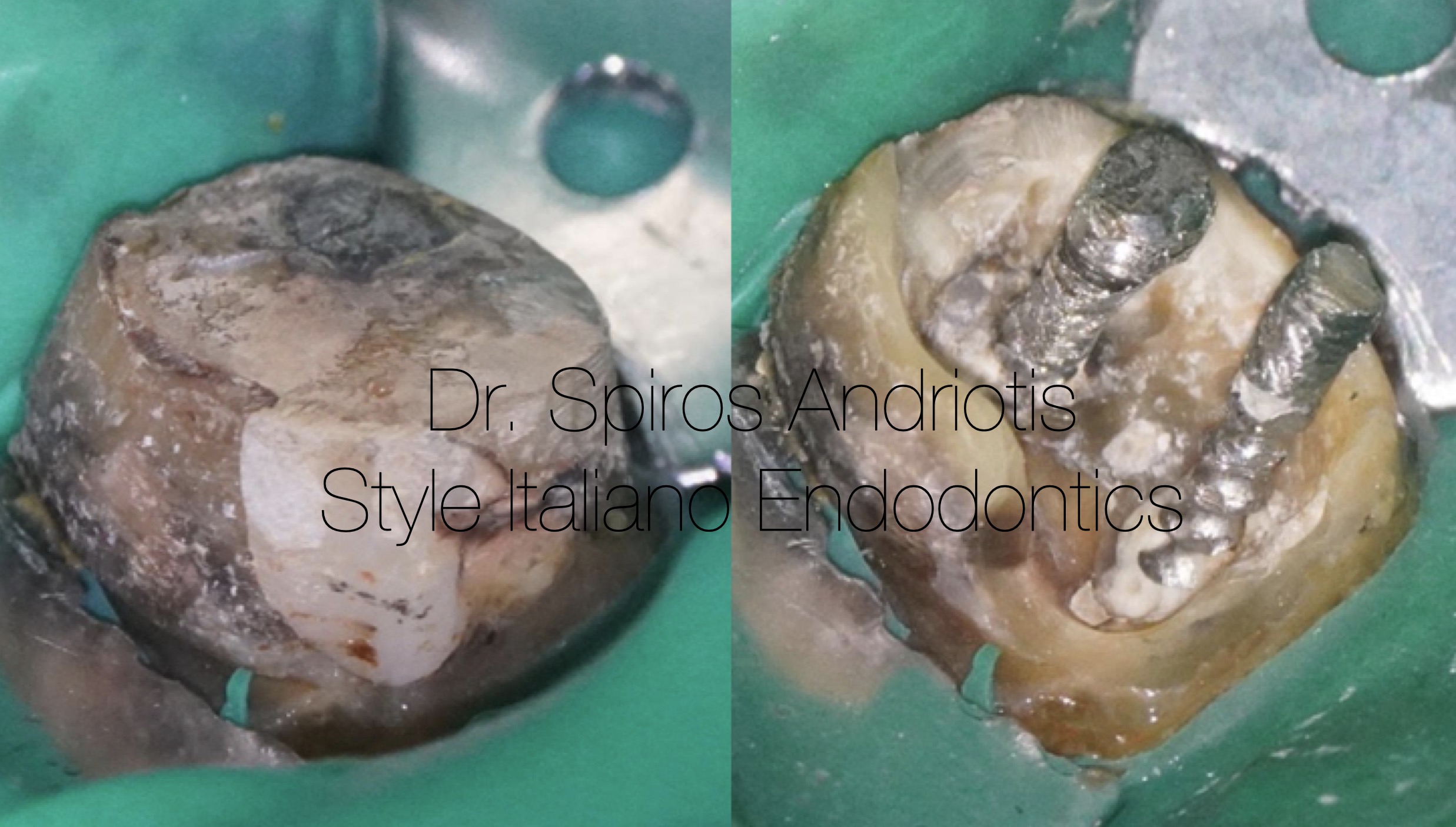
Fig. 2
After the prosthesis removal, any restorative material was removed ina gente way that wouls allow us to create around the post.the posts were simply removed by underscrewing them with a thing plier
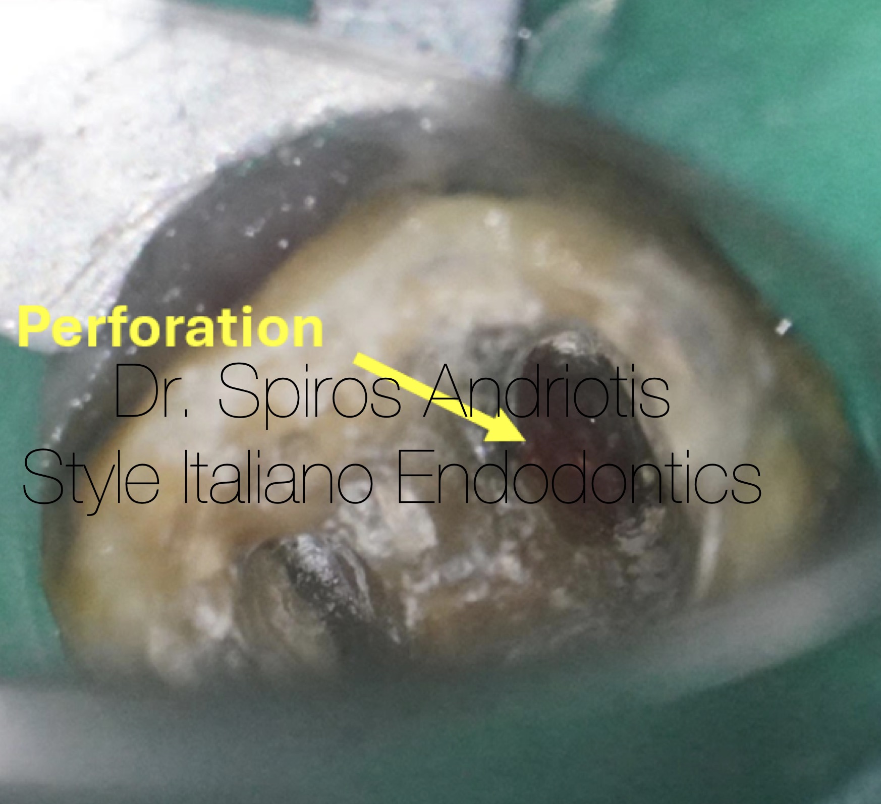
Fig. 3
As soon as both posts were removed, a large perforation was observed by the distal root.The bleeding was controlled by the action of the NaOCl solution as we proceeded with the instrumentation of the canals. All three canals were blocked and calcified apically. An attempt was made to greate a glyde path using directly a rotary instrument 15/0.3 with no success, the dentin presented increases sclerosis. Hand scouting with D-finders #8 in combination with EDTA sollution 17% allowed us to reach the apical constriction after several minutes of negotiations.
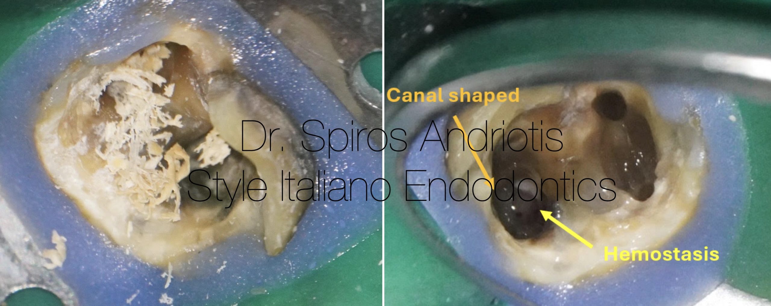
Fig. 4
The working length was estimated with the help of an electronic apex locator at 00 reading using a #10 K hand file.
Rotary instrumentation:
-15/0.3 500 rpm
-20/0.4 500 rpm
-25/0.4 500 rpm (final preparation for the mesial canals)
-40/0.4 500rpm (final preparation for the distal canal)
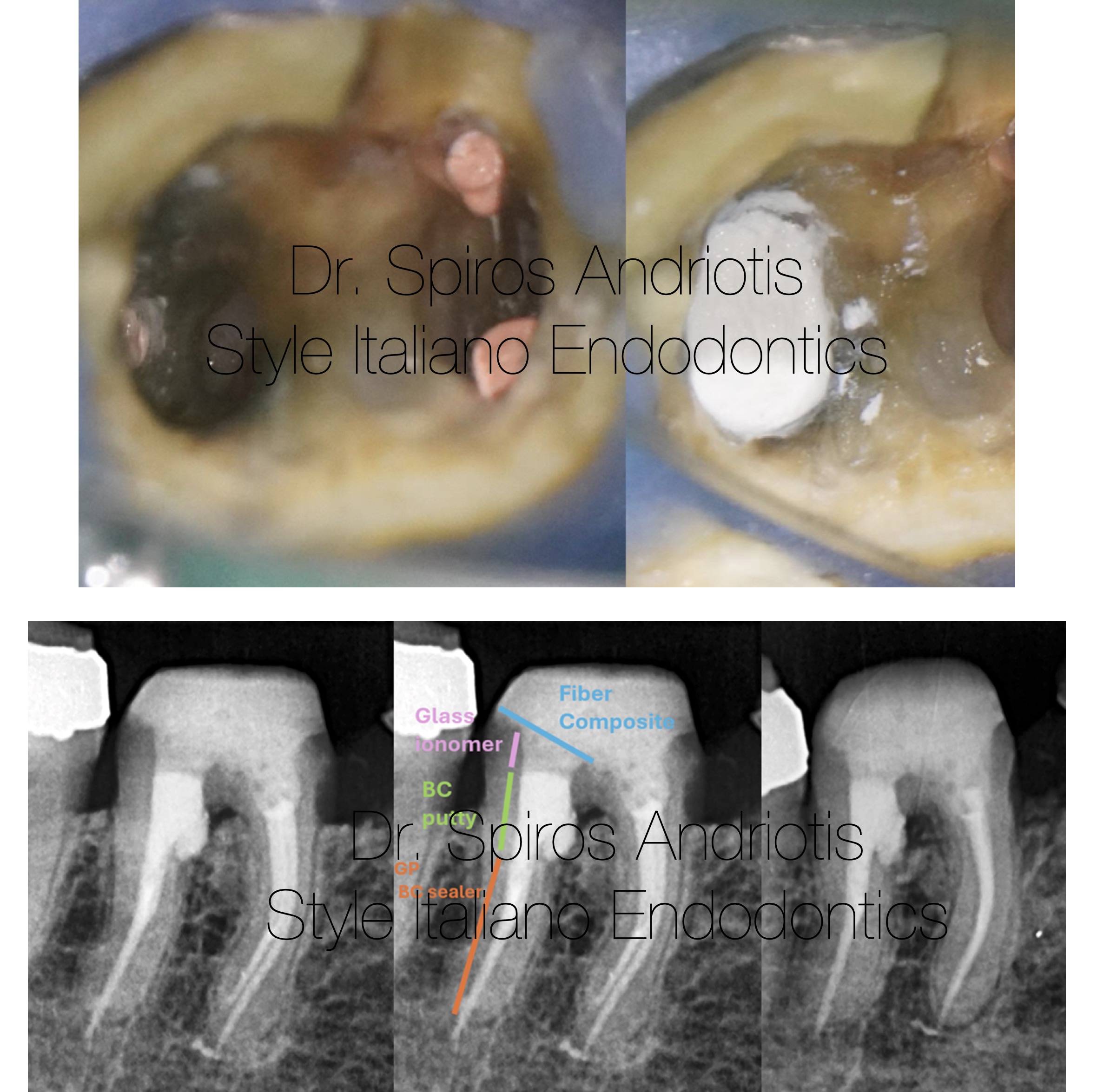
Fig. 5
Final irrigation:
EDTA 17% - Ultrasonic activation
NaoCL 5% - Heating, Ultrasonic activation
The canals were obturated with bioceramic sealer and cold hydraulic condensation. For the distal canal the space of the perforation was filled with bioceramic putty protected by a layer of glass ionomer composite.
The rest of the tooth was restored with fiber composite and the patient was returned to the referrer for indirect restoration.
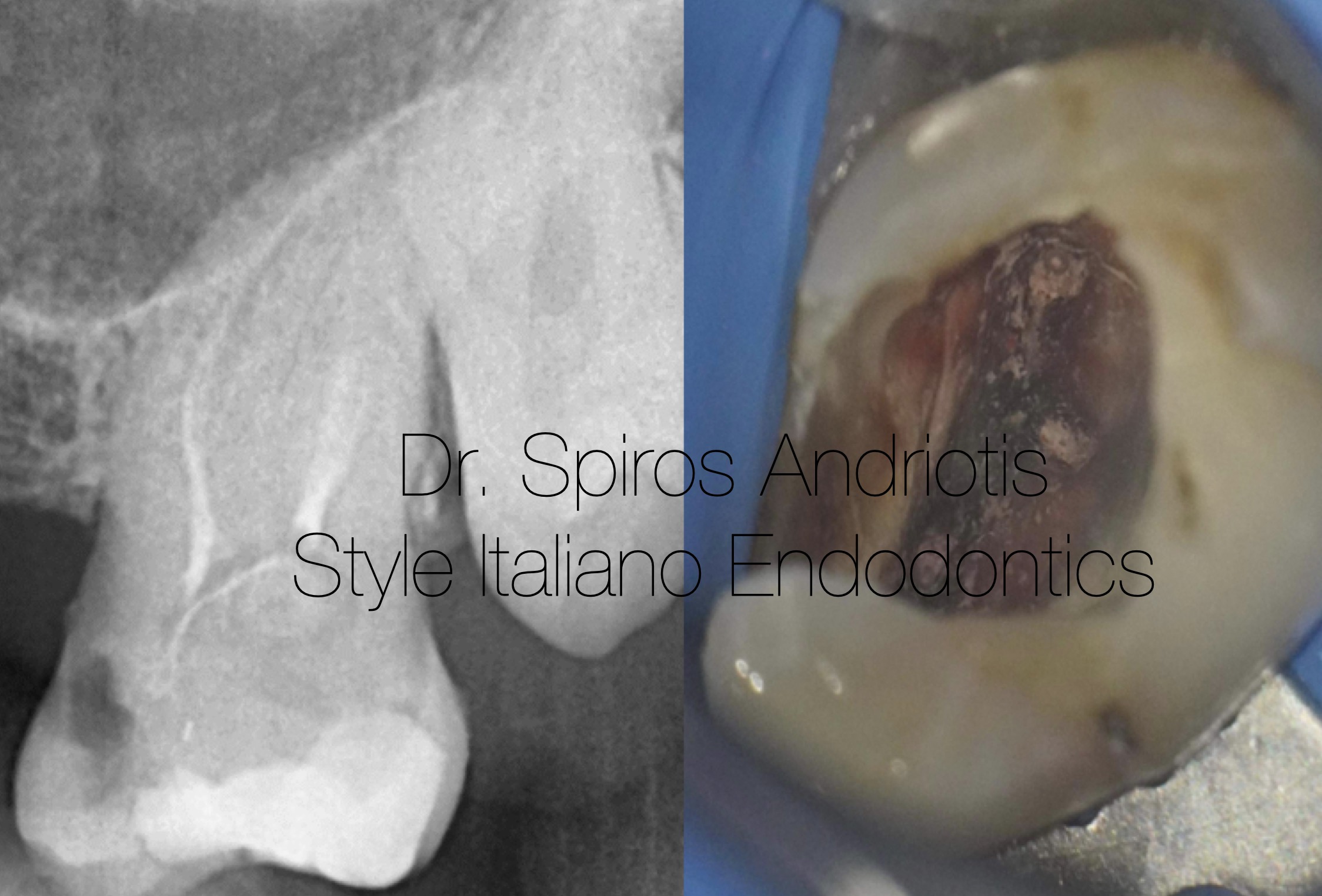
Fig. 6
Case #2:
Male patient, 45 years of age, free medical history.
UL7 with no symptomatology. The patient was referred for evaluation of his endodontic treatment for a tooth soon to be abutment of a bridge prosthesis. The tooth was treated in Ukraine 25 years ago. After the consultation, the patient desired to repeat his previous endodontic treatment and possibly avoid future problems.
Diagnosis: Asymptomatic Apical Periodontitis
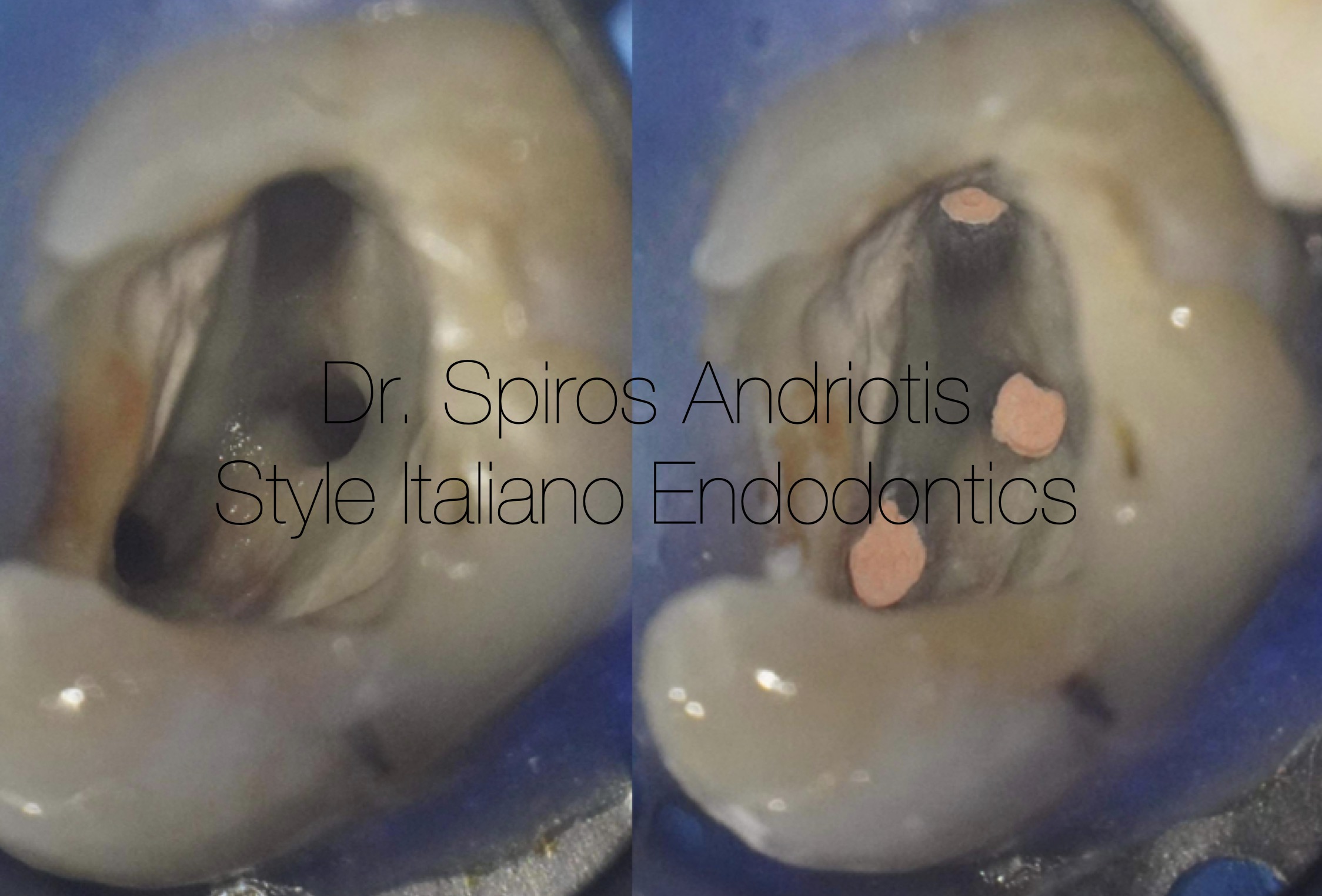
Fig. 7
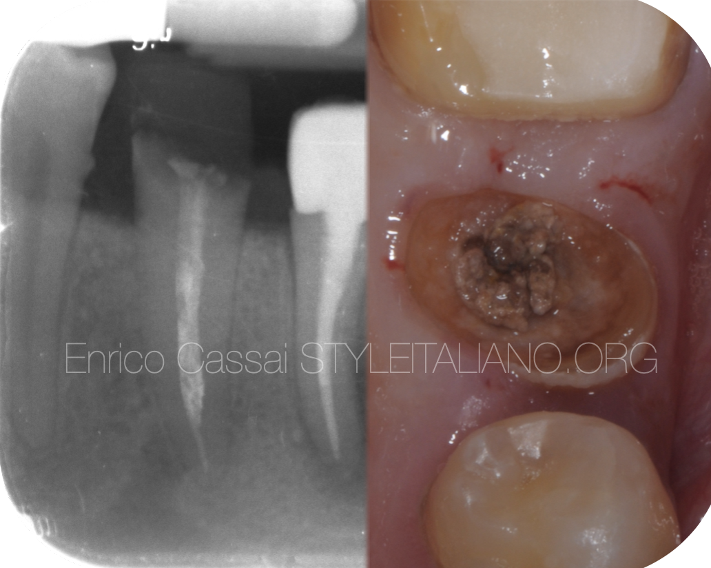
Isolation was placed and any restorative materials were removed. The red color of the previous obturation material made it obvious that it was a “Russian red” case or else Resorcinol-Formaldehyde resin.
The described compound comprises two potentially harmful ingredients: formaldehyde in liquid form and resorcinol as a powder. To ensure this substance is detectable on X-ray images, it might contain radiopaque agents such as zinc oxide or barium sulfate. Adding 10% sodium hydroxide to the mix triggers a polymerization process that creates a very hard, red material that solidifies without any known solvent to dissolve it. This durability, while beneficial in some applications, also poses challenges in terms of safe handling and disposal due to its toxicity and inability to break down.
Ultrasonics were used to remove the resorcinol from the orifices and a reciprocating rotary file (25/0.8) created a pre flaring of the canals.
Continuous rotation between D-finders #10 and the reciprocating instrument facilitated quick and efficient removal of the old obturation material. The same sequence of files were used for this case as well, with 25/0.4 being the final preparation size for all three canals.
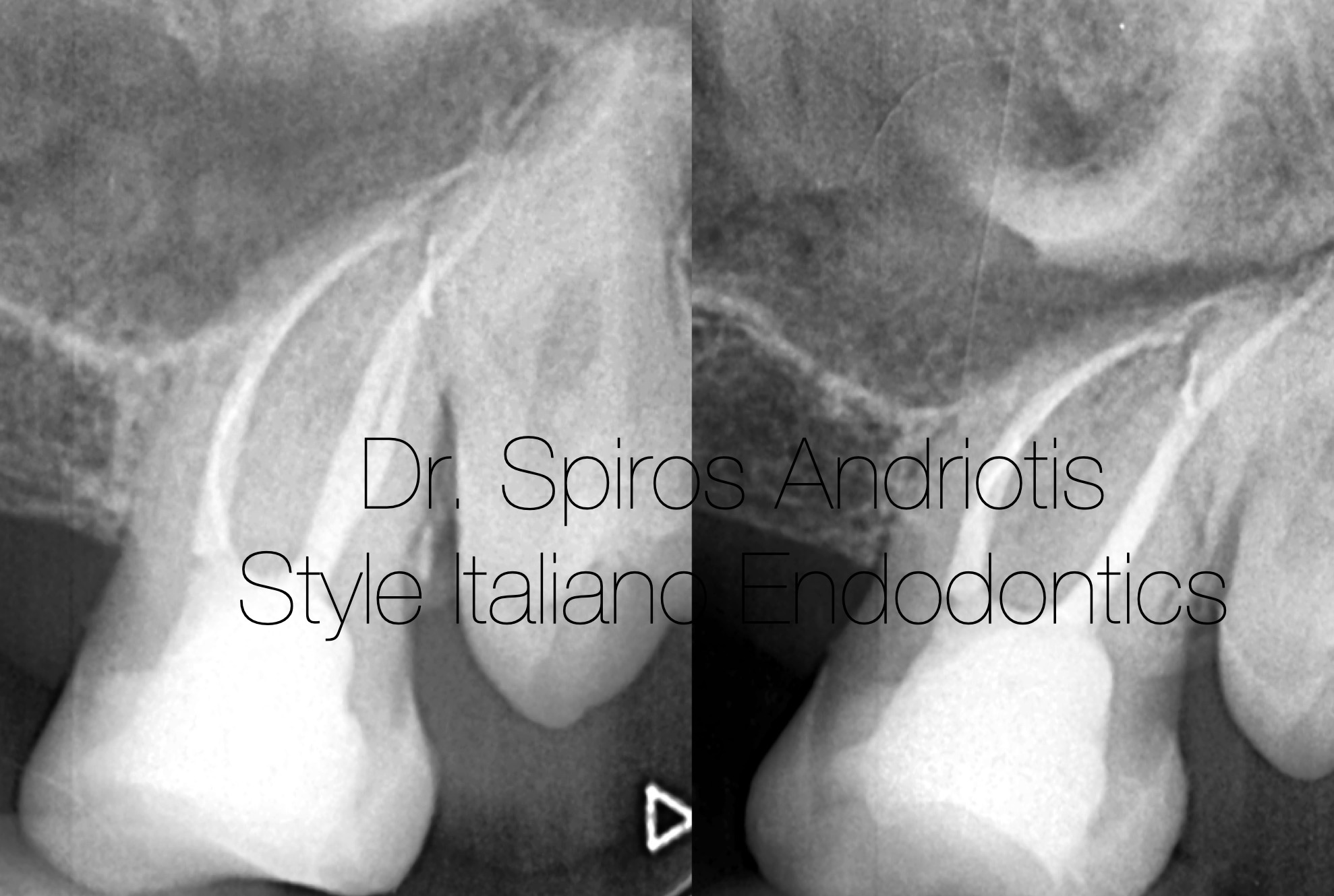
Fig. 8
Final irrigation:
EDTA 17% - Ultrasonic activation
NaoCL 5% - Heating, Ultrasonic activation
Obturation:
Epoxy based sealer and warm vertical compaction.
Restoration:
Fiber composite
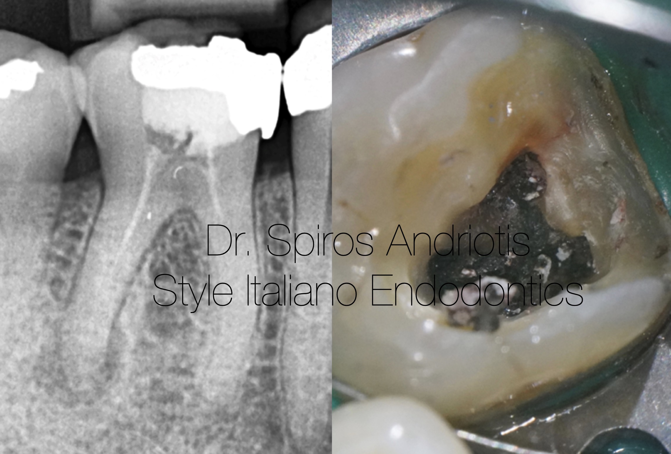
Fig. 9
Case #3:
Male patient, 60 years of age, free medical history.
Severe periodic pain. LR6 previously treated endodontically, amalgam restoration. The patient had undergone the treatment more than 30 years ago in Greece. Severe calcification of the root canal system is observed.
Diagnosis: Symptomatic apical periodontitis
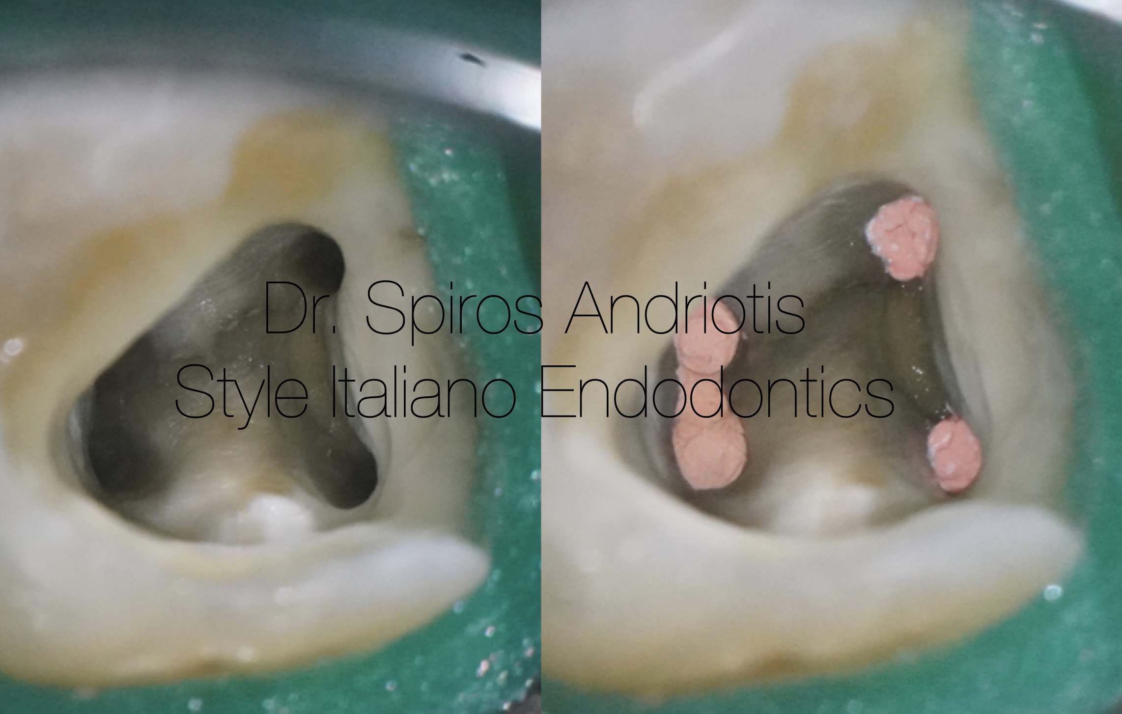
Fig. 10
The root canals appeared not patent with sclerosed dentin obliterating them. A flaring of the canals was performed and irrigations with EDTA 17% were carried out for 10 minutes along with ultrasonic activation. After this the EDTA rinse, space inside the root canals had started to get freed as the calcified tissue was getting softer. Once again, EDTA 17%, RC prep, D-finders hand files and a fair amount of patience helped us to create a reproducible glide path and before the rotary instrumentation.
Final sizes of preparation:
25/0.4 for the mesials
40/0.4 for the distal
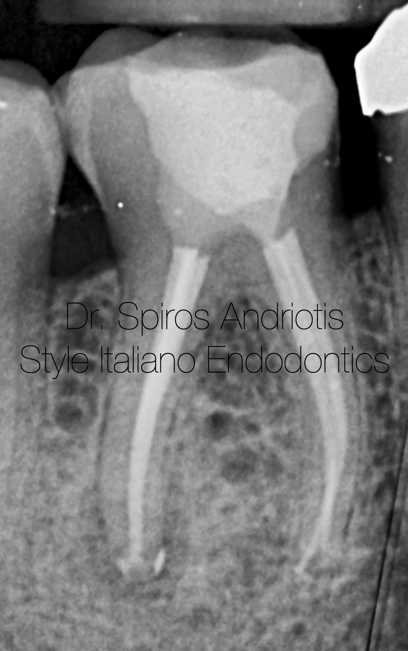
Fig. 11
Final irrigation:
EDTA 17% - Ultrasonic activation
NaoCl 5% - Heating, Ultrasonic activation
Obturation:
Bioceramic sealer, cold hydraulic condensation
Restoration:
Fiber Composite. The patient was returned to the referrer for indirect resoration

Fig. 12
Conclusions
In my opinion, we should not judge older treatments as the knowledge provided at that period was limited. We live in an era of increased involvement and endodontics has never been easier and safer for both the dentist and the patient. The microscope, the isolation, the irrigations, the use of the right materials and the understanding that we do not deal with canals but with an infection are the cornerstone for successful endodontic treatments.
Bibliography
Endodontic Orthograde Retreatments: Challenges and Solutions Alessio Zanza, Rodolfo Reda, Luca Testarelli
Factors that affect the outcomes of root canal treatment and retreatment—A reframing of the principles Kishor Gulabivala, Yuan Ling Ng
Bioceramics in Endodontics: Updates and Future Perspectives Xu Dong, Xin Xu


