The advantage to use Extendo in removing Thermafil Plastic Carrier on retreatment case
21/02/2022
Massimo Giovarruscio
Warning: Undefined variable $post in /var/www/vhosts/styleitaliano-endodontics.org/endodontics.styleitaliano.org/wp-content/plugins/oxygen/component-framework/components/classes/code-block.class.php(133) : eval()'d code on line 2
Warning: Attempt to read property "ID" on null in /var/www/vhosts/styleitaliano-endodontics.org/endodontics.styleitaliano.org/wp-content/plugins/oxygen/component-framework/components/classes/code-block.class.php(133) : eval()'d code on line 2
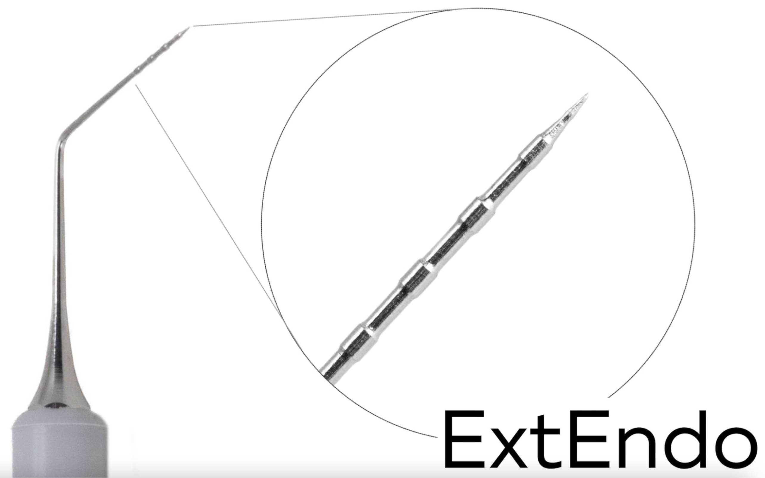
In this article I am going to describe and show how we can use Extendo in daily practice.
It is an instrument of SMART ENDO KIT, from DEPPELER.
The kit has 5 instruments and an endo ruler.
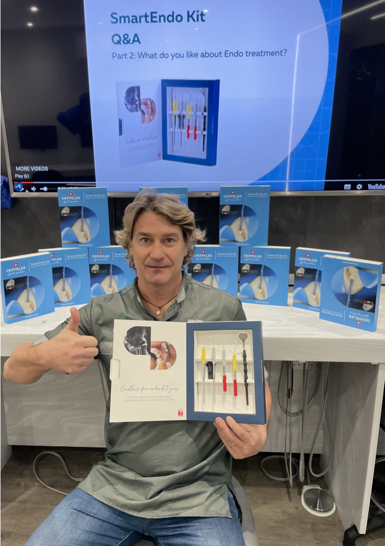
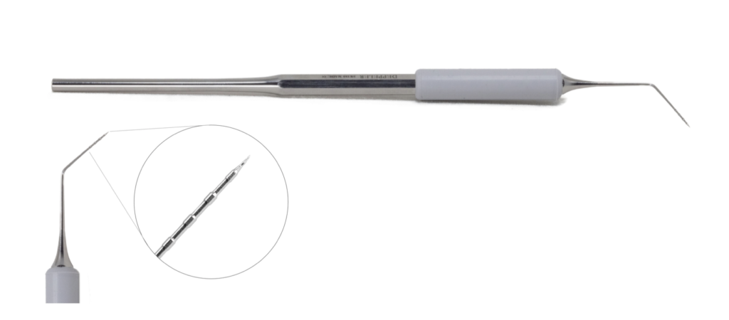
Fig. 1
Extendo is a multi use instrument, it is very useful and it can be used in different endodontic scenario.
In this retreatment case, I have used it to remove Thermafil plastic carrier. And every dentist knows how difficult it can be.
Extendo is a smart choices for this job.
So lets go…
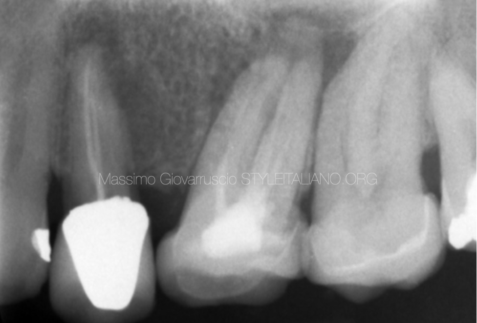
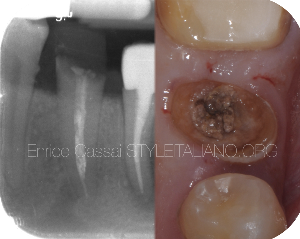
Fig. 2
A 45-year-old patient has been referred to my practice due to a periodical endodontic lesion.
The peri apical radiographic examination taken, revealed an upper premolar root canal treated few years ago. A large apical area is present. It also visible a distal darkness on the root.
On clinical examination, a zirconia crown is fitted on this tooth. The tooth responded painfully during percussion test (TTP). A large buccal sinus is present.
The final diagnosis was a symptomatic apical periodontitis.
Here is a video of the retreatment. The sharp end of ExtEndo makes it easier to remove the plastic carrier, procedure that is usually time consuming.
Once the plastic carrier has been loosened into the root canal, removing it with tweezers is simple.
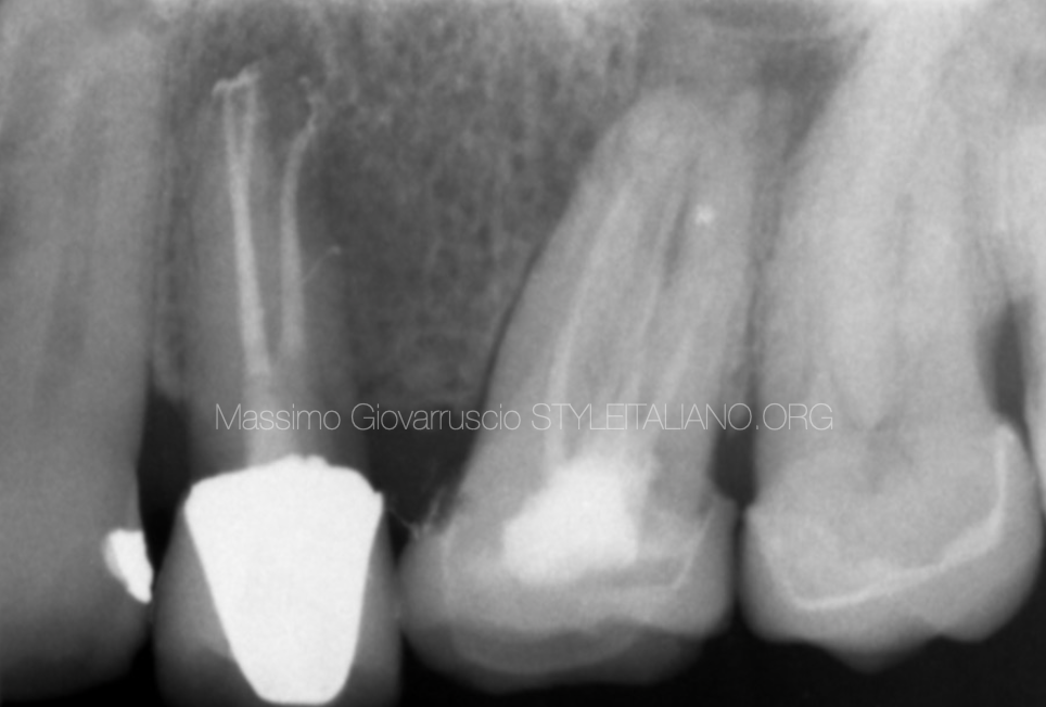
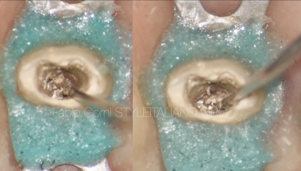
Fig. 3
Canal inspection and location of root canal orifices was performed using Deppeler mirror and Explora, which results in an excellent tool for detecting canals too. Especially during retreatments. The use microscope is highly recommended on complex cases and on difficult anatomies.
The use of a GP solvent (Eugenate Desobturator, PD) was very useful to dislocate the plastic carrier. The sharp tip of ExtEndo creates a space between the root canal walls and the plastic carrier. ExtEndo easily allows to create space between gutta-percha and a plastic carrier. When the gutta-percha has been dissolved properly, the plastic carrier has been removed firmly using Stieglitz tweezer.
All GP material has been removed completely with mechanical instruments (Reciprocating file), leaving only the most apical part to solvent, manual k-files and paper points.
In this way the amount of potentially infected material dissolved by the solvent is minimal, as the materia is spread on the canal side walls by the action of the instruments.
After scouting the canal at the working length, it was possible to clean and shape the entire canal.
Reciprocating instrument has been performed up to the working length. GP master cone has been confirmed with x-ray and the apical gauging has been checked. After that, it was possible to proceed with the final cleaning. 5 minutes NaOCL (30sec sonic activation for each canal) and EDTA 17% (30sec sonic activation for each canal).
After drying the canal with paper pint, the tooth has been obdurated using the continuous wave of condensation.
The access cavity has been restored permanently with dual composite. Th ePost Op Periapical X-Ray shows a 3d Obturation filling with apical delta and lateral portal of exit.
Conclusions
This article showed:
- How to approach a complex anatomy in a premolar tooth
- How to remove a plastic carrier using ExtEndo Deppeler instrument
- How achieve a proper 3D Obturation filling
Bibliography
Scianamblo MJ: Endodontic failures: the retreatment of previuosly endodontically retreated teeth, Revue Odontostomatol (Paris), 1988
Parreira FR et al: Cast prosthesis removal using ultrasonic and thermoplastic resin adhesive, J Endod, 1994
Carr GB: Retreatment. In Cohen S, Burns Rc, editors: Pathways of pulp, 1998 Retreatments, Edra