Retreatment series: Episode 3
22/07/2021
Ibrahim El Naggar
Warning: Undefined variable $post in /var/www/vhosts/styleitaliano-endodontics.org/endodontics.styleitaliano.org/wp-content/plugins/oxygen/component-framework/components/classes/code-block.class.php(133) : eval()'d code on line 2
Warning: Attempt to read property "ID" on null in /var/www/vhosts/styleitaliano-endodontics.org/endodontics.styleitaliano.org/wp-content/plugins/oxygen/component-framework/components/classes/code-block.class.php(133) : eval()'d code on line 2
The Root canal treatments is a way full of challenges, that may be the ANATOMY and also it may be The Dealing with the “PROCEDURAL ERRORS” in this article i will describe how I managed this Treatment Mishap.
I have described in two Episodes before different approaches for Non-Surgical Root Canal Retreatment this time i am discussing a SURGICAL APPROACH.
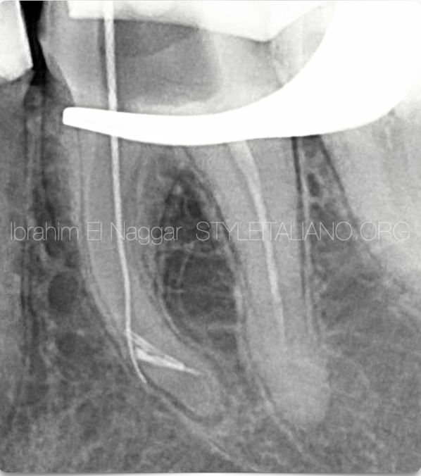
Fig. 1
This was the Pre-Op image from the referral, clearly showing 2 separated Instruments & Stripping on the outer wall during trial for bypassing the fragments
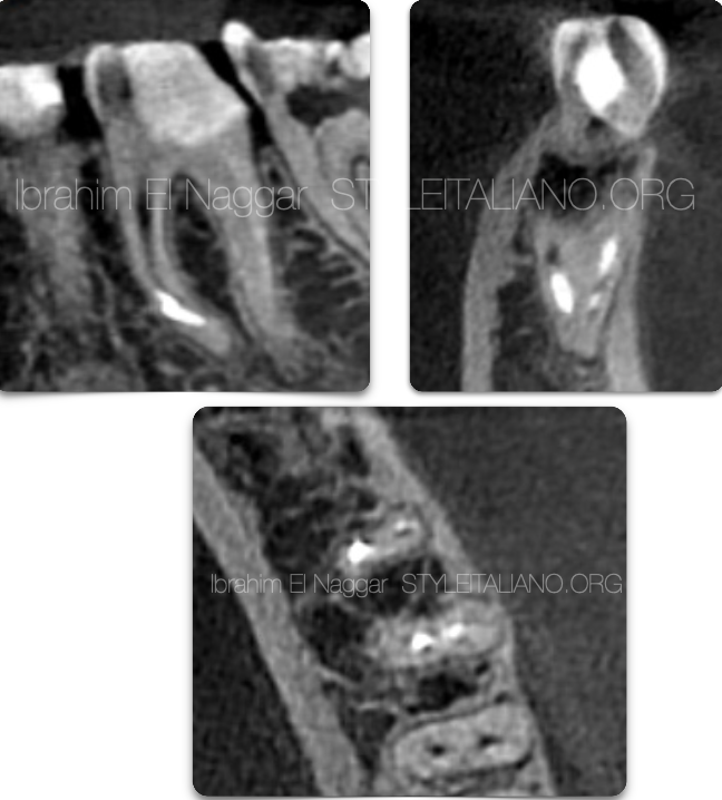
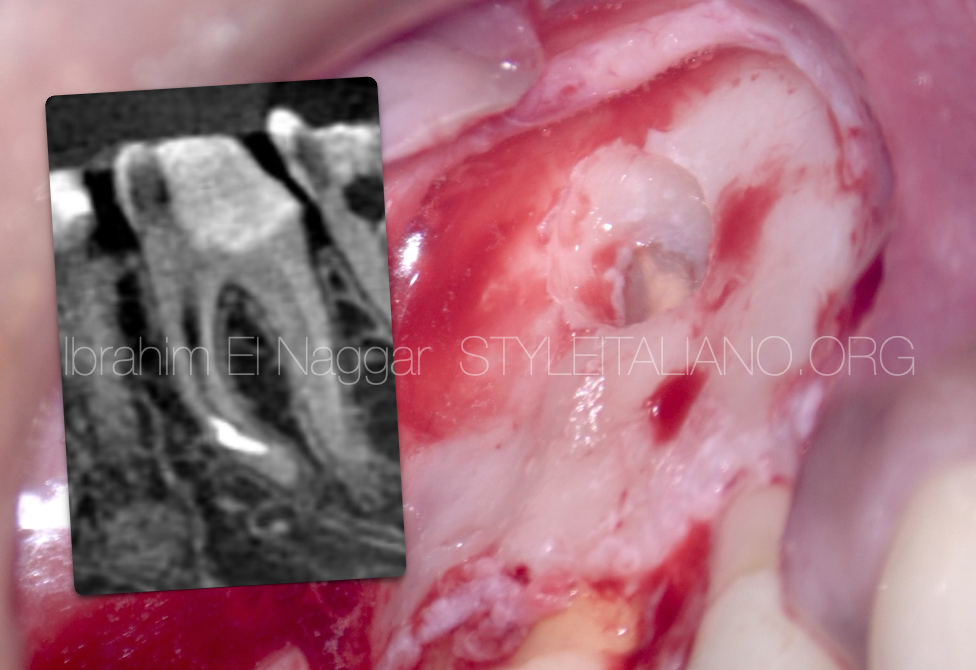
Fig. 2
CBCT was requested an in the SAGITTAL section it showed that the instruments are below the Curve .
Coronal Cut for checking the thickness of the cortical Bone.
Axial cut at the level of planned resection
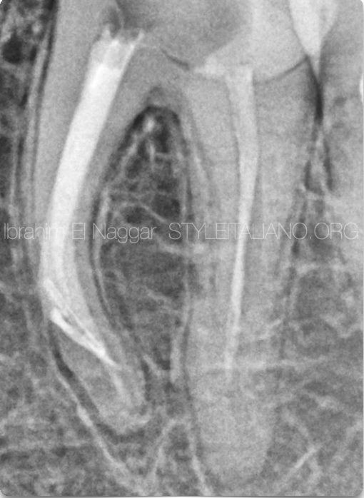
Fig. 3
First of all mesials were filled with SQUIRTING technique before the Surgery.
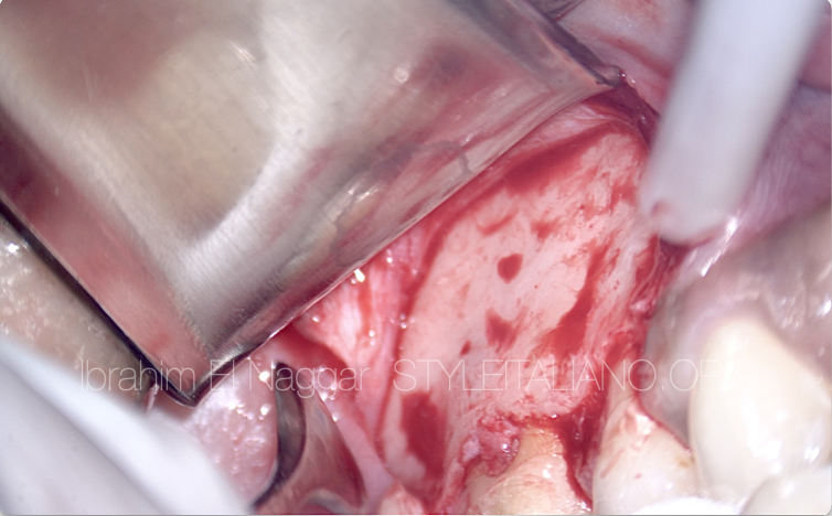
Fig. 4
Full thickness sulcular flap was performed
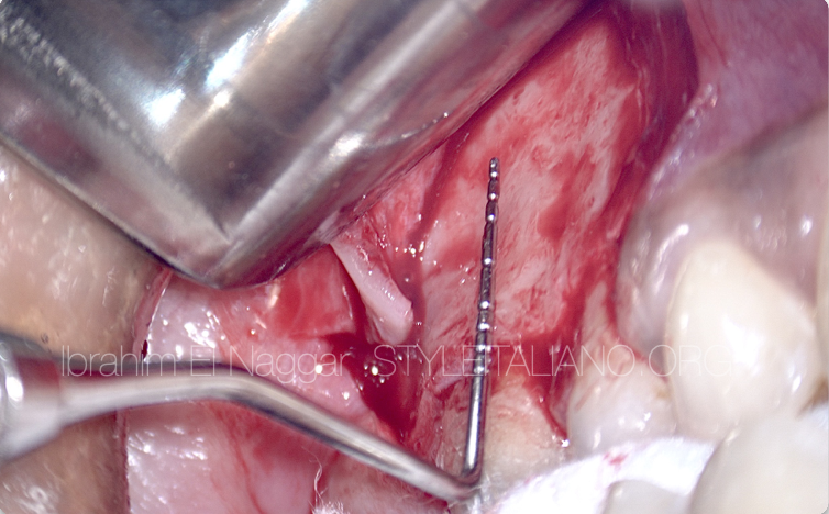
Fig. 5
Measurement using Periodontal Probe for the site of osteotomy
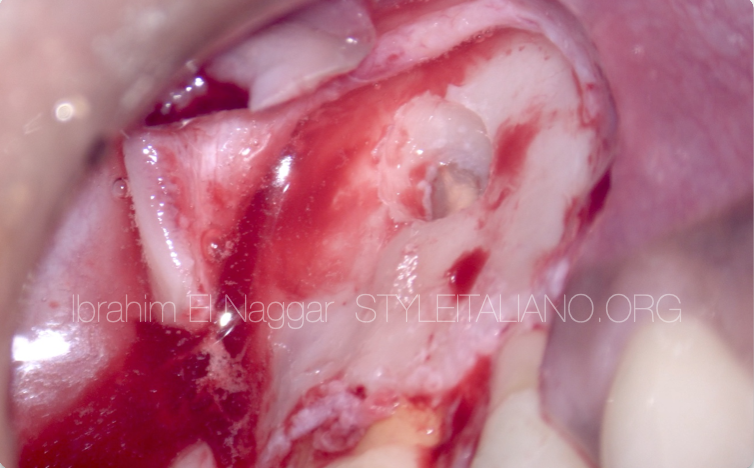
Fig. 6
Osteotomy site done Precisely based on CBCT
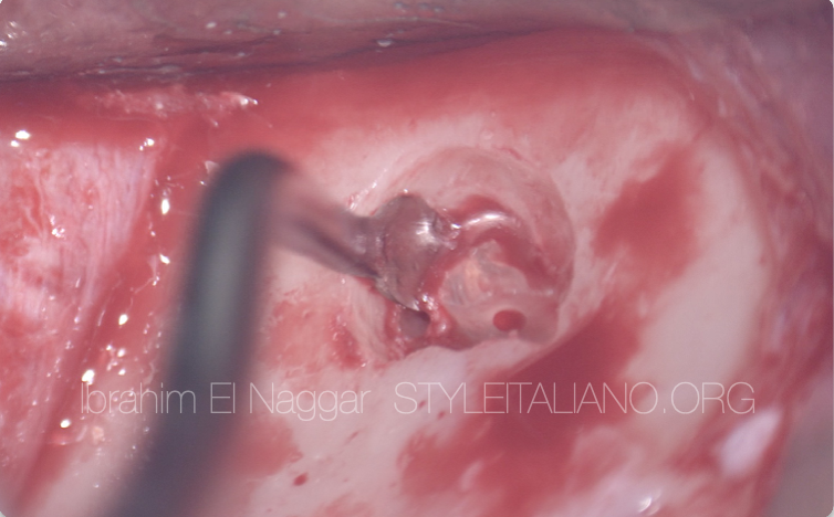
Fig. 7
Root End Resection and removal using the Curette
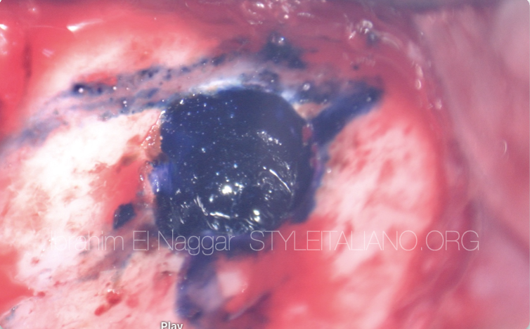
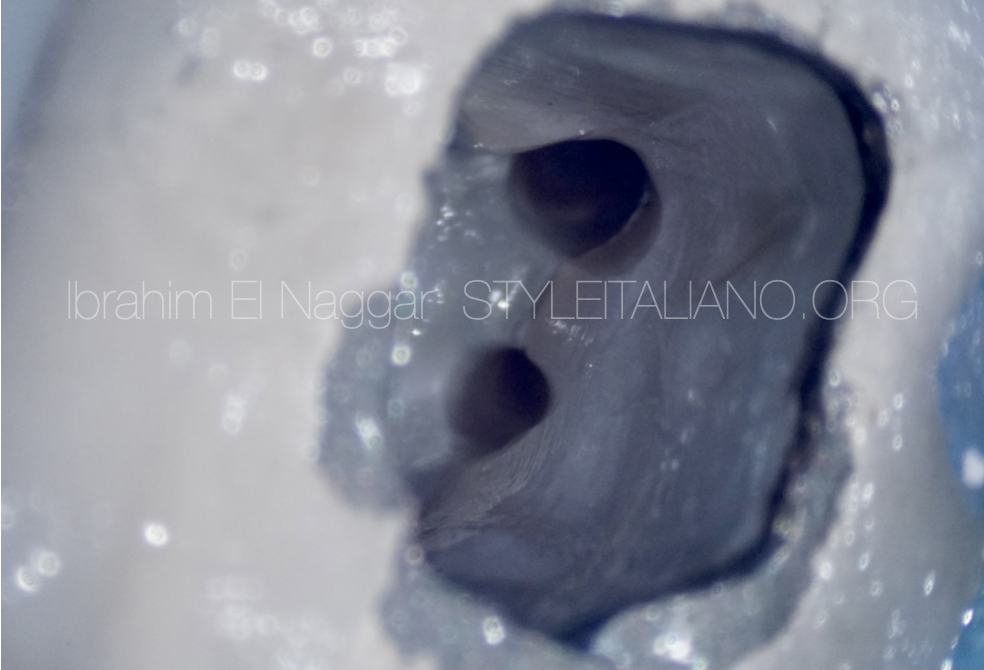
Fig. 8
Anatomy Identification Before the Retro Prep using Methylene Blue
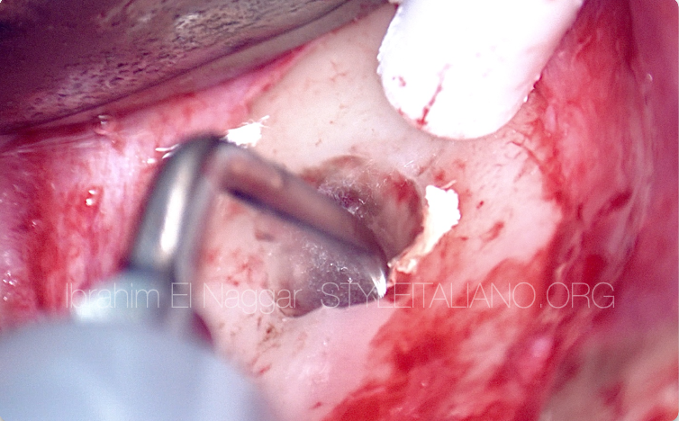
Fig. 9
Retro Prep Using the Ultrasonics
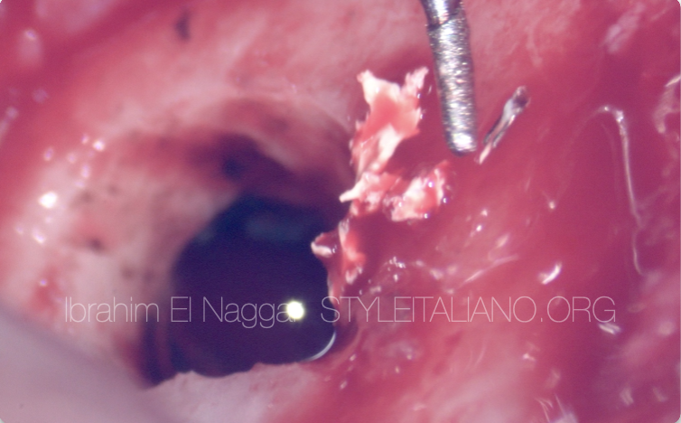
Fig. 10
File Removed
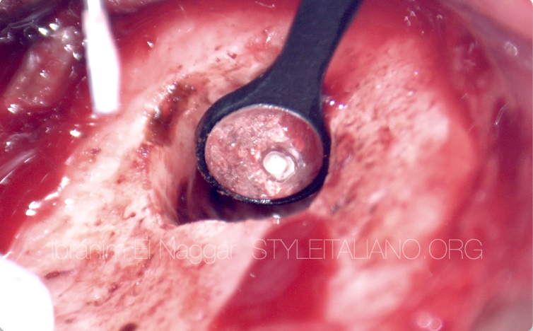
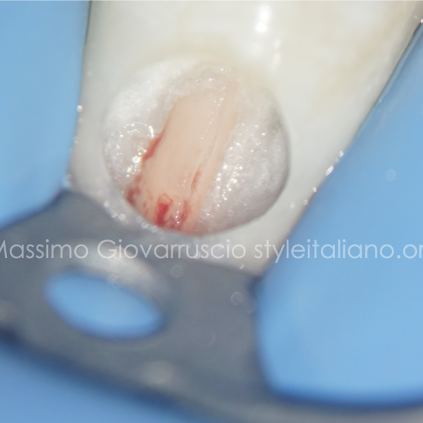
Fig. 11
Retro filling using Bioceramic Putty
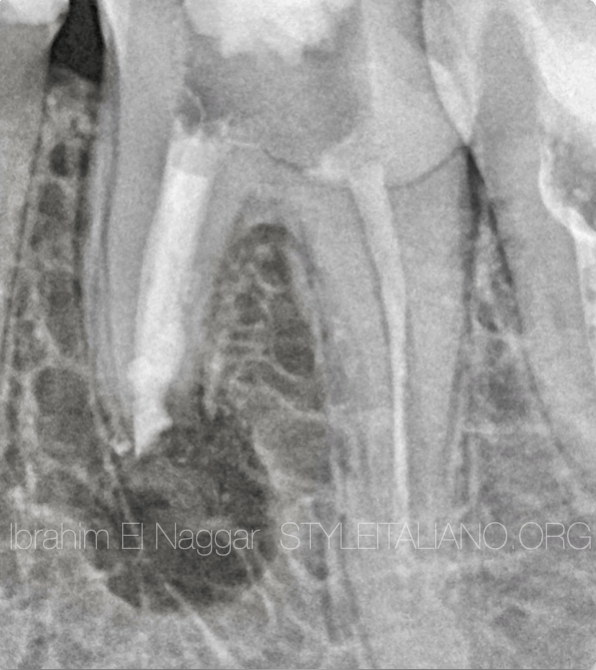
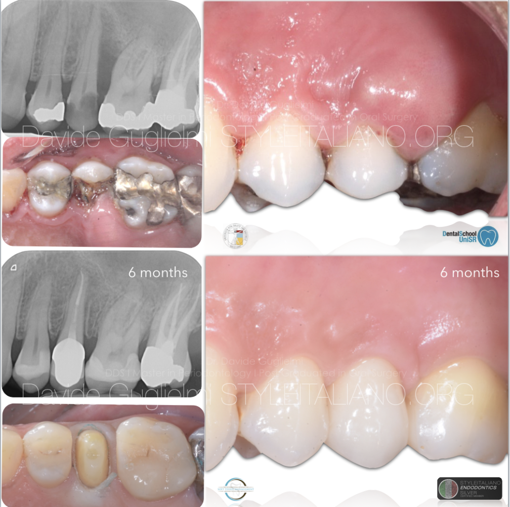
Fig. 12
Post Op X Ray
Conclusions
The Decision making during the therapy to interfere with SURGICAL on NON-Surgical approach is so important in term of success also the using of the right tools including the instrument and the Microscope is so important for this kind of treatment.
Bibliography
Kang M. Jung H.I. Song M. et al.Outcome of nonsurgical retreatment and endodontic microsurgery: a meta-analysis.
Clin Oral Investig. 2015; 19: 569-582
Tsesis I. Rosen E. Taschieri S. et al.Outcomes of surgical endodontic treatment performed by a modern technique: an updated meta-analysis of the literature.
J Endod. 2013; 39: 332-339
von Arx T. Jensen S.S. Hanni S. et al.Five-year longitudinal assessment of the prognosis of apical microsurgery.
J Endod. 2012; 38: 570-579
Saunders W.P. A prospective clinical study of periradicular surgery using mineral trioxide aggregate as a root-end filling.
J Endod. 2008; 34: 660-665
Song M. Shin S.J. Kim E. Outcomes of endodontic micro-resurgery: a prospective clinical study.
J Endod. 2011; 37: 316-320