Middle Mesial Canal How to Detect it
24/03/2022
The Community
Warning: Undefined variable $post in /home/styleendo/htdocs/styleitaliano-endodontics.org/wp-content/plugins/oxygen/component-framework/components/classes/code-block.class.php(133) : eval()'d code on line 2
Warning: Attempt to read property "ID" on null in /home/styleendo/htdocs/styleitaliano-endodontics.org/wp-content/plugins/oxygen/component-framework/components/classes/code-block.class.php(133) : eval()'d code on line 2
Article by: Elias Abboud, Damascus Syria
The mandibular first molar seems to be the tooth that most often requires an endodontic procedure (vital pulp capping, pulpotomy, root canal therapy) therefore, its morphology has received a great deal of attention.
The canals in the mesial root are the mesiobuccal and mesiolingual canals. A middle mesial canal (MMC) sometimes is present in the developmental groove between the other mesial canals
Troughing into the groove between the mesiobuccal and mesiolingual orifices may be indicated to identify this canal
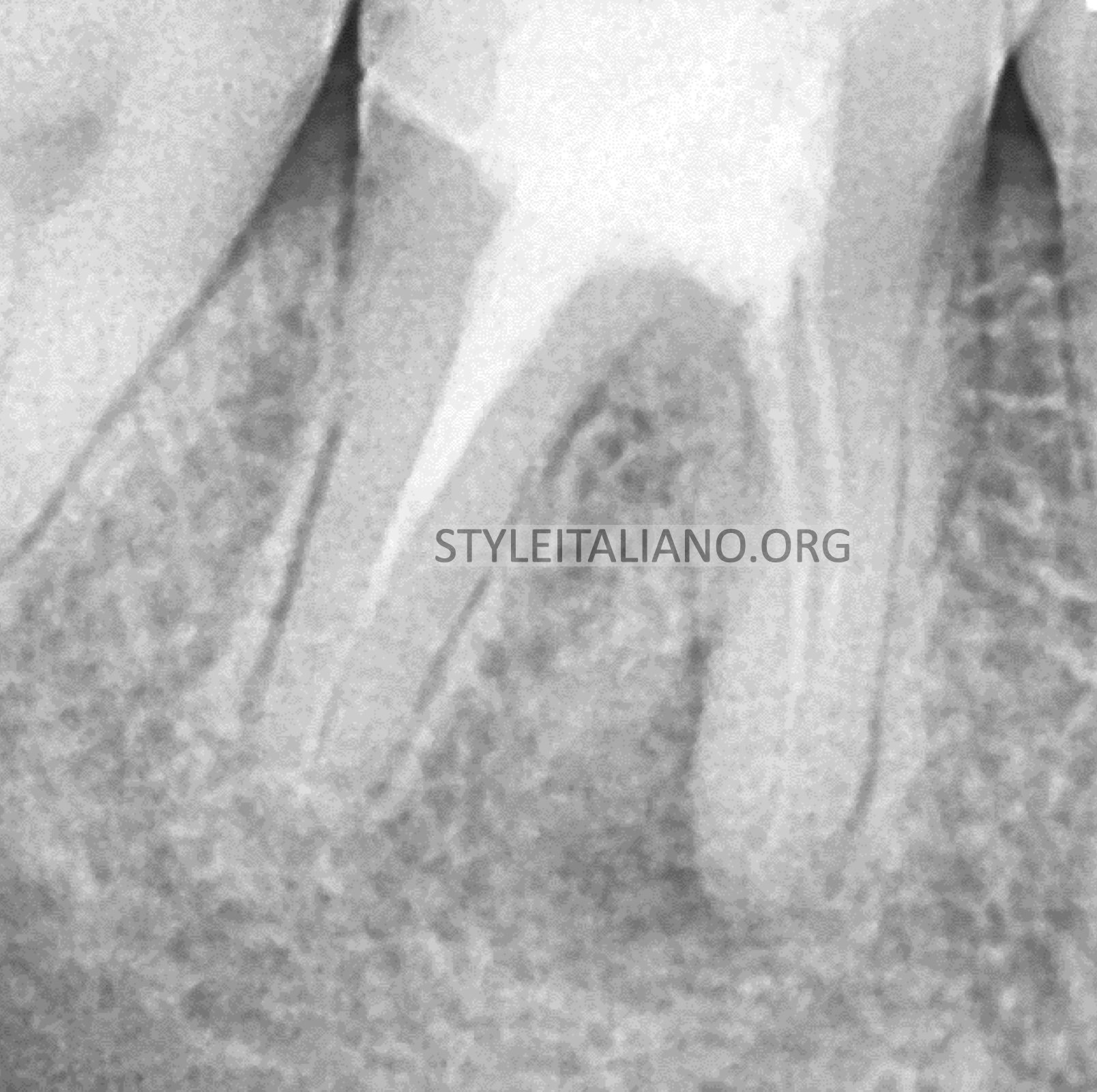
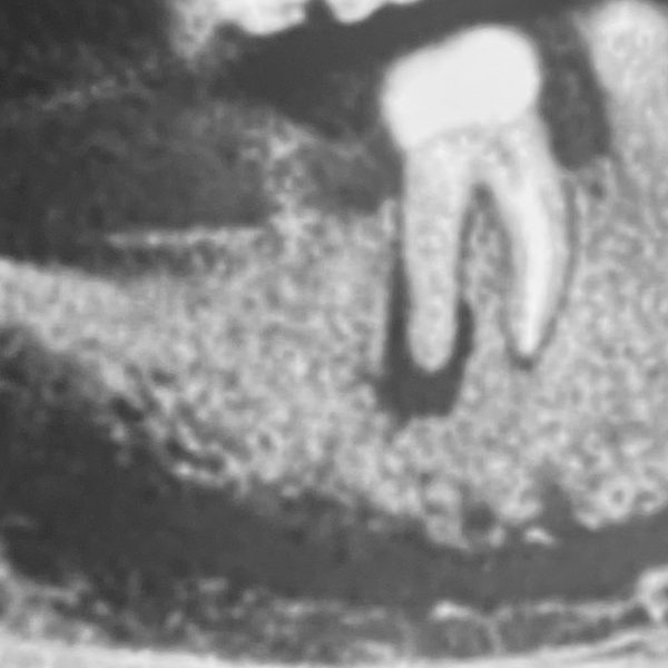
Fig. 1
A Pre Opertative X-Ray show a large lesion with very bad old treatment
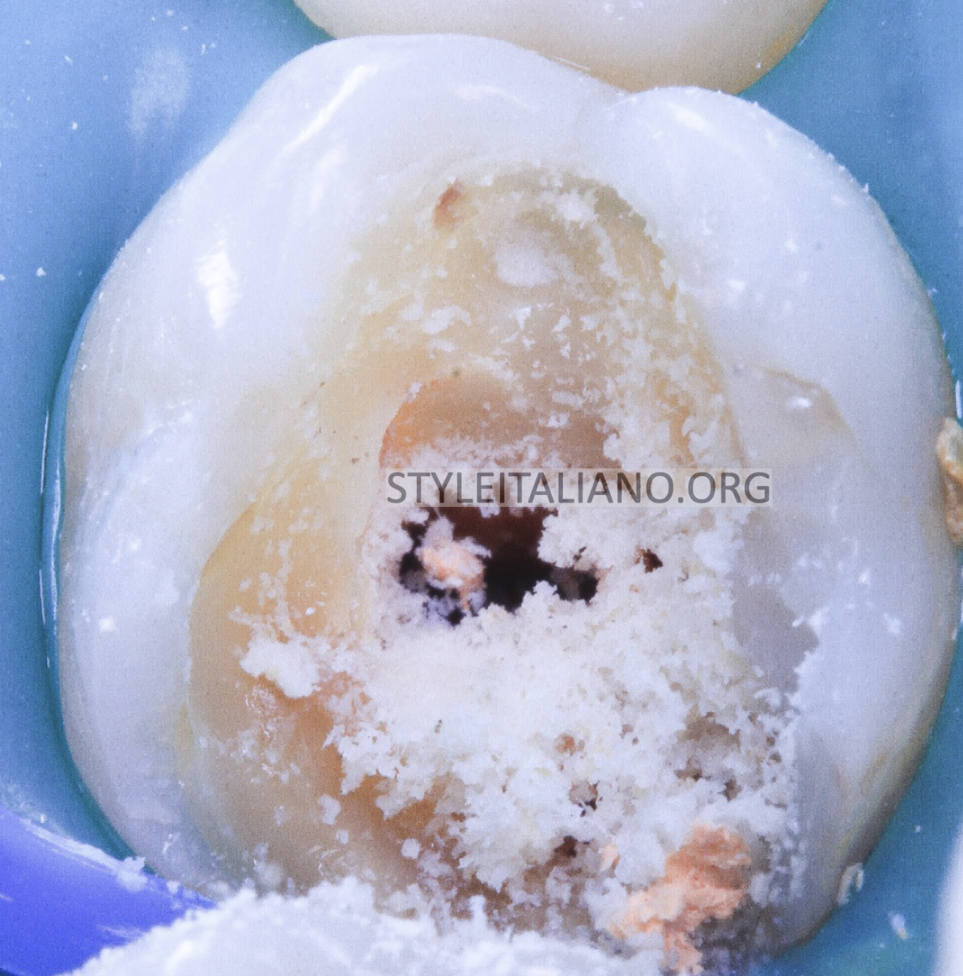
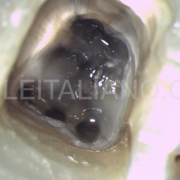
Fig. 2
Starting of the procedure with rubber dam
Removing all caries and old materials before start the cleaning and shapping of the canals
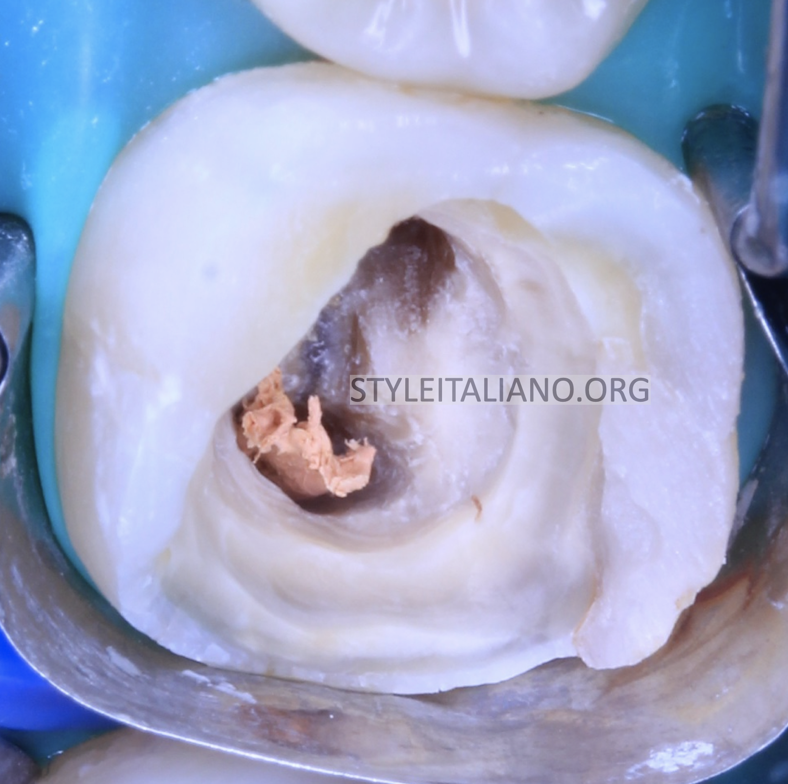
Fig. 3
Build up the missing wall before start to make sure that all irrigation will be saved
In the box of the access cavity
Here we do not need to make perfect contact point because the tooth prepare for a crown
Just rewalling the missed wall
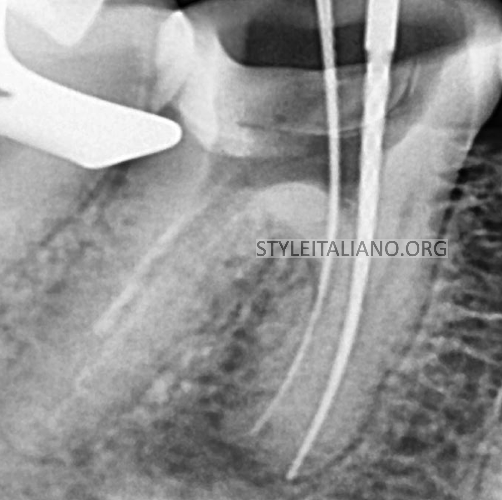
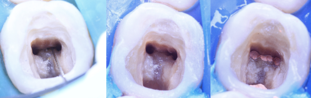
Fig. 4
Start to remove old obturation in the mesial root and reach the apex
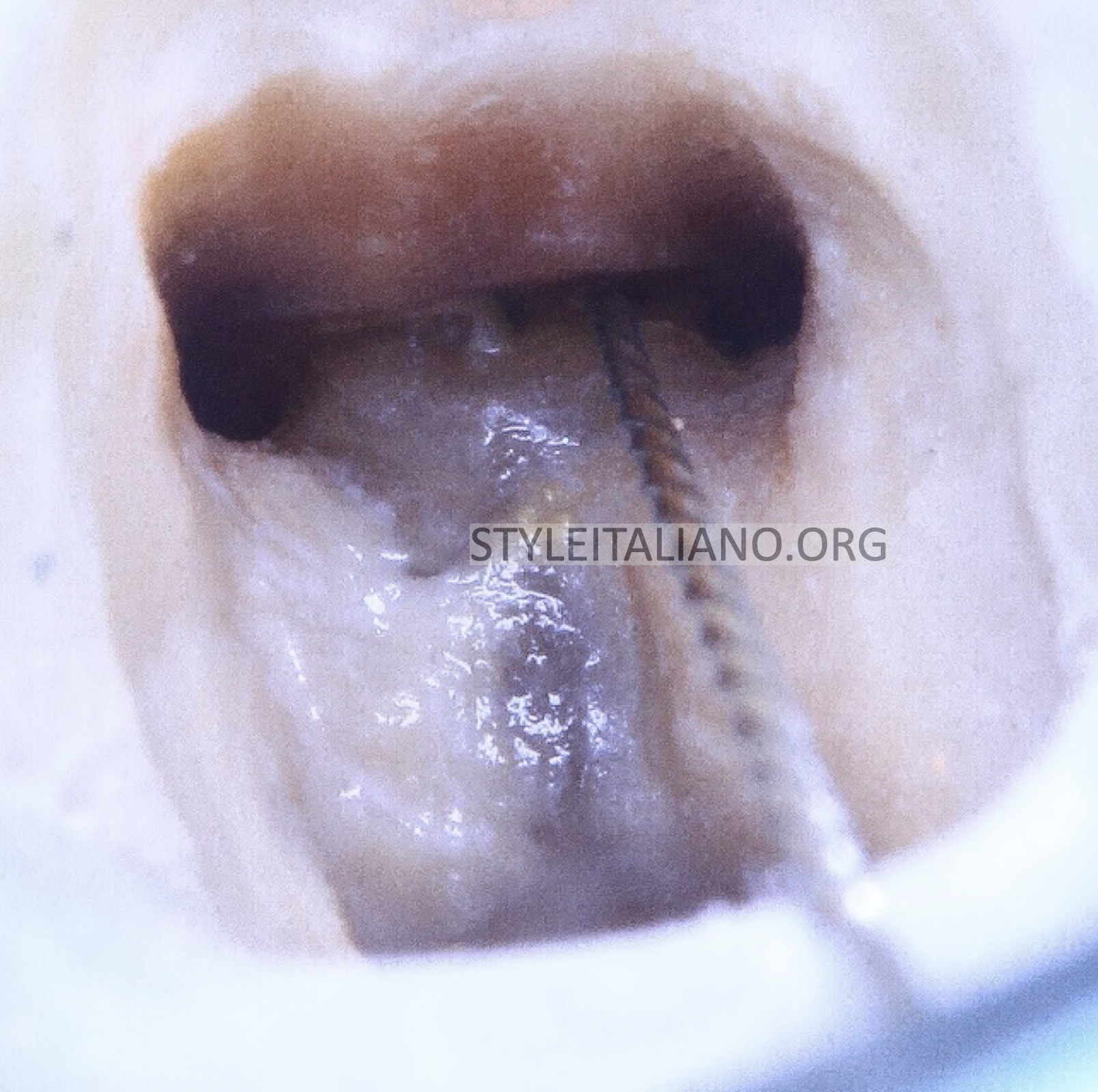
Fig. 5
After cleaning the isthmus between the mesial canals and catching the middle mesial canal
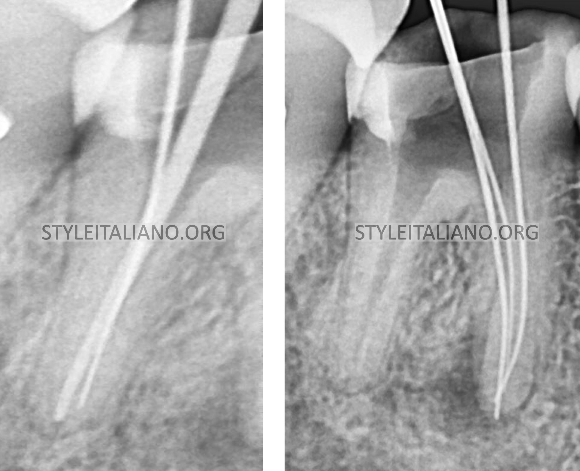
Fig. 6
We notice that MMC were joined with the mesiobuccal canal and detected
Very deep split in the distal root
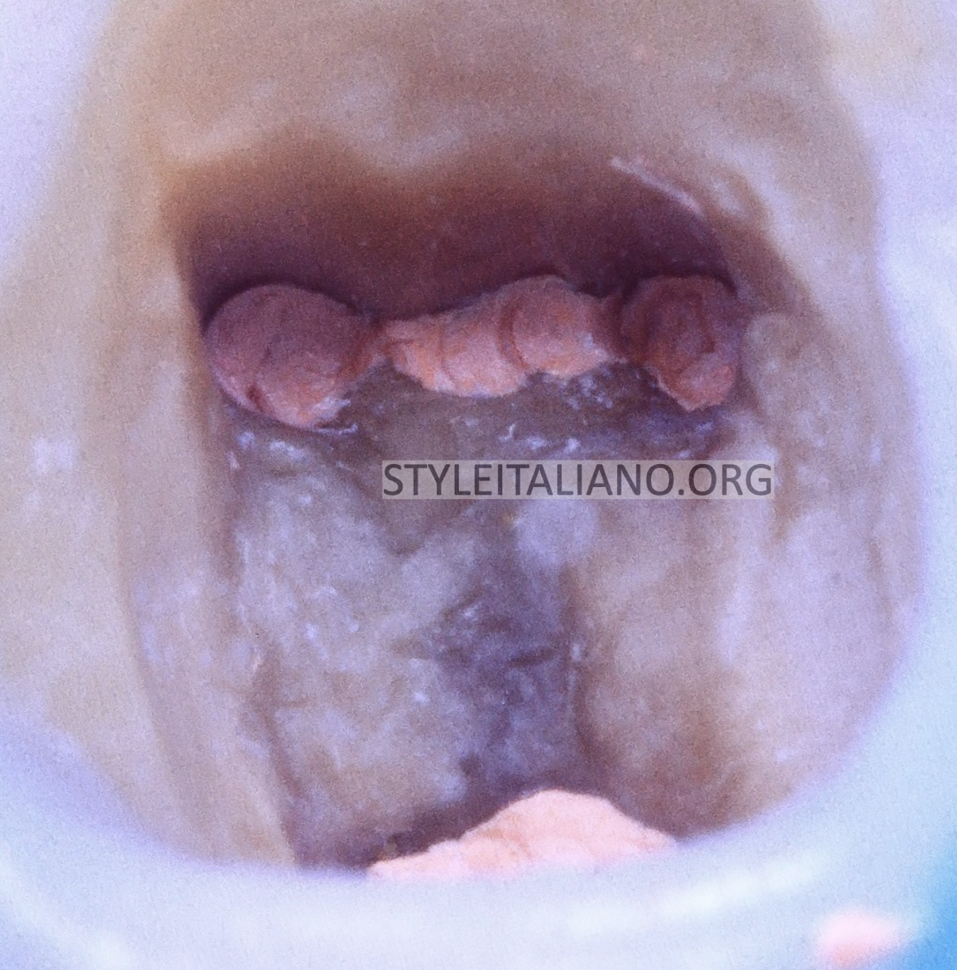
Fig. 7
After obturation by WVC technique and cleaning the access cavity
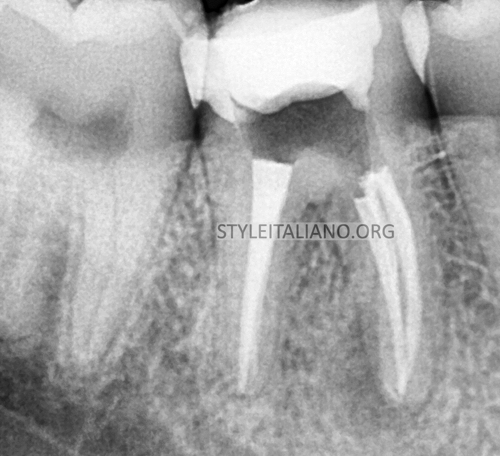
Fig. 8
Post op X-Ray
Conclusions
The anatomy is very important and we have to study the case very well
CBCT can help us to detect secondary canals like MMC with the using of
Magnification and illumination
Bibliography
1- Zhang R, Wang H, Tian YY, et al: Use of cone-beam
computed tomography to evaluate root and canal
morphology of mandibular molars in Chinese individuals,
Int Endod J 2011;44: 990
2- Akbarzadeh N, Aminoshariae A, Khalighinejad N, et al:
The association between the anatomic landmarks of the
pulp chamber floor and the prevalence of middle mesial
canals in mandibular first molars: An in vivo analysis, J
Endod 2017;43: 1797.
3-Bansal R, Hedge S, Astekar M. Morphology and
prevalence of middle canals in the mandibular molars: a
systematic review, J Oral Maxillofac Pathol 2018;22: 216.
4-Navarro LF, Luzi A, Garcia AA, et al: Third canal in the
mesial root of permanent mandibular first molars: review
of the literature and presentation of 3 clinical reports and
2 in vitro studies, Med Oral Patol Oral Cir Bucal 2007;12:
E605.