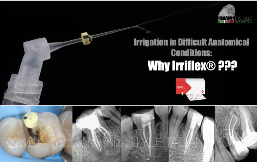
Irrigation in Difficult Anatomical Conditions: Why IrriFlex®?
08/03/2021
Xavier-Fructuós Ruiz
Warning: Undefined variable $post in /var/www/vhosts/styleitaliano-endodontics.org/endodontics.styleitaliano.org/wp-content/plugins/oxygen/component-framework/components/classes/code-block.class.php(133) : eval()'d code on line 2
Warning: Attempt to read property "ID" on null in /var/www/vhosts/styleitaliano-endodontics.org/endodontics.styleitaliano.org/wp-content/plugins/oxygen/component-framework/components/classes/code-block.class.php(133) : eval()'d code on line 2
IrriFlex® has appeared on the market to change our current irrigation protocols. Its main advantages like its flexibility, its ability to reach the apical part of the root canal system adapting to any curvature, its size and its simplicity to use it have made from this needle a revolution in disinfection if compared to traditional stainless steel needles. IrriFlex® is designed to improve our irrigation procedures not only in difficult anatomical conditions but also during our day-to-day cases.
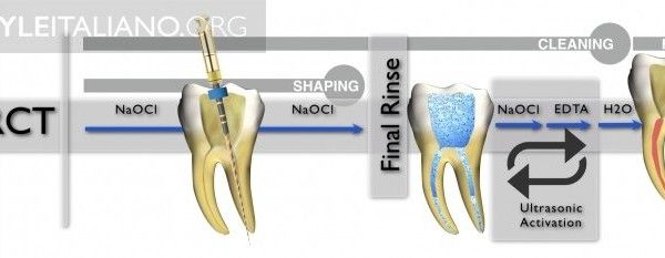
Root canal treatment consists in the mechanical preparation of the root canal system followed by a proper cleaning and disinfection to finally perform a three-dimensional obturation. The main goal of root canal therapy is to eliminate intracanal bacteria or at least achieve its maximum reduction to prevent or heal apical periodontitis.
Although mechanical preparation is an important step during root canal treatment, several articles have reported that most parts of the root canal system remain untouched due to the nature of root canal anatomy. Therefore, as shaping by itself cannot guarantee a total cleaning of the root canal system which is plenty of canal fins and isthmus that can harbor debris remnants and bacteria, irrigation plays a major role to achieve endodontic success.
The most commonly used procedure for root canal disinfection consists in a metal needle and a syringe which are in charge of delivery the irrigant solution. In the coronal part of the root canal space, there is no argue that all needles may work. Nevertheless, the apical portion of teeth represent a greater challenge due to the major presence of anatomical variations (apical ramifications and lateral canals).
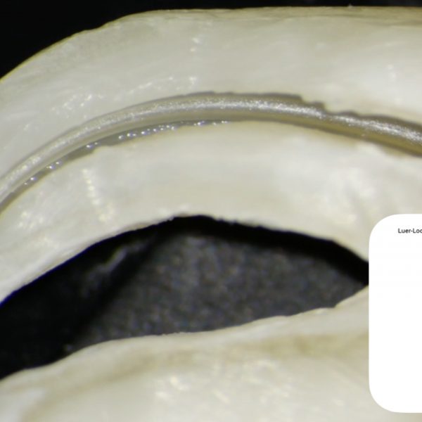
Recently, a new needle called IrriFlex® (Produits Dentaires, Vevey, Switzerland) has appeared on the endodontic market which is called to revolutionize our current irrigation protocols. IrriFlex® is made of polypropylene plastic which allows an easy insertion of the needle into the deep apical areas of the root canal system without pressure, favoring the irrigant delivery.
Irriflex® consists of a 2-side vented needle with a safe end that guarantees a balanced irrigant delivery avoiding an excessive force and the possible extrusion of the irrigant to the periodontal tissues. It is a 30G needle with a 4% constant taper which fits the majority of our root canal preparations after shaping is performed.
In this article, different clinical situations will be described in which IrriFlex® becomes an easier, more comfortable and safer needle compared to traditional metal needles and which improves our irrigation procedures.
Apical curvatures
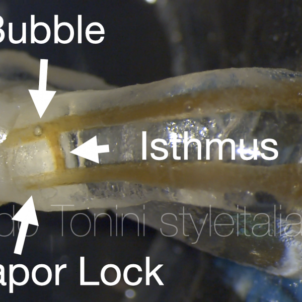
One of the major disadvantages of using metal needles is the impossibility to deliver the irrigant in the apical portion of the root canal. What’s more, as stainless steel needles are not able to penetrate in the most apical areas and follow accented curves, the `vapor lock´ effect is increased. The `Vapor Lock´ is a physiological effect that implies an air entrapment in the apical part of the root canal while performing irrigation in the coronal and middle thirds, which avoids contact from the irrigant with the apical root canal. IrriFlex® can overcome this problem because it easily reaches the apical part of the root, mostly adapting to any shape and delivering the irrigant up to working length if desired, which may increase our disinfecting procedure.
Traditional metal needle vs IrriFlex® plastic needle
The following in-vitro video with resin blocks shows how a conventional stainless steel needle is unable to reach the apical third and get stuck into the curvature, while IrriFlex® perfectly follows the anatomy reaching the apical part.
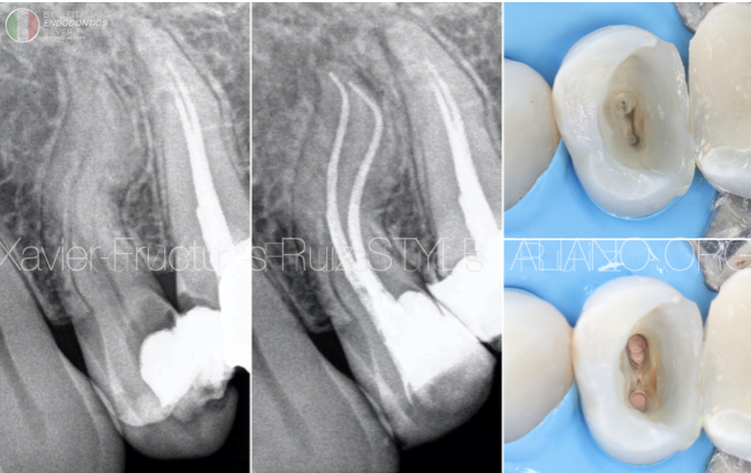
Fig. 1
Double curvatures
If IrriFlex® represents a great advantage when delivering the irrigant in presence of apical curvatures its efficacy in cases of double curvatures is incredibly higher if compared to regular metal needles. The following case shows a maxillary first premolar with a double curvature which was instrumented with Trunatomy system up to 26.03. IrriFlex® was used to irrigate the narrow root canals up to desired working length due to its flexibility.
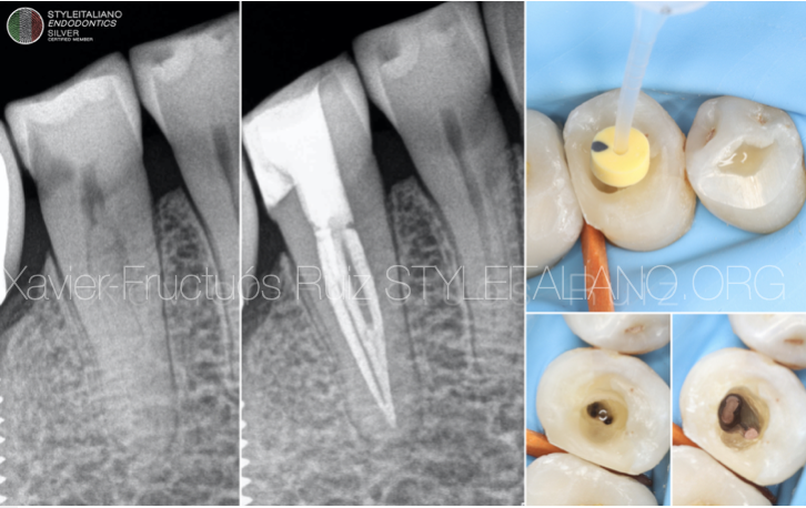
Fig. 2
Deep splits
One of the main advantages of IrriFlex® is the possibility of pre-bend the needle as desired in cases where multiple canals are encountered as for example in three-rooted maxillary premolars or mandibular premolars with deep splits. The following case shows a mandibular premolar with a deep split in the coronal third where 3 root canals are found. IrriFlex® permits to easily irrigate the different root canals just pre-bending the needle which is extremely soft and allows its constant manipulation without being damaged.
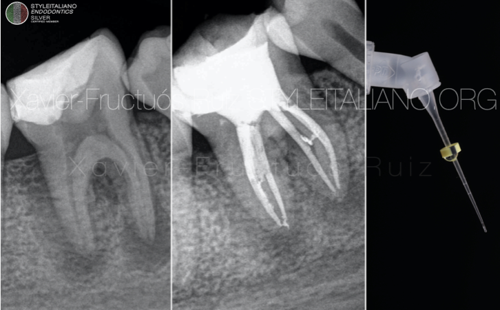
Fig. 3
Anatomical variations: isthmuses and lateral canals
Cleaning and disinfection of root canal inter-communications is one of the most challenging procedures during root canal treatment. Isthmuses and lateral canals represent a potential space for the reservoir of microbes and the accumulation of debris produced from mechanical preparation of the root canal system. The following case shows a first mandibular molar in which after instrumentation, continuous irrigation with IrriFlex® was performed. After root canal obturation, observe how the anatomical complexity of the root canal system is revealed showing the presence of a lateral canal at the mesial root and the presence of an isthmus at the distal root.
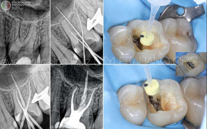
Fig. 4
Second mesiobuccal canals (MB2)
Another main advantage of IrriFlex® is that it easily penetrates into a narrow, curvy and complex canal as it is the 2nd mesiobuccal canal of maxillary molars. Achieving continuous patency in such canals is essential because MB2 has been described as a difficult canal to be scouted, negotiated and finally instrumented. Therefore, IrriFlex® allows the clinician to perform a constant irrigation having the canal free of detritus which may impede a proper instrumentation and thus irrigation into the apical part. The following case describes a regular root canal treatment in a first maxillary molar with the presence of a narrow MB2 canal that merges with the main mesiobuccal canal in the middle third, which was continuously irrigated with an IrriFlex® needle.
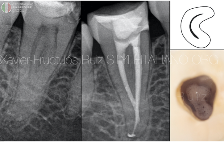
Fig. 5
C-shaped canal system
Second mandibular molars are the most prevalent teeth where c-shaped anatomy is encountered. The anatomical configuration consists in the presence of isthmuses and fins that connect the main root canal. This morphology presents a great challenge in all steps of root canal treatment specially during root canal disinfection.
In such cases, IrriFlex® is without any doubt a useful tool which can explore the C-shaped anatomy performing a continuous delivery of the irrigant while activating it with agitation as if manual dynamic activation was performed with a gutta-percha point.
The following case shows the initial and post-operative radiograph of a root canal treatment in a mandibular second molar with a C1 configuration which was instrumented with Wave One Gold 20.07 and where continuous irrigation with NaOCl 4.25% using IrriFlex® was performed. Check the status of the pulp chamber after performing the agitation of the irrigant.
Conclusions
IrriFlex® has come to stay and change our current irrigation protocols during root canal treatment. The use of this flexible needle implies a series of advantages when compared to traditional metal needles such as an easy insertion into the apical third of the root canal system adapting itself to any curvature present while delivering the irrigant in a safe way. Furthermore, IrriFlex® allows the clinicians to pre-bend the needle according to the desired anatomy of teeth and favours a continous agitation of the irrigant avoiding the undesired `Vapor Lock´ effect while removing all debris and remnants from the root canal system.
Bibliography
1. Boutsioukis C, Verhaagen B, Versluis M, Kastrinakis E, Wesselink PR, van der Sluis LWM. Evaluation of Irrigant Flow in the Root Canal Using Different Needle Types by an Unsteady Computational Fluid Dynamics Model. J Endod. 2010;36:875-79
2. Fan B, Cheung GS, Fan M, Gutmann JL, Fan W. C-shaped canal system in mandibular second molars: Part II--Radiographic features. J Endod. 2004;30:904-8.
3. Gazzaneo I, Vieira GCS, Pérez AR, et al. Root Canal Disinfection by Single- and Multiple-instrument Systems: Effects of Sodium Hypochlorite Volume, Concentration, and Retention Time. J Endod. 2019;45:736-41
4. Gu L sha, Kim JR, Ling J, Choi KK, Pashley DH, Tay FR. Review of Contemporary Irrigant Agitation Techniques and Devices. J Endod. 2009;35(6):791-804
5. Zuolo ML, Carvalho MC, De-Deus G. Negotiability of second mesiobuccal canals in maxillary molars using a reciprocating system. J Endod. 2015;41:1913-17
6. Tomaszewska IM, Skinningsrud B, Jarzębska A, Pękala JR, Tarasiuk J, Iwanaga J. Internal and external morphology of mandibular molars: An original micro-CT study and meta-analysis with review of implications for endodontic therapy. Clin Anat. 2018;31:797-811