Deep bifurcation in the palatal canal of an upper molar
01/10/2020
Omran Abdllatif Issa
Warning: Undefined variable $post in /var/www/vhosts/styleitaliano-endodontics.org/endodontics.styleitaliano.org/wp-content/plugins/oxygen/component-framework/components/classes/code-block.class.php(133) : eval()'d code on line 2
Warning: Attempt to read property "ID" on null in /var/www/vhosts/styleitaliano-endodontics.org/endodontics.styleitaliano.org/wp-content/plugins/oxygen/component-framework/components/classes/code-block.class.php(133) : eval()'d code on line 2
This clinical case shows the need of being very careful while we approach the palatal canal in Upper first molars, especially in the apical part .
Of course some studies have reported the presence of two canals in palatal root, but in this case there is one canal with apical bifurcation.
In this case I rely on two points to scout :
1- radiographic interpretation. I carefully read the x-ray during taking working length: the most important thing watching my K file being "not centered" sets my doubt higher that there is an apical variation.
2- Scouting. I insert a pre-curved 15 K-file and scout till I feel a slight catch, then I passively insert another k-file and take an x-ray.
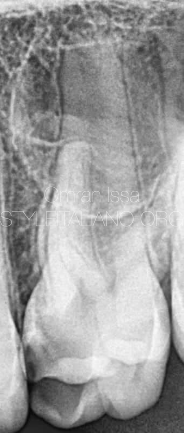
Fig. 1
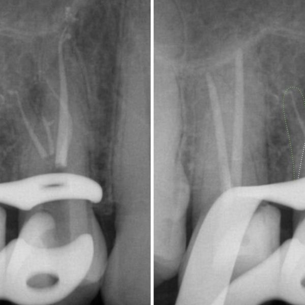
From preoperative x-ray the variation in apical area of palatal root is quite noticed
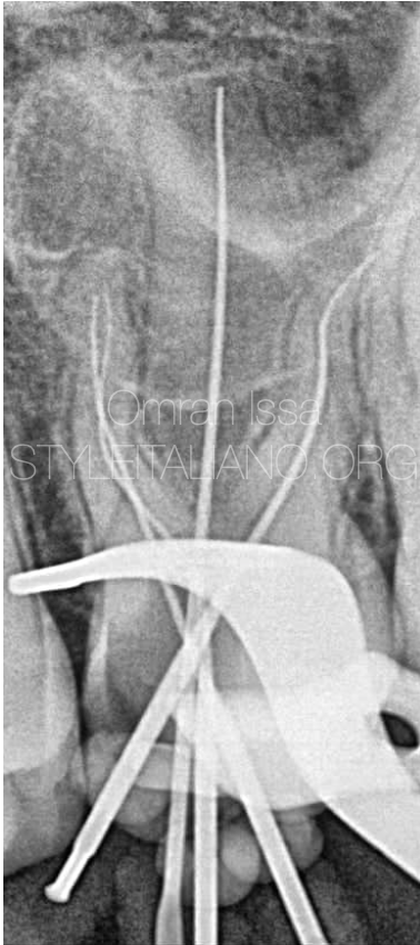
Fig. 2
Working length is taken by EAL and only one K file inserted in palatal canal
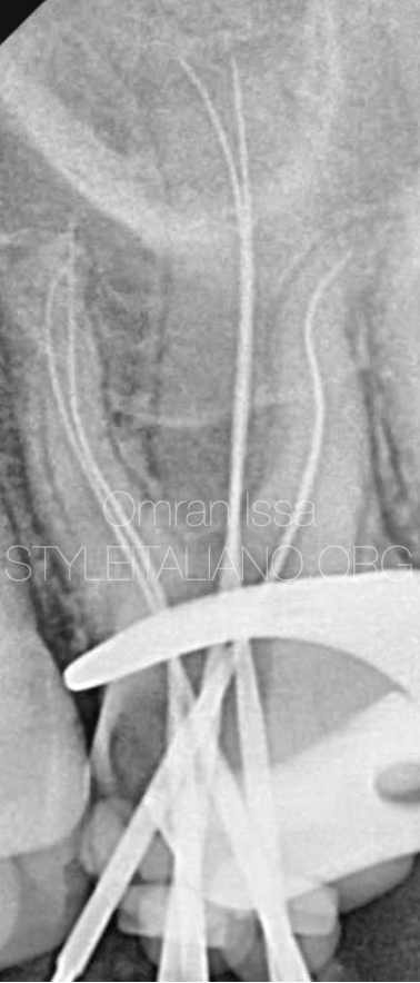
Fig. 3
Scouting the deep bifurcation by inserting another K file in the split
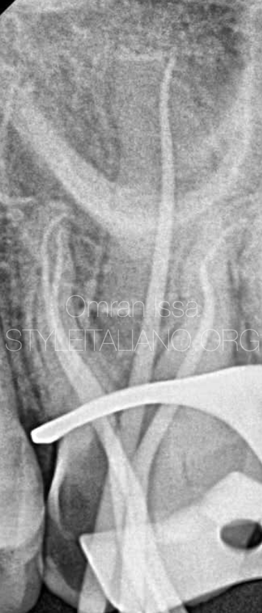
Fig. 4
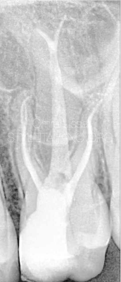
The presence of an apical curvature in obvious in both mesials and distal canals while cone fit is achieved
Fig. 5
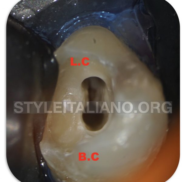
Microscopic view show the apical bifurcation of palatal canal. The picture was captured by zooming 25x.
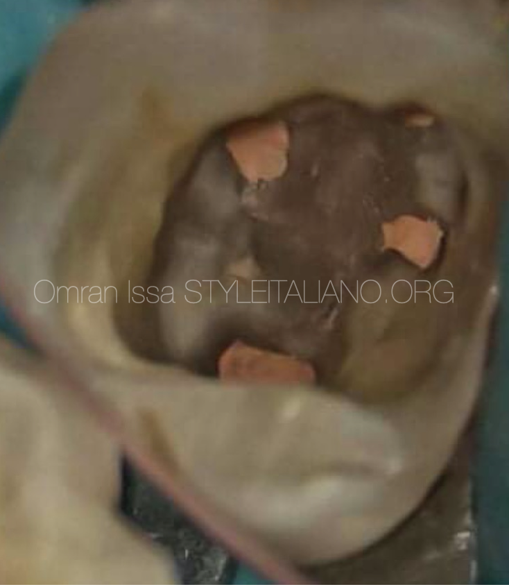
Fig. 6
Intra oral photo shows obturation of all canals, as we notice the location of MB2 canal is right opposite to the DB canal
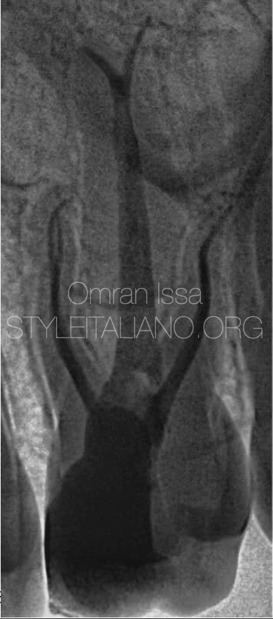
Fig. 7
The final x-ray shows obturation in invert
Conclusions
Radiographic interpretation is very important before any endodontic approach.
In particular, it is very helpful in cases in which the anatomy presents some peculiarity. Nevertheless, taking different angles of x-ray may be handy as well.
Bibliography
Nosrat A, Verma P, Hicks ML, Schneider SC, Behnia A, Azim AA. Variations of Palatal Canal Morphology in Maxillary Molars: A Case Series and Literature Review. J Endod. 2017;43(11):1888-1896. doi:10.1016/j.joen.2017.04.006