Daily Endo-Restorative
04/03/2021
Gabriele Ragucci
Warning: Undefined variable $post in /var/www/vhosts/styleitaliano-endodontics.org/endodontics.styleitaliano.org/wp-content/plugins/oxygen/component-framework/components/classes/code-block.class.php(133) : eval()'d code on line 2
Warning: Attempt to read property "ID" on null in /var/www/vhosts/styleitaliano-endodontics.org/endodontics.styleitaliano.org/wp-content/plugins/oxygen/component-framework/components/classes/code-block.class.php(133) : eval()'d code on line 2
As dentists 70% of the cases that come to us daily in our clinics are routine cases., cases too often underestimated where avoidable mistakes are made, making them complex
In my work I share and apply a multidisciplinary approach, dealing not only with the endodontic part but also with the restorative and in some cases surgical part this is a great help to me in making the best decisions in the patient's interest
The cause of conventional root canal treatment failure may be attributed to multiple variables. Although conventional practices in root canal treatment display a clinical success of 68–85% [1], among others, missing root canal anatomy, leading to residual intracanal infection [1], and compromised mechanical integrity, leading to fracture [2], have been considered as important causes of endodontic treatment failure.
Today's case is a lower molar with large caries that will require root canal treatment and indirect restoration, this just wants to be an example of how by following the correct protocols, we can obtain predictable results.
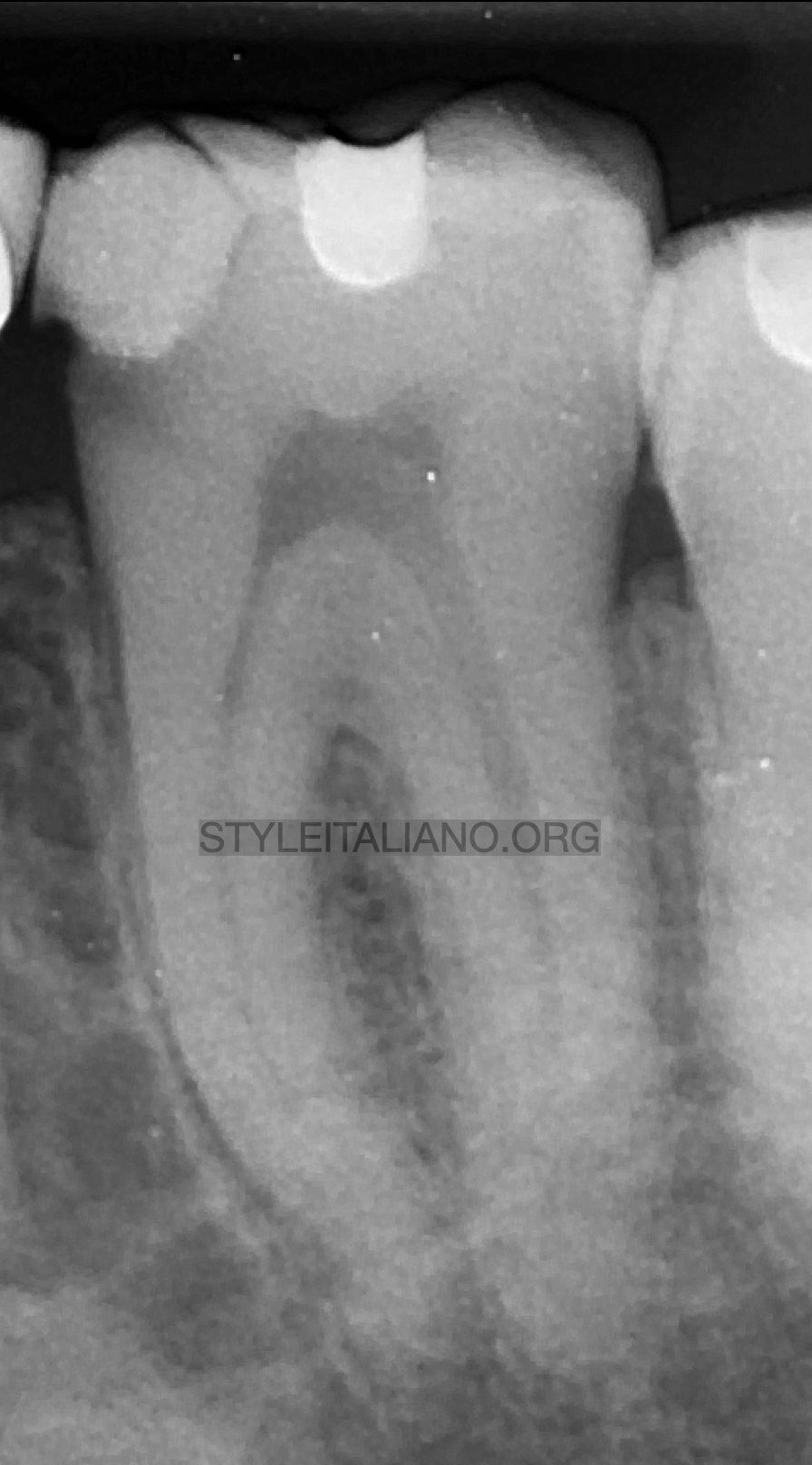
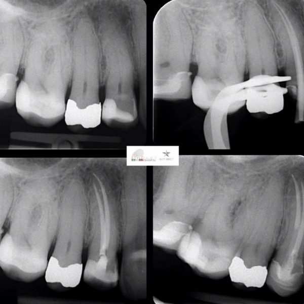
Fig. 1
Pre-Op Xray the show us a big decay in the mesial part of the tooth.
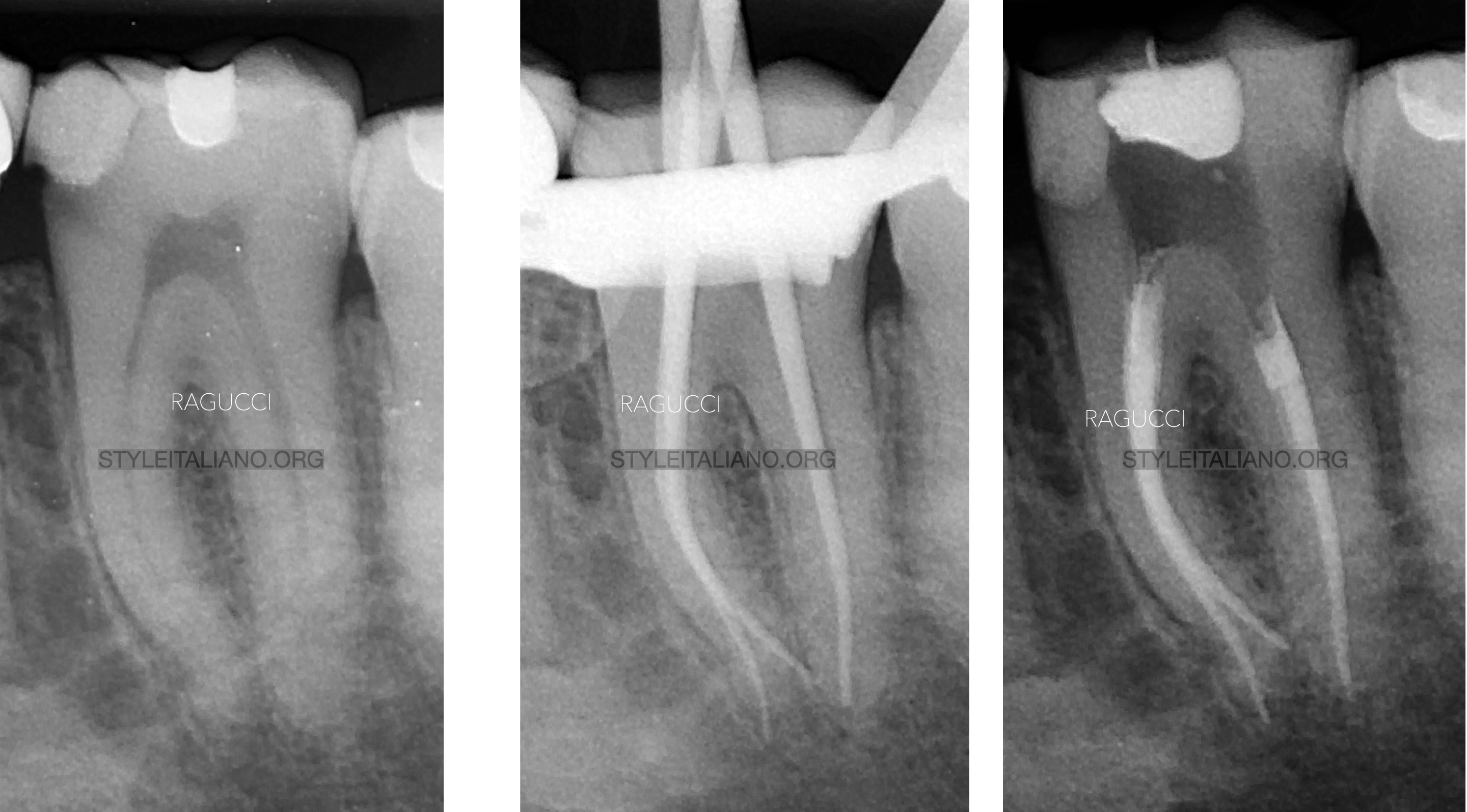
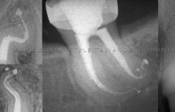
Fig. 2
Endodontics steps initial, cone-fit, final
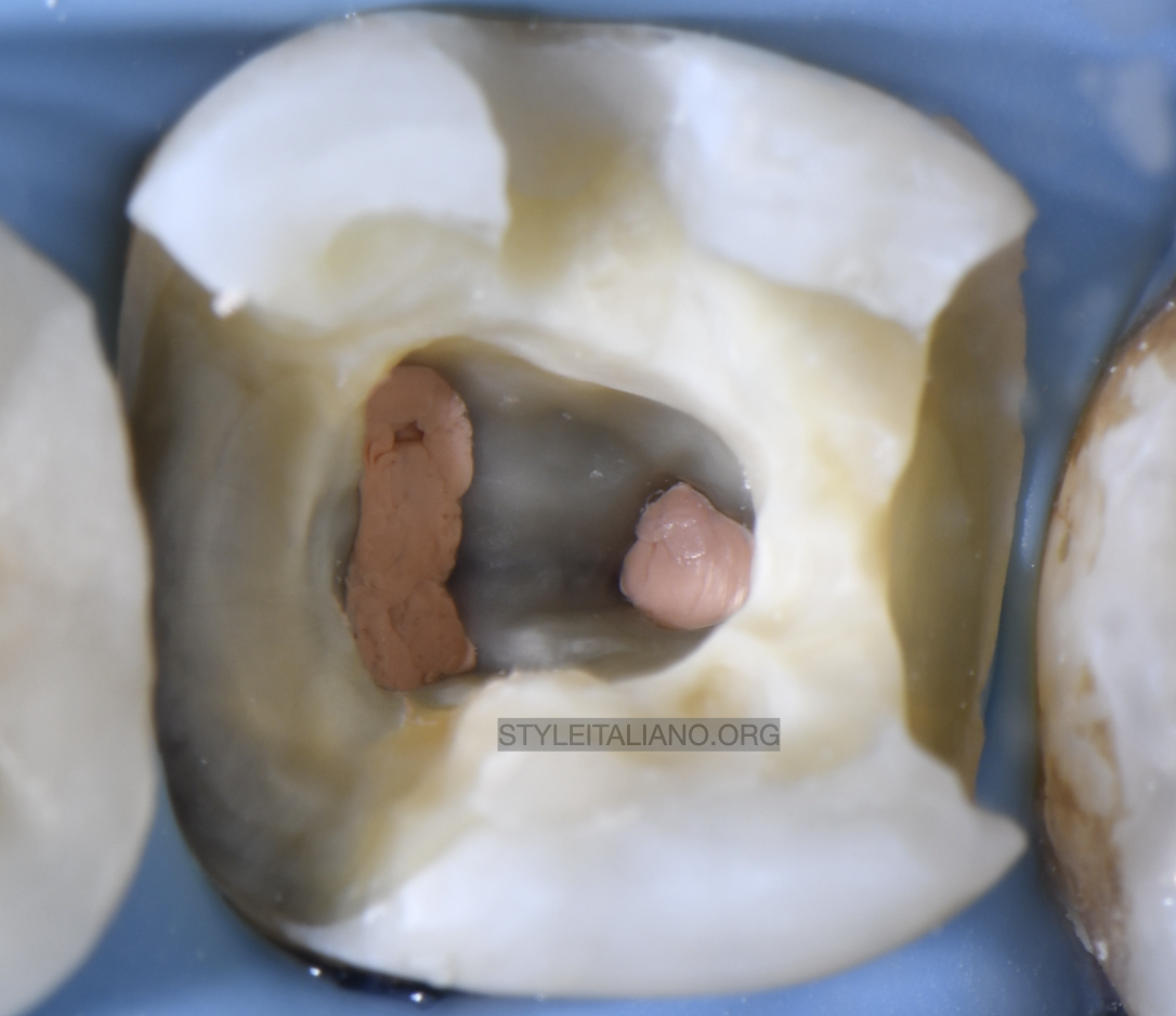
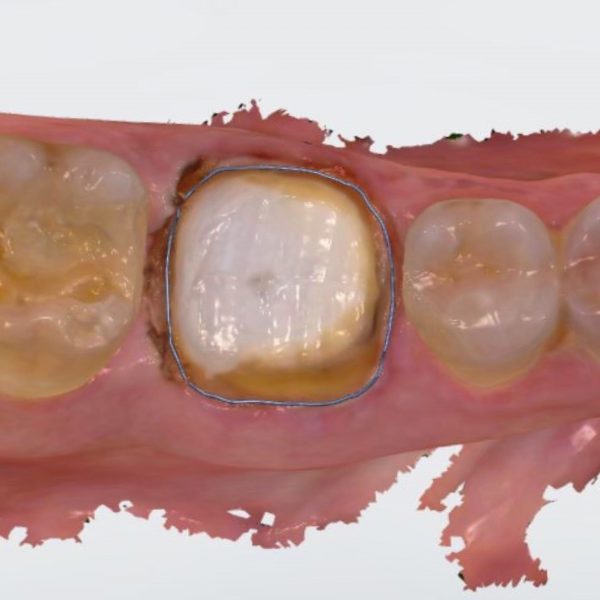
Fig. 3
Structural deficit affecting the central and lateral units of the middle and occlusal third of the cuspal residue with and without association of structural deficit of the distal marginal ridge and related occlusal views: covering total cerclage cusp.
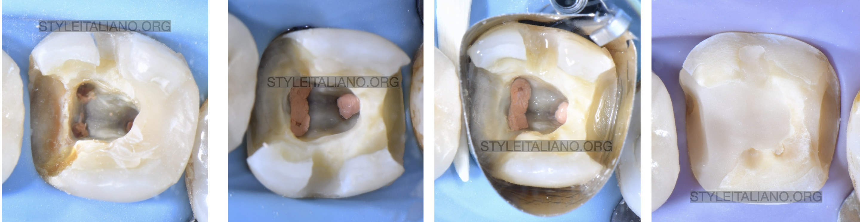
Fig. 4
Restorative steps
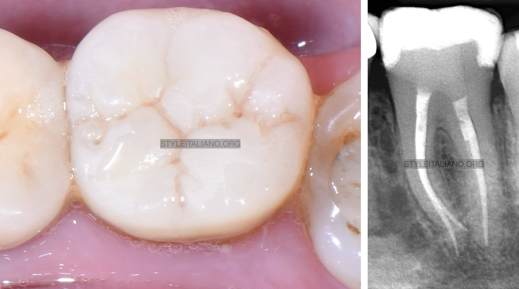
Fig. 5
Final restoration and Rx
Conclusions
Correct and simple protocols allow us predictable results, too often we see clear examples of iatrogenic dentistry, which makes simple cases complex.
Bibliography
- Ng Y, Mann V, Rahbaran S, Lewsey J, Gulabivala K. Outcome of primary root canal treatment: systematic review of the literature—Part 1. Effects of study characteristics on probability of success. Int Endod J. 2007;40:921–39.
- Kishen A. Mechanisms and risk factors for fracture predilection in endodontically treated teeth. Endod Top. 2006;13:57–83.
- Fichera G Re D Restauri estetico-adesivi indiretti: modello per diagnosi di con figurazione cavitaria.