Use of Itena Dentoclic Glass Fiber Post in the Restoration of endodontically treated tooth
18/02/2022
Davide Guglielmi
Warning: Undefined variable $post in /home/styleendo/htdocs/styleitaliano-endodontics.org/wp-content/plugins/oxygen/component-framework/components/classes/code-block.class.php(133) : eval()'d code on line 2
Warning: Attempt to read property "ID" on null in /home/styleendo/htdocs/styleitaliano-endodontics.org/wp-content/plugins/oxygen/component-framework/components/classes/code-block.class.php(133) : eval()'d code on line 2
Endodontically treated teeth (ETT) are more susceptible to biomechanics failures compared to vital teeth, mostly due to the amount of internal tooth structure that is removed during endodontic treatment and the loss of coronal hard tissue.
The prognosis of ETT is based not only on the quality of the endodontic treatment, but also on the sub- sequent restorative techniques.
The best way to restore ETT has been extensively discussed but it is still controversial, concerning the best type of final restoration. Conventional restoration modality involves fabricating a full-coverage crown with a metal-post, as this was believed to provide better protection and reinforcement of the remaining tooth structure. However, complete crown restoration usually also requires extensive tooth preparation and new occlusal schemes. In addition, the loss of anatomic structures such as cusps, ridges and the pulp chamber roof may decrease the strength of the remaining tooth.
Furthermore the preservation of an healthy tooth structure must always remain the main goal of restorative dentistry.
The bio-mechanical analysis of the residual healthy tooth substance, the proven reliability of bonding techniques and the availability of aesthetic, high performance restoration materials have lead to a review of the treatment paradigm.
Therefore, with recent advances in bonding technologies, adhesively bonded ceramic inlay or on-lay restorations have been suggested in several studies.
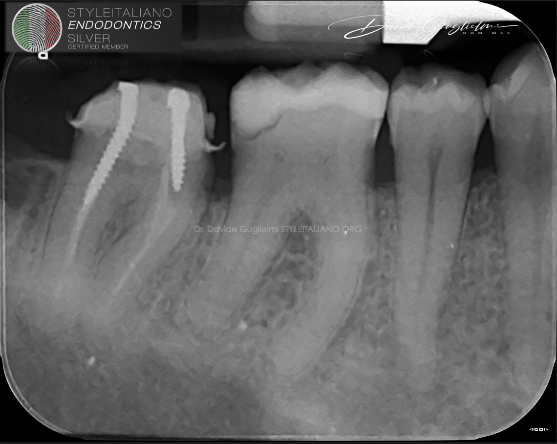
Fig. 1
Tooth 4.7 was symptomatic on percussion.
The periapical preliminary X-ray showed a poor endodontic primary treatment with short obturation in the distal root and in both mesial canals.
Furthermore a radiolucency was noticed on both roots.
Nonetheless, a leaking cast post and core on the lingual limit of the restoration was present.
A diagnosis of symptomatic apical periodontitis was established.
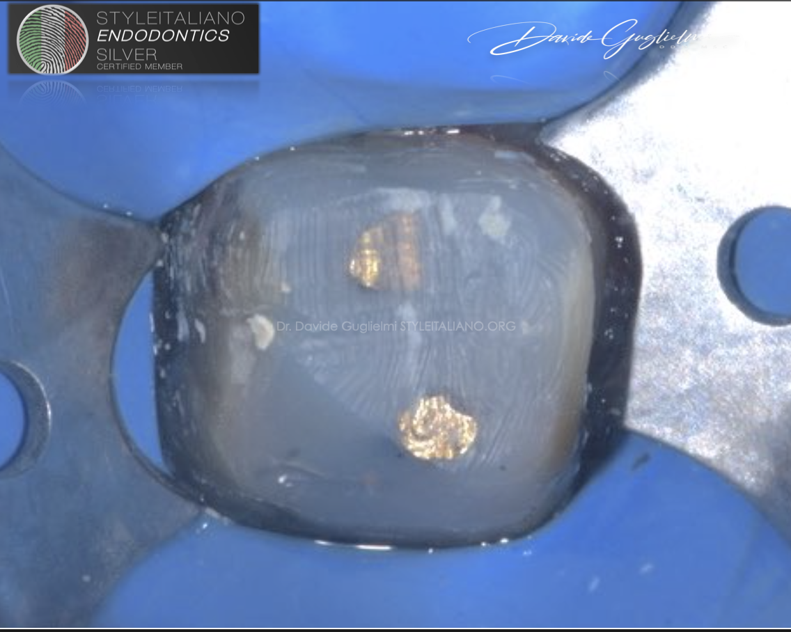
Fig. 2
A Non Surgical root canal retreatment (NSRCrT) was planned prior to a full coverage crown.
The tooth was isolated with a 2A clamp and a NicTone medium-weight rubber dam.
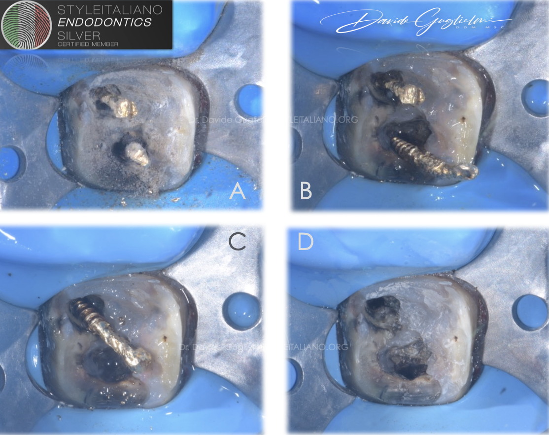
Fig. 3
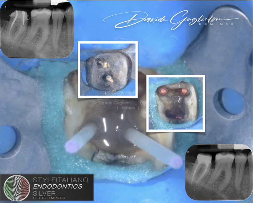
Screws were disassembled using ET18 ultrasonic tip (A).
The tip worked in counterclockwise direction around both distal (B) and mesio-lingual (C) posts in order to remove them.
This technique allowed the unscrewing of the posts with maximum preservation of the dental tissue.
The posts removed (D).
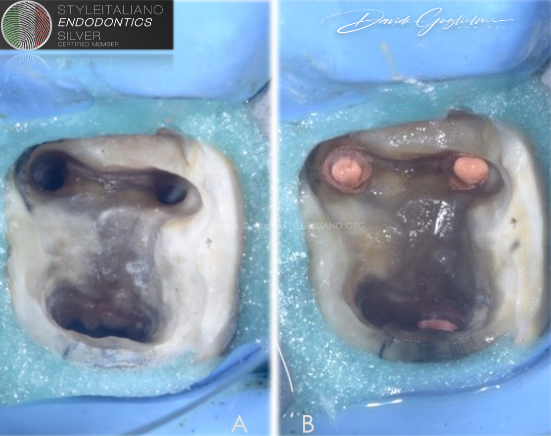
Fig. 4
An additional isolation was also obtained by injecting the liquid dam around the preparation.
The pulp chamber and the four root canals after being cleaned and shaped. (A)
The pulp chamber aspect after 3D obturation and cleaning with ultrasonic tips under irrigation. (B)
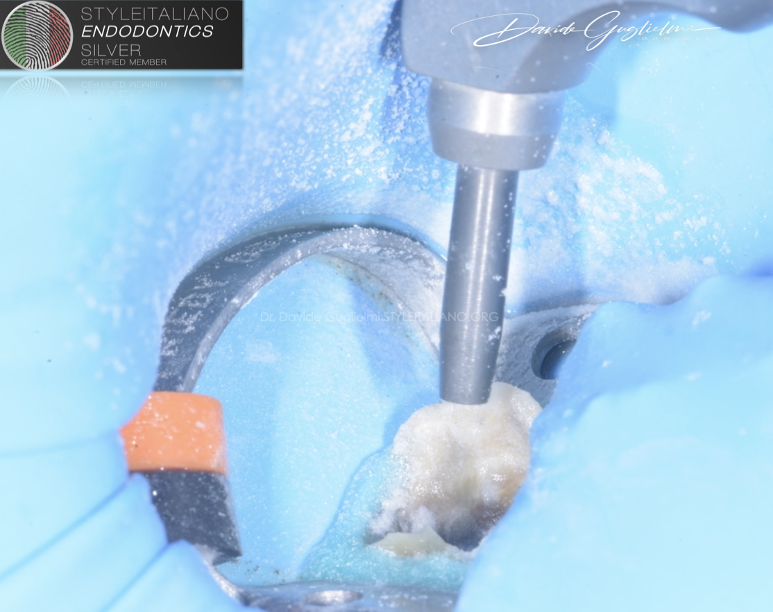
Fig. 5
Dentine prepared by using of micro-etcher sandblaster (aluminium and silicon oxide powder_particle size 20-80 micron).
The Micro-etcher Sandblaster was used to clean the pulp chamber and to improve the adhesion of the bonding agents.
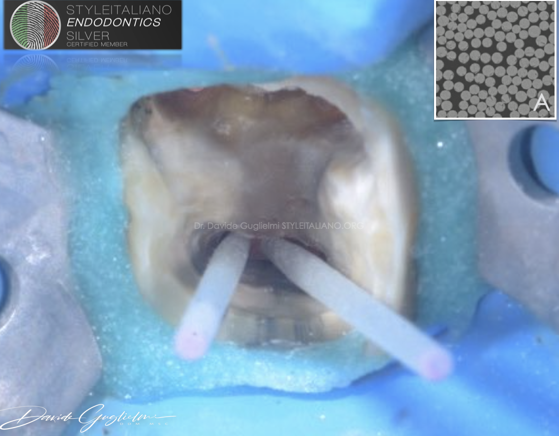
Fig. 6
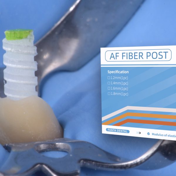
Fiber posts fitting.
The post must have a passive fitting, without prepare the root canal without any bur or drill.
It’s mandatory to preserve as much as possible of the remaining dentine.
The Itena Dentoclic Glass Fiber Post are biocompatible, with no risk of corrosion or coloration and compatible with Dentoclic drills.
Dentoclic glass-fiber posts are made of 80% unidirectional parallel oriented glass fibers embedded in 20% of epoxy-resin.
Fig. A: Microscopic observation of a Dentoclic glass-fiber cross section (SEM x500)
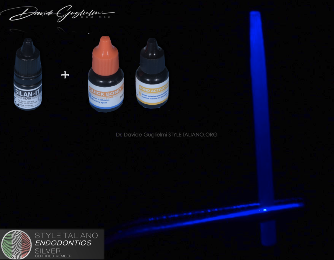
Fig. 7
The posts were both prepared with alcohol and bonding as suggested by the producer of the sealer (Itena). The Itena Dentoclic Glass Fiber Post have excellent mechanical properties for a reduced risk of fracture due to:
- an excellent resistance to flexion (836Mpa);
- its many longitudinal glass-fibers;
- elastic behavior similar to dentin.
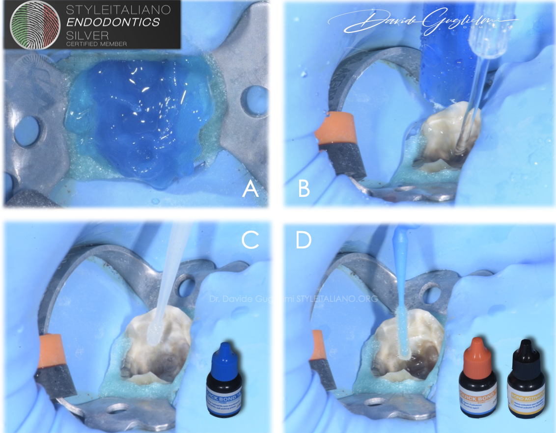
Fig. 8
Pulp chamber, the root canal orifices and the post space were etched with Dentoetch, (37% ortho-phosphoric acid ) that was characterized by a perfect viscosity with provided quick and easy rinse (B).
Afterward the tooth was dried with air and paper point and then primer (C) and bonding Quick Bond (D) were applied into the root canals as suggested by the producer (Itena).
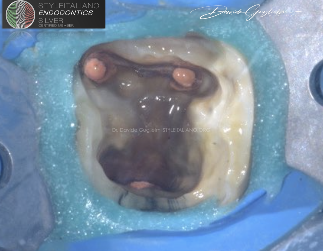
Fig. 9
Pulp chamber condition after bonding passages.
These ensure excellent and reliable adhesion to enamel and dentin, due to a remarkable shear bond strength.
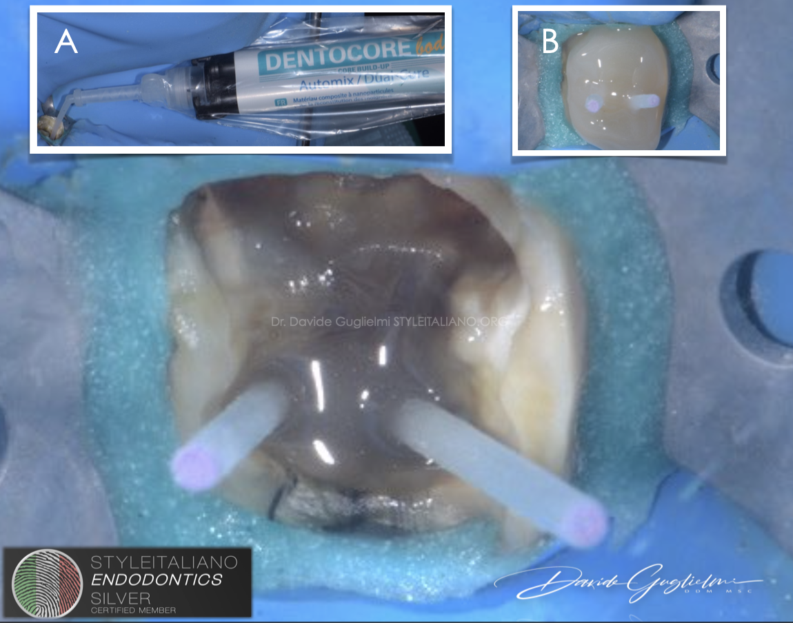
Fig. 10
Nano - filled composite dual cure material placement (Dentocore Itena) (A).
A recent study demonstrated that hardness of this material is close to the dentine one.
Furthermore it has a low shrinkage rate, despite being a very fluid material useful to avoid bubbles in the post cementation.
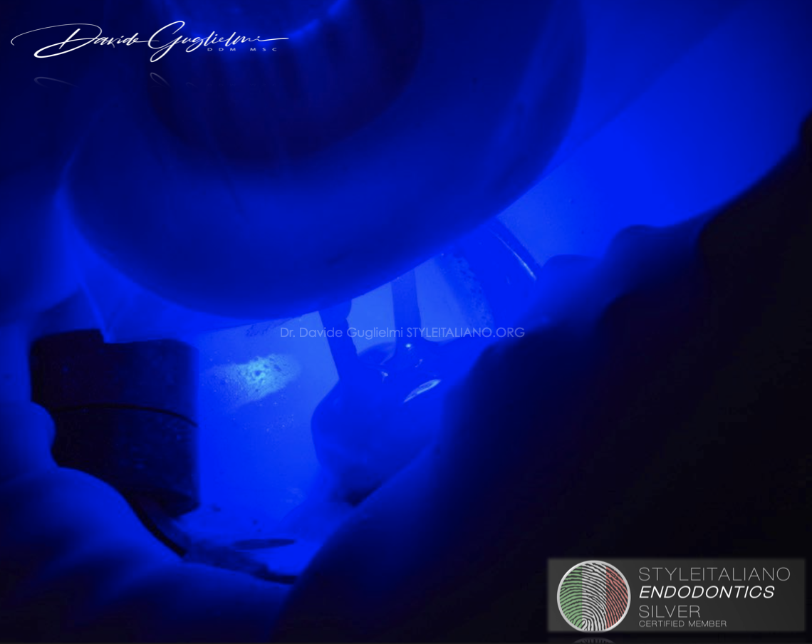
Fig. 11
The Dentocore is a dual curing Material.
Nevertheless it is suggested to cure this resin composite material for at least 40 seconds.
Due to properties of the posts composition, excellent distribution of the light in the whole material is guaranteed.
Furthermore it allows polymerization of the core build-up material on all of the post surfaces.
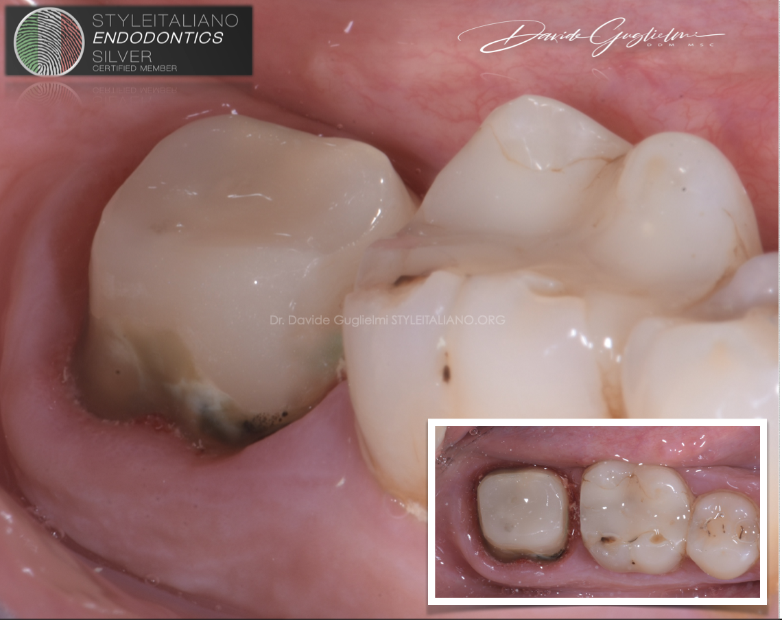
Fig. 12
Side and occlusal views
of the clinical situation after the build up preparation.
Due to both clinical and radiographic carious infiltrations, the tooth 4.6 must be treated with an indirect restorative therapy.
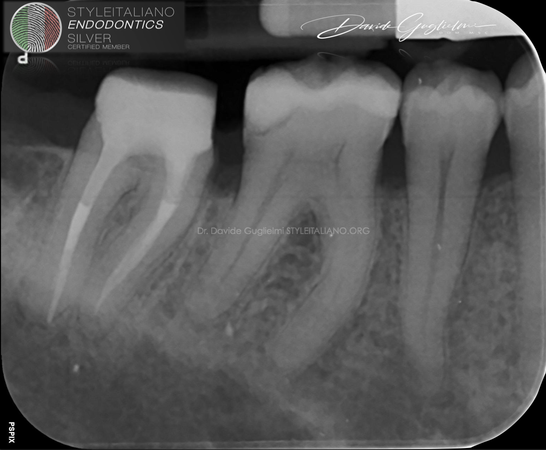
Fig. 13
Post operative x-ray.
Conclusions
Based on author knowledge and experience, the Itena DentoClic products seems to be safe and effective for core build-up and post cementation.
However high-quality clinical trials are needed.
Bibliography
Shu, X., Mai, Q.-Q., Blatz, M., Price, R., Wang, X.-D., & Zhao, K. (2018). Direct and Indirect Restorations for Endodontically Treated Teeth: A Systematic Review and Meta-analysis, IAAD 2017 Consensus Conference Paper. The Journal of Adhesive Dentistry, 20(3), 183–194.
Fichera, G. (2005). Restaurativa post endodontica con perni in fibra.indicazioni e tecnica operativa. Il Dentista Moderno, 23–57.
Keçeci AD, Heidemann D, Kurnaz S. Fracture resistance and failure mode of endodontically treated teeth restored using ceramic onlays with or with- out fiber posts-an ex vivo study. Dent Traumatol 2016;32(4):328–335.
Mannocci F, Qualtrough AJE, Worthington HV, Watson TF, Pitt Ford TR. Randomized clinical comparison of endodontically treated teeth restored with amalgam or with fiber posts and resin composite: Five-year results. Oper Dent 2005;30:9–15.
Figueiredo FE, Martins-Filho PR, Faria-E-Silva AL. Do metal post-retained restorations result in more root fractures than fiber post-retained restorations? A systematic review and meta-analysis. J Endod. 2015 Mar;41(3):309-16. doi: 10.1016/j.joen.2014.10.006. Epub 2014 Nov 11. PMID: 25459568.
Correlation between the mechanical properties and structural characteristisc of different fiber posts systems – Novais et al. – Brazilian Dental Journal - 2016


