UL6 with unusual anatomy
15/06/2025
Fellow
Warning: Undefined variable $post in /home/styleendo/htdocs/styleitaliano-endodontics.org/wp-content/plugins/oxygen/component-framework/components/classes/code-block.class.php(133) : eval()'d code on line 2
Warning: Attempt to read property "ID" on null in /home/styleendo/htdocs/styleitaliano-endodontics.org/wp-content/plugins/oxygen/component-framework/components/classes/code-block.class.php(133) : eval()'d code on line 2
The incidence of MB2 and MB3 in upper first molars is low (Ronald Ordinola-Zapata et al., 2020), however one of the most common reasons for endodontic failures is missed anatomy (Tabassum and Raza Khan, 2016). It is therefore very important to investigate every potential groove, and locate all canals, as demonstrated in this case.
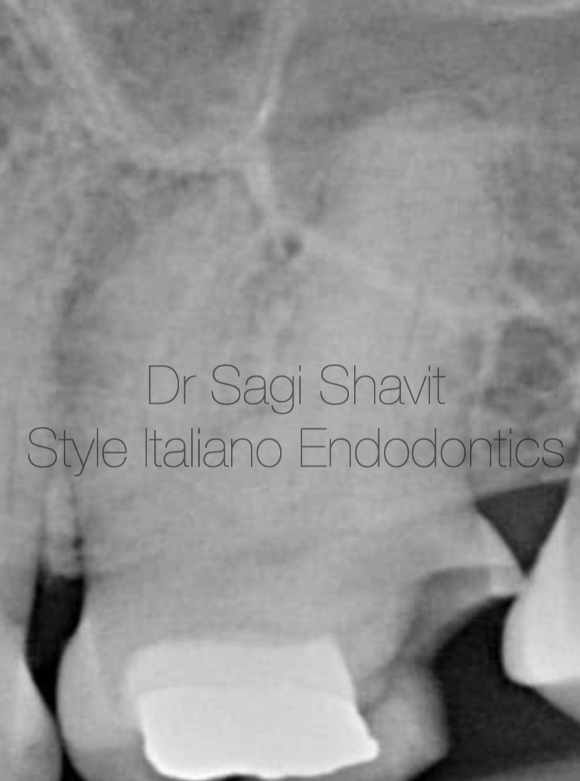
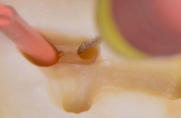
Fig. 1
This UL6 (26) presented with irreversible pulpitis, and pulp canal obliteration.

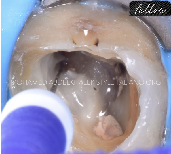
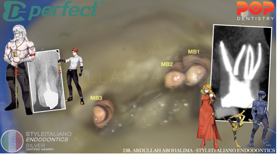
Fig. 2
An investigation of a groove in the MB root, using fine ultrasonic tip reveals the presence of MB2 and MB3 canals.
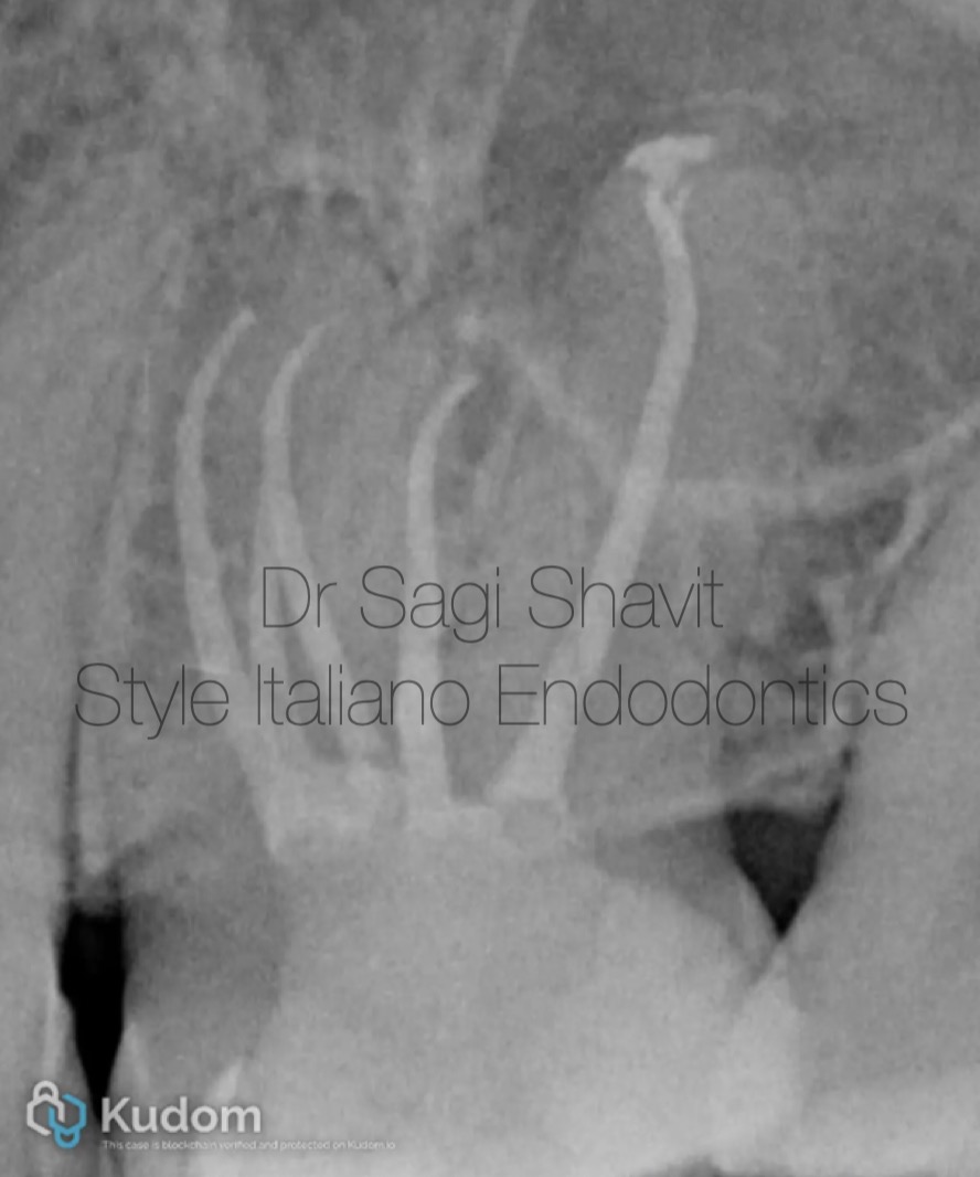
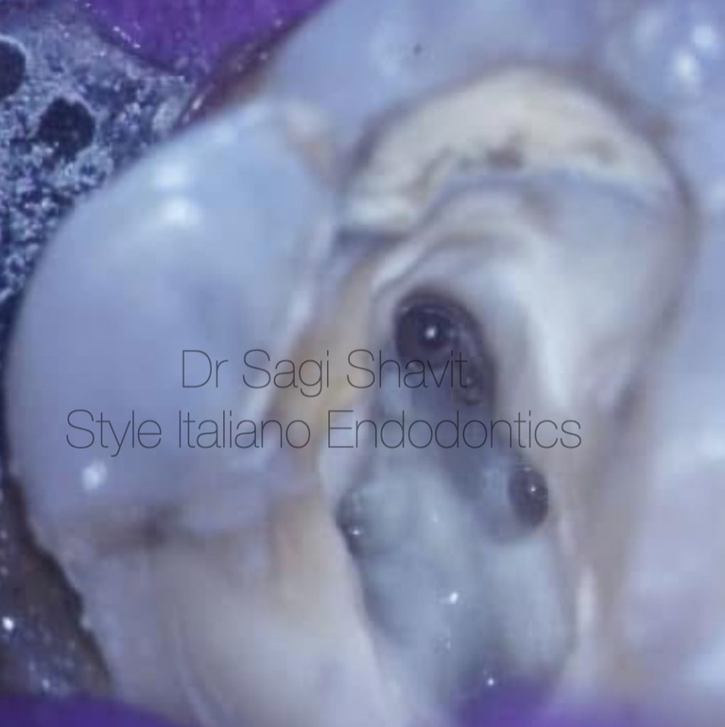
Fig. 3
The canals were instrumented using Hyflex EDM files (glide file, 05/20 and OneFile).
All canals were irrigated with 17% EDTA, 5.25% NaOCl using passive ultrasonic irrigation.
Obturation was done using bioceramic ceramic, and warm vertical compaction.
The access cavity was sealed with composite and the patient was instructed to returned to his general practitioner for a crown.
Fig. 4
About the author:
Sagi Shavit
Graduated as DMD Bucharest 2009 - UMF “Carol Davila” Bucharest, Romania
MSc in Endodontology - Chester University
PG diploma in implantology - City of London Dental School
Conclusions
Anatomy is always a challenge; magnification, ultrasounds help the clinician to find the hidden anatomy.
Bibliography
Reference List:
- Ordinola-Zapata R, Martins JNR, Plascencia H, Versiani MA, Bramante CM. The MB3 canal in maxillary molars: a micro-CT study. Clin Oral Investig. 2020 Nov;24(11):4109-4121. doi: 10.1007/s00784-020-03284-7. Epub 2020 May 7. PMID: 32382930.
- Tabassum S, Khan FR. Failure of endodontic treatment: The usual suspects. Eur J Dent. 2016 Jan-Mar;10(1):144-147. doi: 10.4103/1305-7456.175682. PMID: 27011754; PMCID: PMC4784145