Three-rooted maxillary premolars: an uncommon anatomical variation.
24/09/2026
Warning: Undefined variable $post in /home/styleendo/htdocs/styleitaliano-endodontics.org/wp-content/plugins/oxygen/component-framework/components/classes/code-block.class.php(133) : eval()'d code on line 2
Warning: Attempt to read property "ID" on null in /home/styleendo/htdocs/styleitaliano-endodontics.org/wp-content/plugins/oxygen/component-framework/components/classes/code-block.class.php(133) : eval()'d code on line 2
Three-rooted maxillary premolars represent an uncommon but clinically important anatomical variation. The maxillary first premolar most frequently presents with two roots, while the occurrence of three separate roots is considerably less common. In a CBCT study of 350 maxillary first premolars, Olczak et al. reported three roots in 2.6% of teeth, with two-rooted morphology accounting for 69.1% of the sample.
The prevalence of three-rooted anatomy varies between populations and between the first and second maxillary premolars. A large multicenter CBCT study including data from different geographic populations reported a prevalence of 1.8% for three-rooted maxillary first premolars and 0.4% for second premolars. Similarly, an Iraqi CBCT study identified three roots in 1.2% of first premolars and 0.7% of second premolars. These findings demonstrate that although the variation is rare, it should be considered during endodontic diagnosis and treatment.
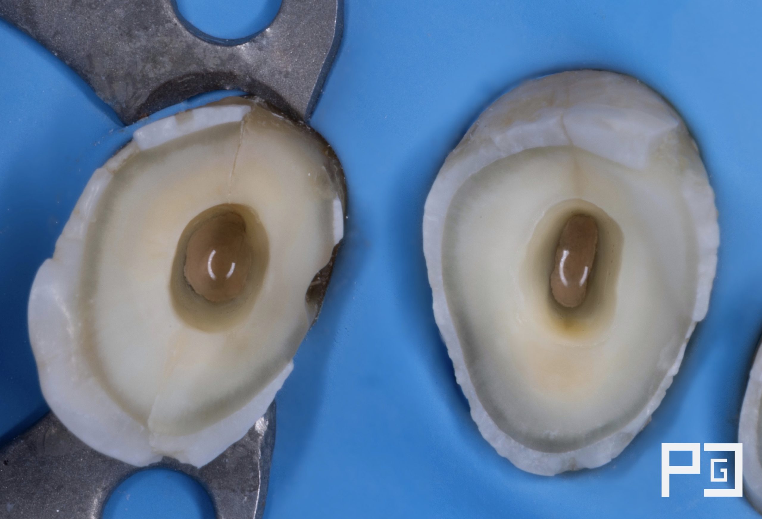
Three-rooted premolars commonly present with three separate canals, frequently following a mesiobuccal, distobuccal and palatal arrangement. The additional root and canal can be difficult to identify on conventional periapical radiographs because of superimposition. CBCT provides three-dimensional visualization of the roots and canals and can therefore be particularly valuable in cases where unusual anatomy is suspected.
The clinical importance of recognizing this morphology lies in the prevention of missed canals. Failure to identify and adequately treat an additional canal may leave infected tissue within the root canal system and contribute to persistent apical disease. Recent case reports of three-rooted maxillary premolars have emphasized the usefulness of CBCT, dental operating microscopes and magnification for detecting additional roots and establishing predictable access to the entire canal system.
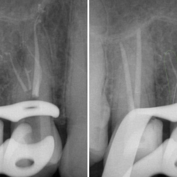
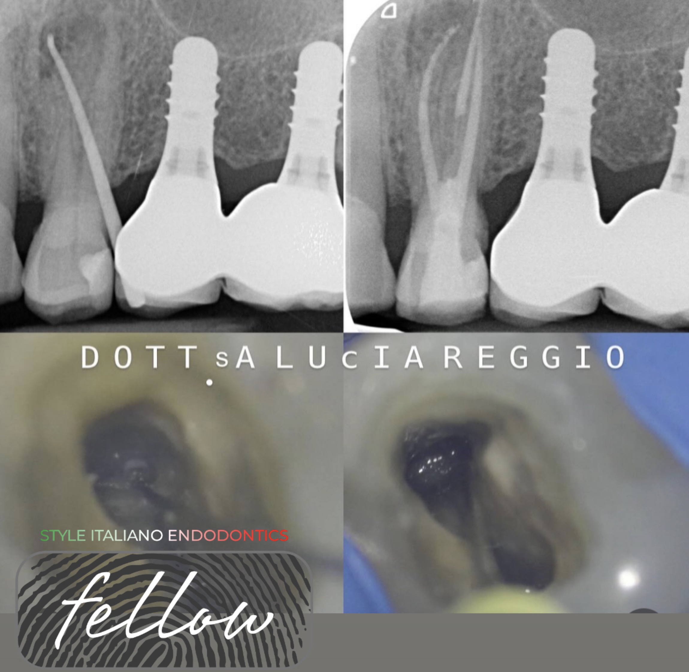
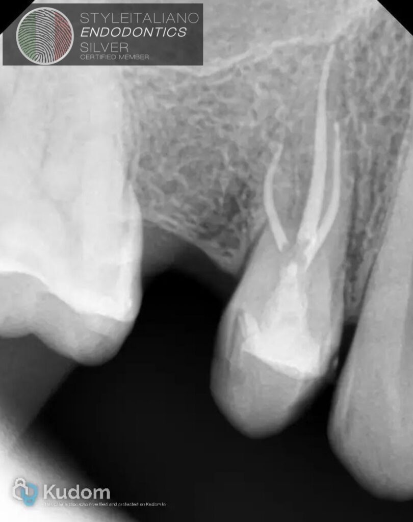
The three radiographic images presented demonstrate the clinical relevance of this anatomical variation, showing premolars with clearly distinguishable root structures and canal systems. Such cases highlight why detailed assessment of root morphology should precede endodontic treatment, particularly when radiographic signs suggest an atypical anatomy.
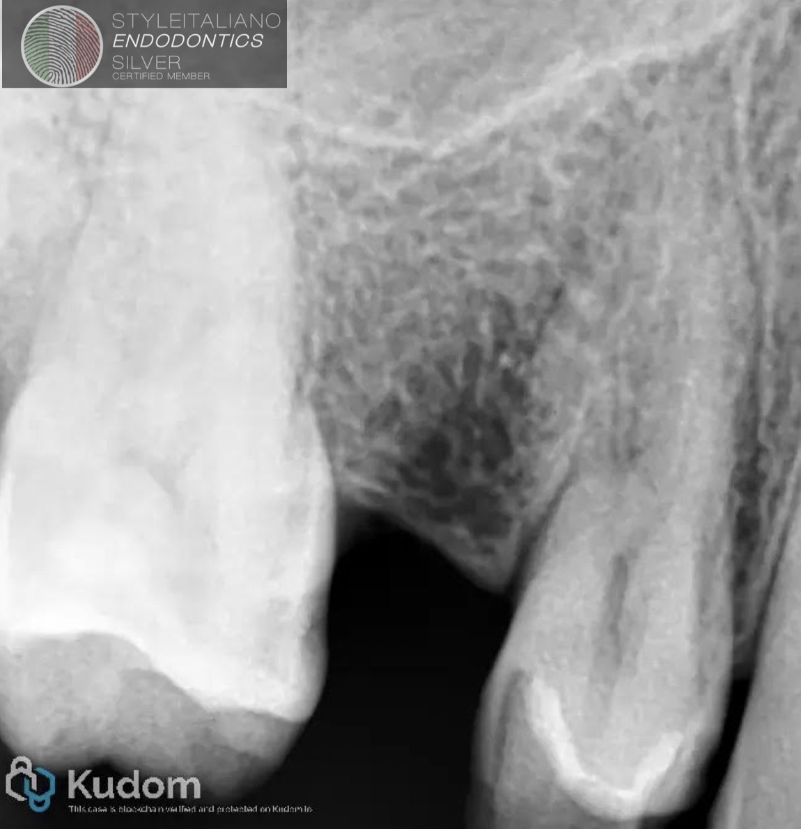
Fig. 1
Pre op x ray
Dg;Acute pulpits 14
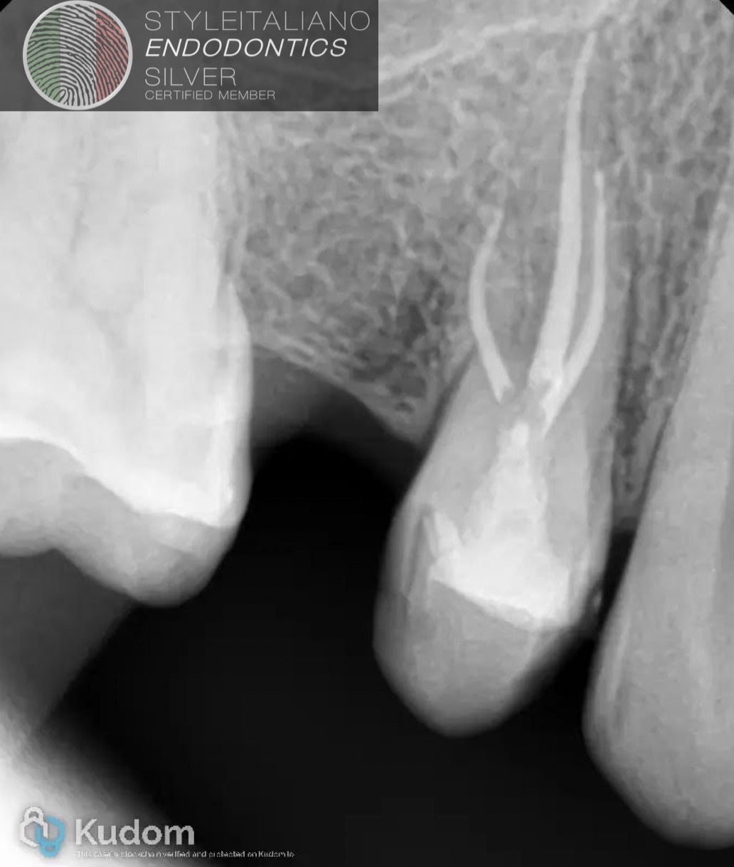
Fig. 2
Post op
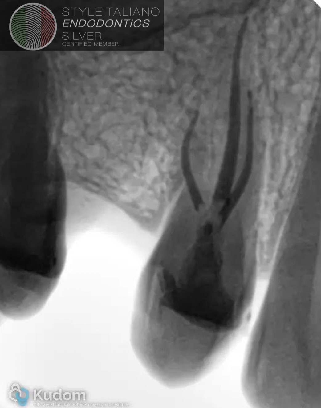
Fig. 3
Inverted X-xray
Conclusions
Root canal anatomy is highly variable, and even apparently straightforward teeth may conceal unexpected anatomical complexities. Recognizing these variations is therefore a key step in everyday endodontic practice.
Careful evaluation of pre-operative radiographs may provide the first clues to unusual canal configurations, additional roots, canal divisions or unexpected curvatures. However, radiographic interpretation is only the beginning. Access cavity design, inspection of the pulp chamber floor, magnification, canal scouting and working length determination all provide additional information that progressively reveals the true anatomy of the tooth.
For this reason, every step of the treatment should also be considered a diagnostic step. Observing, interpreting and combining these findings allows the clinician to adapt the treatment strategy to the anatomy encountered rather than forcing the anatomy to follow a predetermined protocol.
Ultimately, successful endodontic treatment depends not only on shaping, cleaning and filling the root canal system, but first and foremost on recognizing the anatomy that needs to be treated.
Bibliography
- Olczak K, Pawlicka H, Szymański W. Root form and canal anatomy of maxillary first premolars: a cone-beam computed tomography study. Odontology. 2022;110:365–375.
- Worldwide Assessment of the Root and Root Canal Characteristics of Maxillary Premolars: A Multi-center Cone-beam Computed Tomography Cross-sectional Study With Meta-analysis. Journal of Endodontics. 2023.
- Characterization of the root and canal anatomy of maxillary premolar teeth in an Iraqi subpopulation: a cone beam computed tomography study.
- Evaluation of root canal morphology of maxillary premolars: a cone-beam computed tomography study. Australian Endodontic Journal. 2018.
- Ali A, Saleh AR, Elmsmari F, Baruwa AO. Endodontic management of maxillary premolars with three roots: a case series. Frontiers in Oral Health. 2026