The Sandwich Technique: A Convenient and Predictable method for Root Perforation Repair
14/06/2020
Ahmed Shawky
Warning: Undefined variable $post in /home/styleendo/htdocs/styleitaliano-endodontics.org/wp-content/plugins/oxygen/component-framework/components/classes/code-block.class.php(133) : eval()'d code on line 2
Warning: Attempt to read property "ID" on null in /home/styleendo/htdocs/styleitaliano-endodontics.org/wp-content/plugins/oxygen/component-framework/components/classes/code-block.class.php(133) : eval()'d code on line 2
Root perforations are defined as communications between the root canal space and the periodontium. These communications may be caused by a pathological process (as: external or internal resorption) or as a result of an iatrogenic error (as: misdirection/improper size of the post space drill, drilling through the chamber floor or improper root canal instrumentation)1. According to the best available evidence, root perforations are accompanied by a 22%-31% drop in the success rate of non-surgical root canal retreatment, especially when coupled with apical periodontitis2,3. However, with the advancements in bioactive repair materials and the techniques of their application as well as the use of dental operating microscope, the success rate of root perforation repair increases dramatically. This article aims to present a case with strip perforation that occurred as a result of improper post space preparation and its management using a Bioceramic material applied with the sandwich technique.
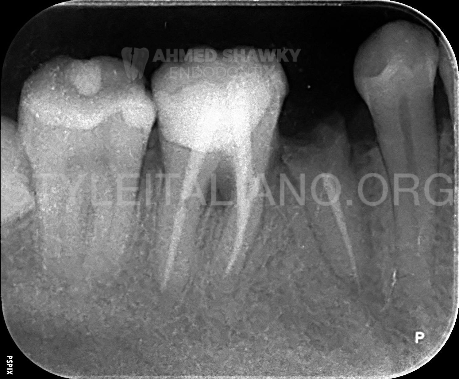
Fig. 1
Preoperative Evaluation:
A patient was referred from a general practitioner with a previously endodontically treated mandibular first molar which was sensitive to biting.
Upon clinical examination, the tooth was sensitive to percussion with a history of recurrent mild swelling of the buccal gingival margin. Periodontal probing revealed a 5 mm probing defect at the furcation buccally.
Radiographic examination revealed the presence of a good coronal resin composite restoration and two metallic screw-retained posts in both roots. The previous endodontic treatment was acceptable in terms of length of fill, but the density of fill was regarded as questionable. A moderately-sized furcal radiolucency was evident on the 2D radiographic image.
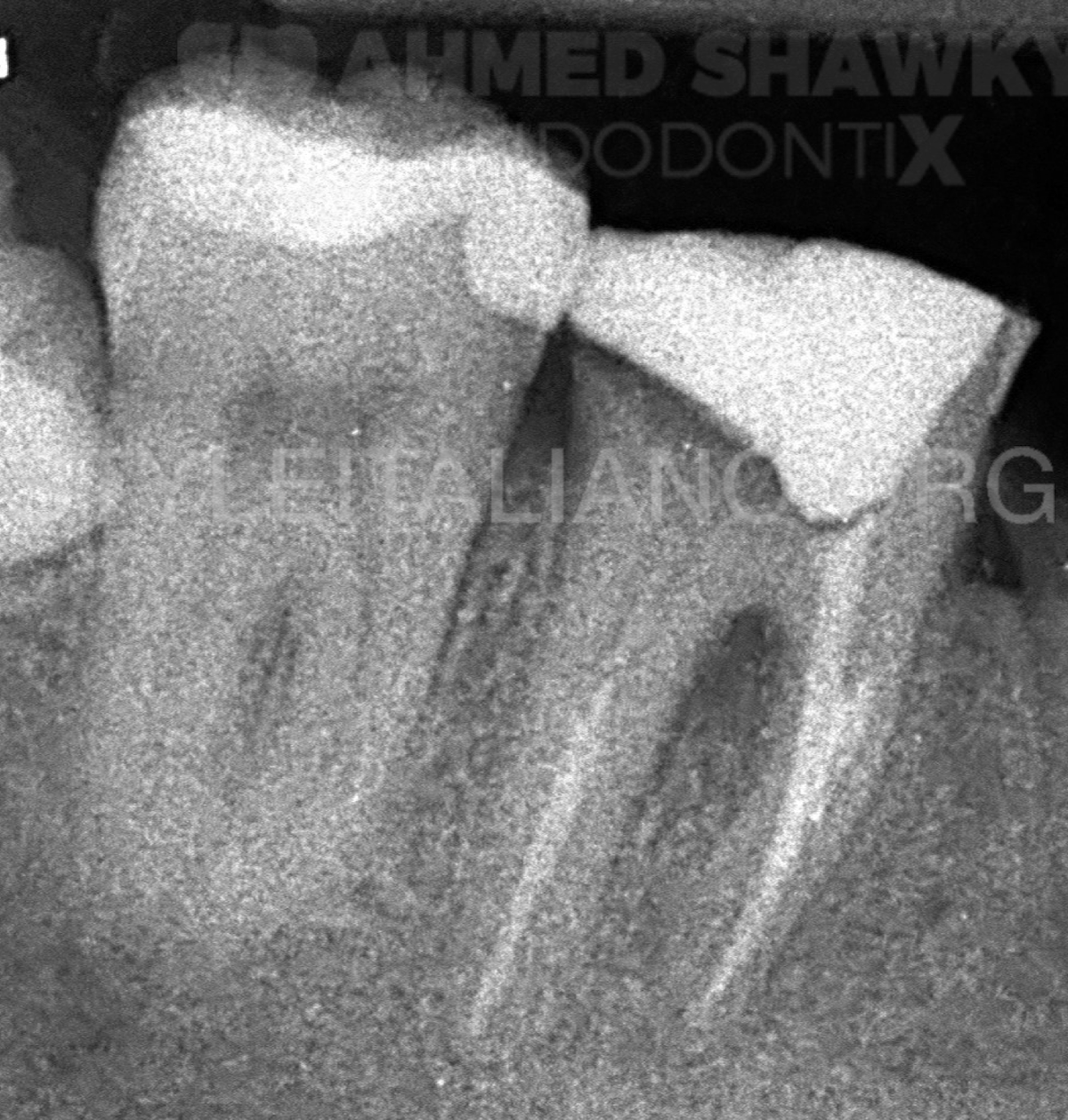
Fig. 2
Disassembly:
During the first visit, removal of the coronal composite restoration followed by removal of the screw retained posts from both the distal and the mesiobuccal canals using ultrasonic tips in the presence of a coolant operated with CCW maneuver.
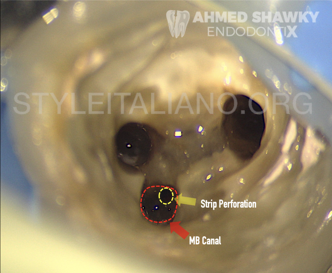
Fig. 3
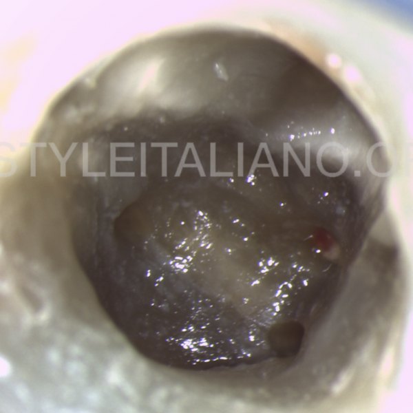
It was noted that the post in the MB canal was exceedingly large. After removal of the posts, removal of the gutta percha was performed using Rotary instruments concurrently with PUI activation of irrigants to remove remnants of filling materials within the root canals.
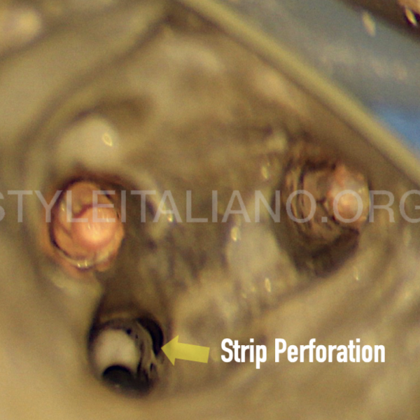
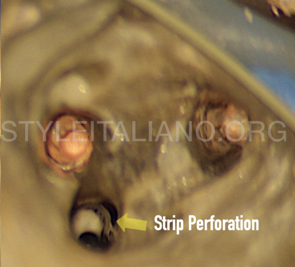
Upon removal of filling materials from the root canals, a strip perforation was visualized under magnification located on the distal wall of the mesiobuccal canal which was probably due to the use of a large post drill.
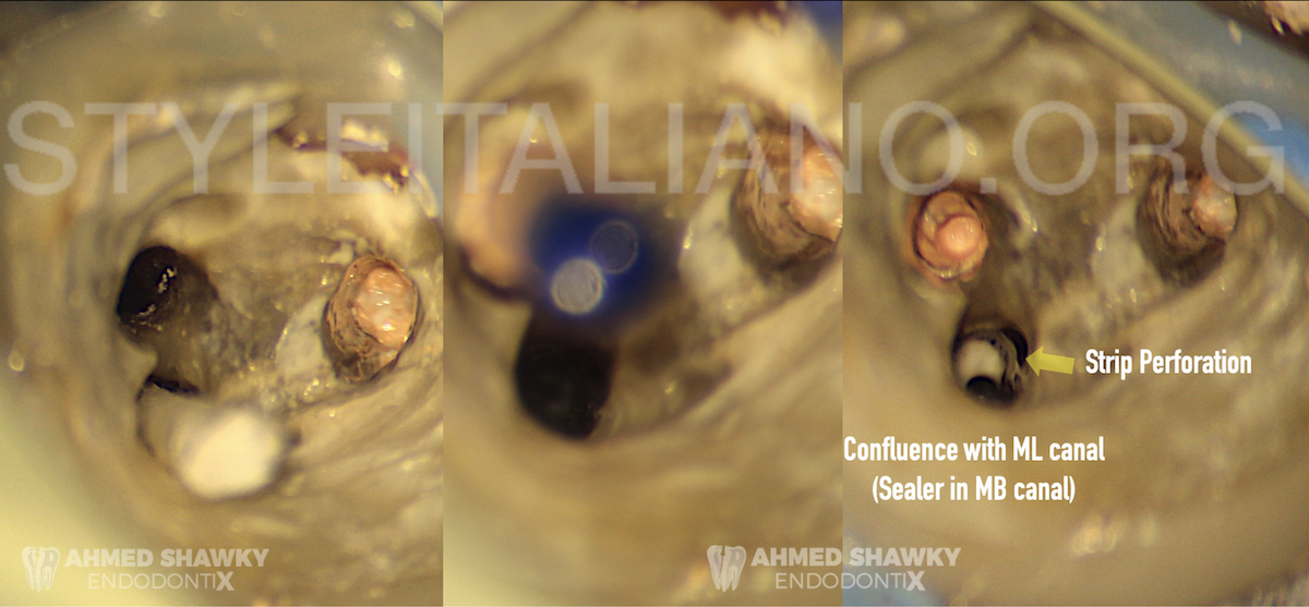
Fig. 4
Obturation:
The mesiolingual and distal root canals were obturated using continuous wave of compaction (CWC) with resin sealer. During obturation of the mesiolingual canal, confluence with the mesiobuccal root canal was observed under magnification.
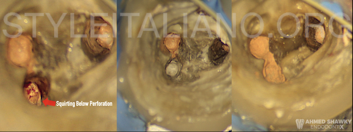
Fig. 5
Perforation Repair using Sandwich Technique:
Simply, the Sandwich Technique involves down-packing the gutta-percha below the level of the perforation followed by placing the Bioceramic repair material at the perforation segment above the previously packed gutta percha. The repair material is then sandwiched coronally with gutta percha. In this way, the repair unit is considered as a “Sandwich” composed of a Bioceramic or a calcium silicate material between two layers of gutta percha.
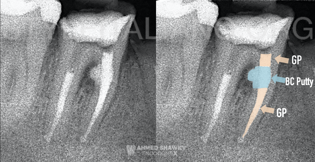
Fig. 6
In this particular case of strip perforation, as a result of confluence, the area apical to the perforation was filled by squirting technique sequentially and packed in a continuous wave fashion.
Hemostasis at the perforation site was achieved using sterile paper points. The repair material used was a bioceramic putty material due to its favorable consistency and setting time (12 minutes). Packing the Bioceramic material in the perforation area was done with a fine microbrush above which gutta percha was dispensed through a back-filling device.
Conclusions
The Sandwich technique represent a reliable and valid method for repair of iatrogenic and pathologic perforations affecting the body of roots.
The current advances in Bioceramic materials allows clinicians to achieve predictable sealing of root perforations over the conventional application techniques.
Bibliography
1. Estrela C, Decurcio D de A, Rossi-Fedele G, Silva JA, Guedes OA, Borges ÁH. Root perforations: A review of diagnosis, prognosis and materials. Braz Oral Res. 2018;32:133-146. doi:10.1590/1807-3107bor-2018.vol32.0073
2. Torabinejad M, Corr R, Handysides R, Shabahang S. Outcomes of Nonsurgical Retreatment and Endodontic Surgery: A Systematic Review. J Endod. 2009;35(7):930-937. doi:10.1016/j.joen.2009.04.023
3. de Chevigny C, Dao TT, Basrani BR, et al. Treatment Outcome in Endodontics: The Toronto Study-Phases 3 and 4: Orthograde Retreatment. J Endod. 2008;34(2):131-137. doi:10.1016/j.joen.2007.11.003