The Importance of Access Cavity Design in Endodontic Treatment
16/09/2026
Warning: Undefined variable $post in /home/styleendo/htdocs/styleitaliano-endodontics.org/wp-content/plugins/oxygen/component-framework/components/classes/code-block.class.php(133) : eval()'d code on line 2
Warning: Attempt to read property "ID" on null in /home/styleendo/htdocs/styleitaliano-endodontics.org/wp-content/plugins/oxygen/component-framework/components/classes/code-block.class.php(133) : eval()'d code on line 2
Access cavity design is far more than simply gaining entry to the pulp chamber , it is the foundation that shapes every step of endodontic treatment. The design of the access cavity directly influences canal location, visibility, instrument positioning, and the overall control of the root canal treatment.
A properly designed access cavity should provide sufficient visualization and a smooth, controlled pathway to the canals while preserving maximum tooth structure. When access is inadequate, instruments may be forced to negotiate unfavorable angles, increasing torsional stress, cyclic fatigue, and the risk of procedural errors including the worst nightmare which is instrument separation.
That’s why , understanding the relationship between access cavity design, canal anatomy, and shaping protocol is essential for reducing unnecessary stress on instruments and improving the predictability and safety of RCT.
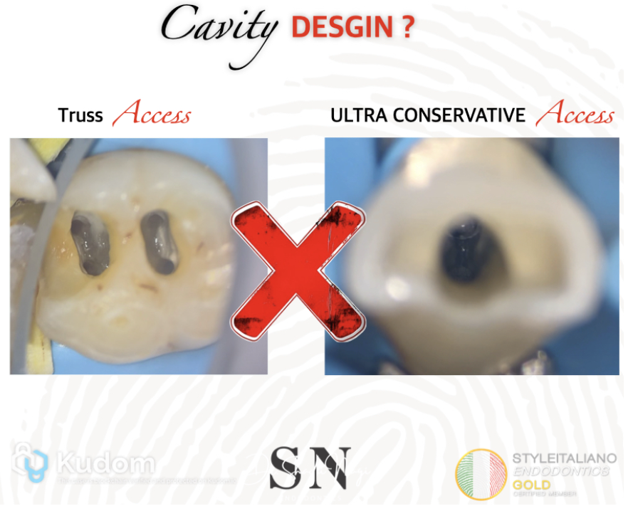
Fig. 1
Different access cavity designs including conventional, ultra conservative, truss, and ninja access preparations have been evaluated in MANY scientific studies. Interestingly, many of these studies have reported similar fracture resistance among the different access cavity designs.
That’s why , the key is not simply to create the smallest possible access cavity. Instead, our goal should be to achieve a conservative access cavity that preserves as much sound tooth structure as possible while remaining wide enough to provide adequate visibility, straight-line access, proper canal orifice location, effective cleaning and shaping, and safe instrumentation during shaping.
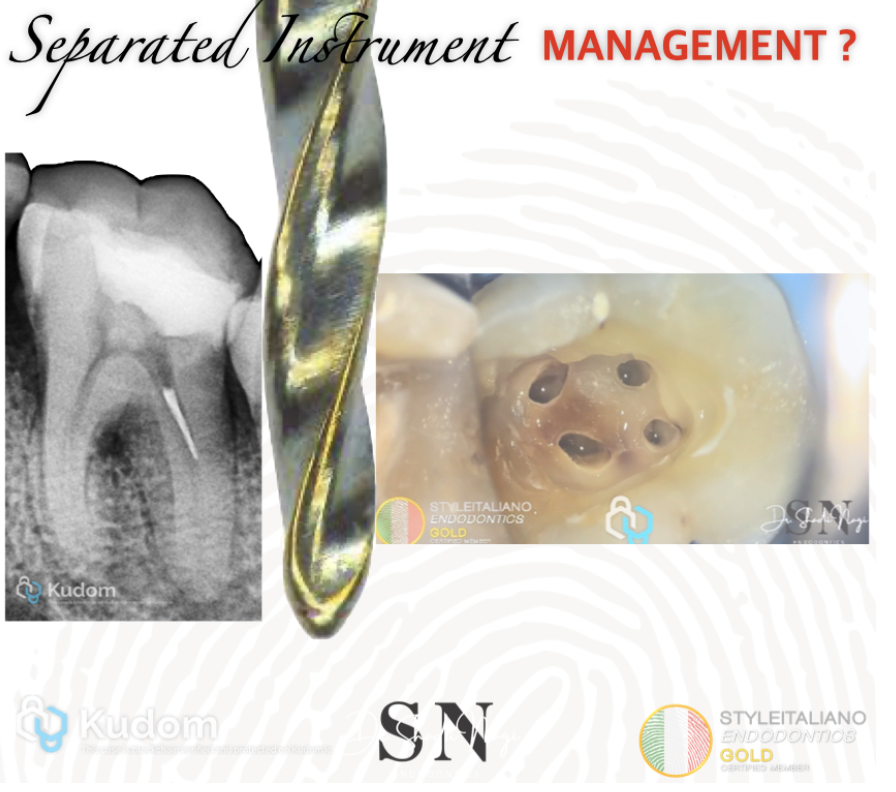
Fig. 2
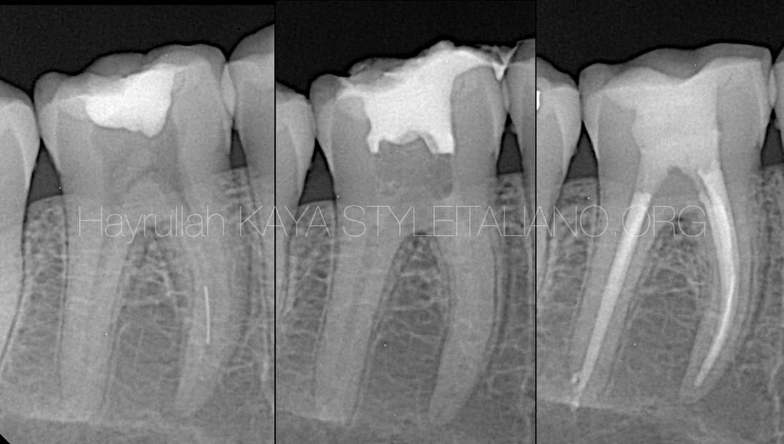
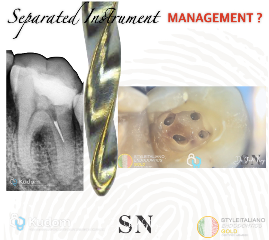
This case was referred to my clinic due to a separated instrument in the coronal portion of the distal canal.
After removal of the temporary filling, the access cavity was carefully evaluated under microscope . It became evident that the initial access preparation had not been correctly deroofed and did not provide proper access to the canals.
The wrong access cavity design resulted in restricted and unfavorable instrumentation access, which likely contributed to excessive stress on the file and led to instrument separation. This case highlights the importance of complete deroofing and establishing a properly designed access , conservative yet sufficiently extended access cavity to provide straight-line access and reduce procedural complications such as instrument fracture or separation.
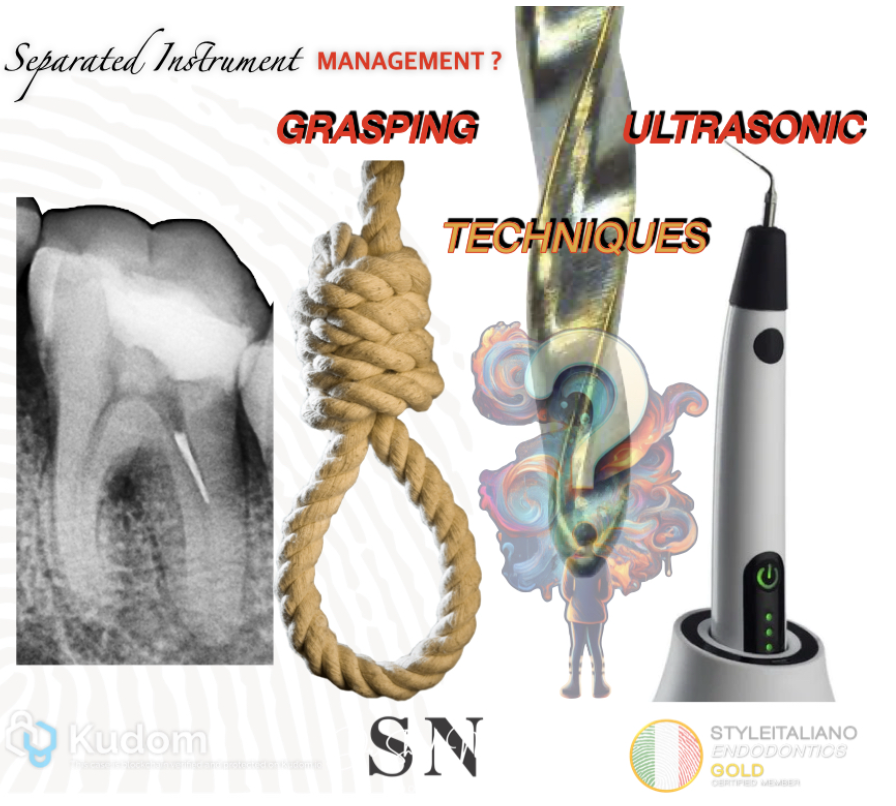
Fig. 3
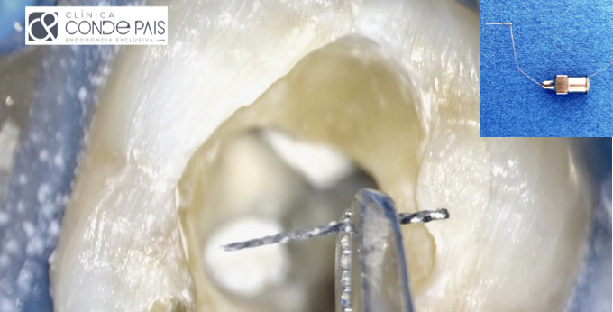
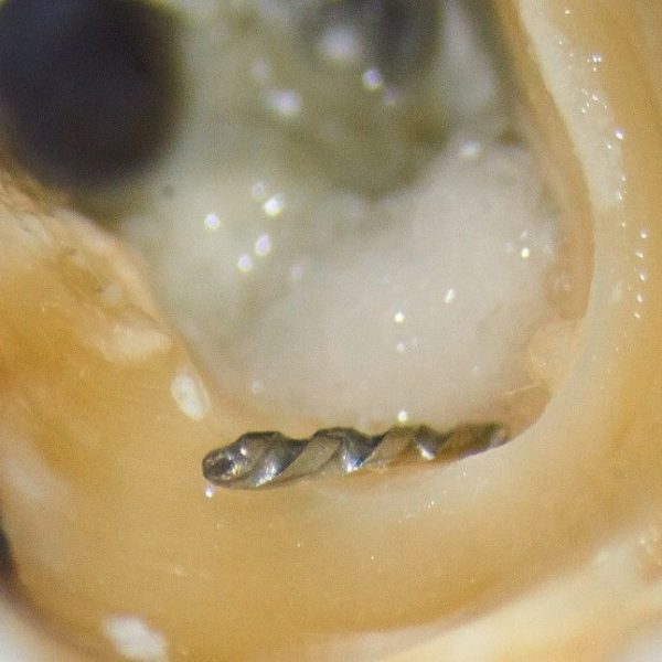
We have two main techniques can be considered for the management of separated instruments. Ultrasonic retrieval is preferred when the fractured instrument is short, typically less than 4 mm, whereas grasping techniques are more appropriate when the separated instrument is longer than 4 mm
In this case, the separated instrument was less than 4 mm in length; that’s why , an ultrasonic retrieval technique was selected. However, before attempting separated file removal, it was essential to modify and refine the access cavity with the use of ultrasonic to achieve adequate straight-line access and direct visualization of the separated instrument. This access modification allowed controlled positioning of the ultrasonic tip while minimizing unnecessary removal of sound dentin and reducing the risk of further complications.
full case video
Conclusions
Successful separated instrument retrieval depends not only on the retrieval technique, but also on adequate access cavity design and proper assessment of the fractured instrument . In this case, the instrument separation was associated with wrong access preparation, highlighting the importance of a conservative yet sufficiently wide access cavity that provides direct visualization and controlled instrument access during shaping.
After modifying the access cavity to establish a more favorable pathway, an ultrasonic retrieval technique was selected based on the short length and accessible position of the separated file . Careful ultrasonic movements allowed successful removal of the separated instrument while preserving the maximum amount of sound dentin and minimizing unnecessary dentin sacrifice. This case emphasizes that proper access modification, magnification, controlled ultrasonic activation, and a minimally invasive attempt are essential for predictable separated instrument management while maintaining the structural integrity of the tooth .
Bibliography
- Barbosa AFA, Silva EJNL, Coelho BP, et al.
The influence of endodontic access cavity design on the efficacy of canal instrumentation, microbial reduction, root canal filling and fracture resistance in mandibular molars.
International Endodontic Journal. 2020;53(12):1666–1679.
DOI: https://doi.org/10.1111/iej.13383 - Özyürek T, Ülker Ö, Özsezer Demiryürek E, Yılmaz F.
The Effects of Endodontic Access Cavity Preparation Design on the Fracture Strength of Endodontically Treated Teeth: Traditional Versus Conservative Preparation.
Journal of Endodontics. 2018;44(5):800–805.
DOI: https://doi.org/10.1016/j.joen.2018.01.020 - Mrinalini M, Gupta A, Soi S, Abraham D, Bukhari SH.
Endodontic Access Cavity Design and Fracture Resistance: A Systematic Review and Meta-Analysis of Conventional vs. Newer Access Cavity.
Cureus. 2024;16(9):e68796.
DOI: https://doi.org/10.7759/cureus.68796 - Madani M, Salehi M, Raiesi O, et al.
The management of separated endodontic instruments: A clinical approach.
Endodontology.
DOI: https://doi.org/10.4103/endo.endo_90_21 - Evaluation of Dentine Structure Loss after Separated File Retrieval by Three Different Techniques: An Ex-vivo Study.
Journal of Clinical and Diagnostic Research.
Full Text: https://pmc.ncbi.nlm.nih.gov/articles/PMC10244914/