Separated file dilemma
04/11/2023
Shadi Nagi
Warning: Undefined variable $post in /home/styleendo/htdocs/styleitaliano-endodontics.org/wp-content/plugins/oxygen/component-framework/components/classes/code-block.class.php(133) : eval()'d code on line 2
Warning: Attempt to read property "ID" on null in /home/styleendo/htdocs/styleitaliano-endodontics.org/wp-content/plugins/oxygen/component-framework/components/classes/code-block.class.php(133) : eval()'d code on line 2
During the root canal treatment the possibility of instruments separation inside the canal is always there and the most common causes are root canal anatomy, improper use, inadequate access, manufacturing defects and insufficient knowledge about the root canal morphology, etc.
And it creates a great stress on the practitioners, so when the separation happens the best option is the removal under magnification.
There are many techniques for retrieval including ultrasonic, loop technique, tube technique and others.
If orthograde methods are non applicable, micro apical surgery is always an option.
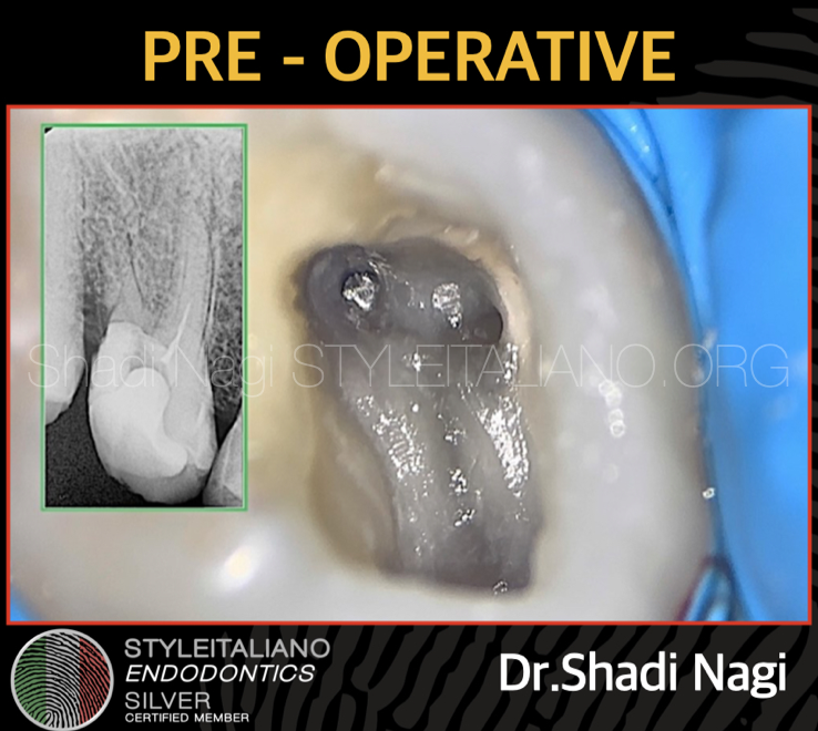
Fig. 1
Case no 1
36 years old female patient referred to our clinic for management of separated file at the coronal one third in maxillary first molar.
Notice here from the pre operative radiograph that there is a narrow canal in the mesial root .
Common reasons for such kind of separation would be errors during the instrumentation by not doing coronal passive pre flaring , glide path and patency followed by correct shaping protocol.
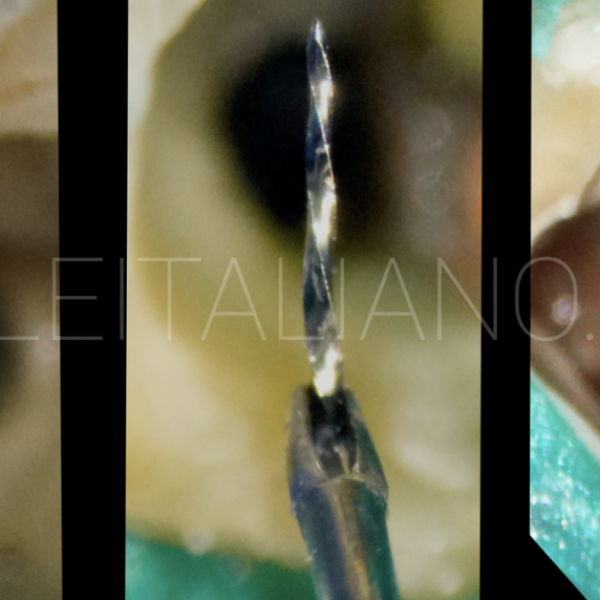
This is a video of a separated file removal with minimal dentin loss
With the use of thin ultrasonic tip at the inner wall of the root to disengage the file and retrieve it .
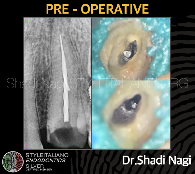
Fig. 2
Case no 2
34 years old male patient referred to our clinic for management
of two separated files in maxillary canine.
Liquid dam was applied for the secondary isolation.
In such a cases when the separated file more than 4.5 mm it’s better to use grasping methods and here tube technique was used for retrieval.
When the the separated file is long - highly engaged the ultrasonic technique only is not effective because the possibility of secondary fracture.
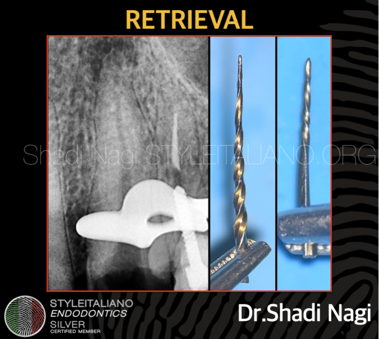
Fig. 3
So here retrieval done to the long one ‘14mm’ using tube technique
after exposing 2mm of the file with ultrasonic.
The second one ‘7mm’ file came out during preparation phase with ultrasonic.
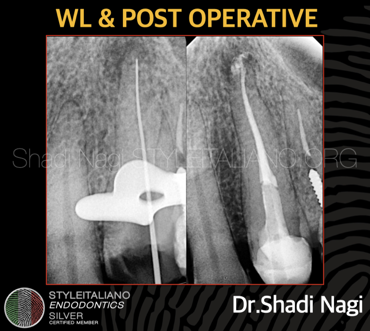
Fig. 4
The working length and post operative radiograph
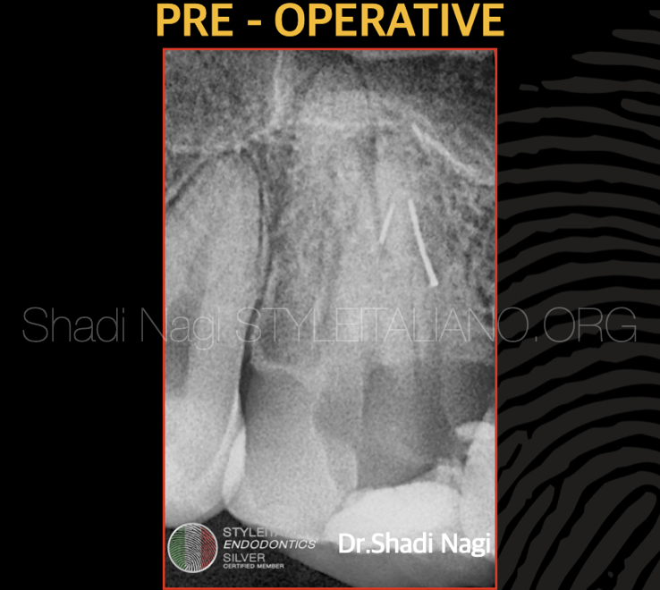
Fig. 5
Case no 3
31 years old male patient referred to our clinic for management of two separated files in maxillary second molar at the apical one third of the DB and MB canals which make the case more difficult.
There was a previous attempt to retrieve the files by the referring dentist using ultrasonics without magnification.
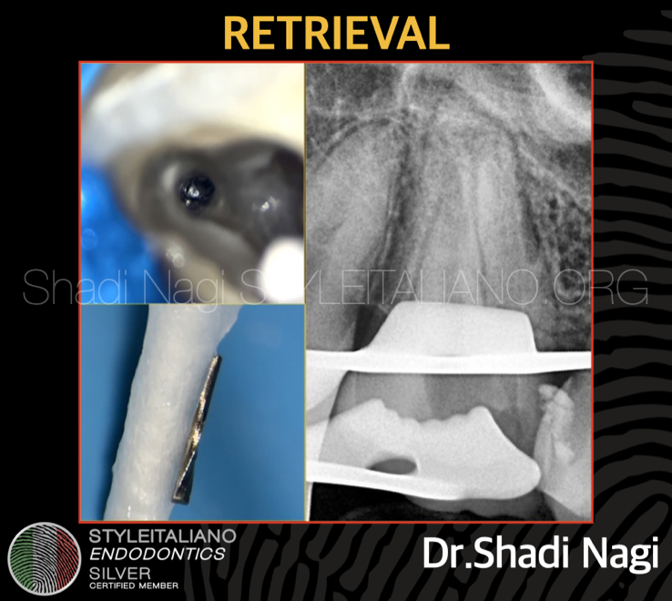
Fig. 6
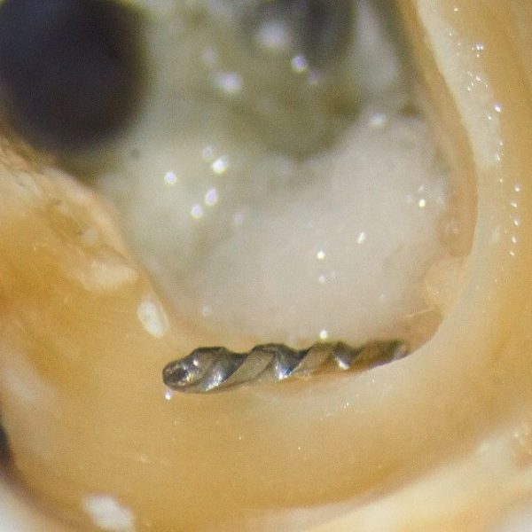
The two separated files retrieved with the use thin ultrasonic at the inner wall of the root under magnification.
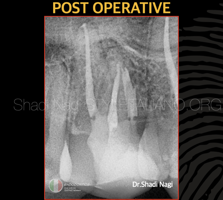
Fig. 7
Post operative radiograph.
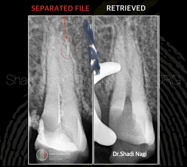
Fig. 8
Case no 4
27 years old female patient referred to our clinic for management of separated file at the apical one third in maxillary second premolar.
From the pre operative radiograph noticed that There is a possibility of extra canal anatomy.
The separated file retrieved with the use of thin ultrasonic at the inner wall of the root.
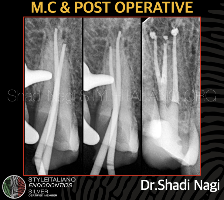
Fig. 9
Master cone fit and post operative radiograph .
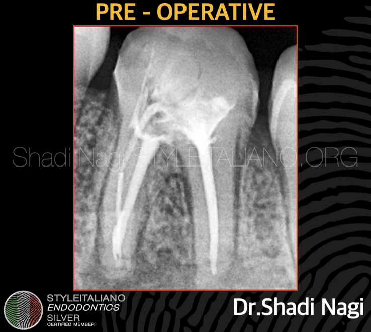
Fig. 10
Case no 5
22 years old female patient referred to our clinic for management of two separated files in mandibular first molar.
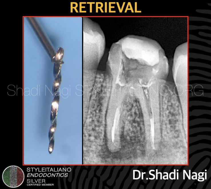
Fig. 11
The long separated file retrieved using loop technique .
The loop technique should circle the exposed separated instrument up to a length allowing tighten the loop between the flutes in the under cut for firm grasping .
The other file was at the apical one third beyond the curvature so decided to bypass it.
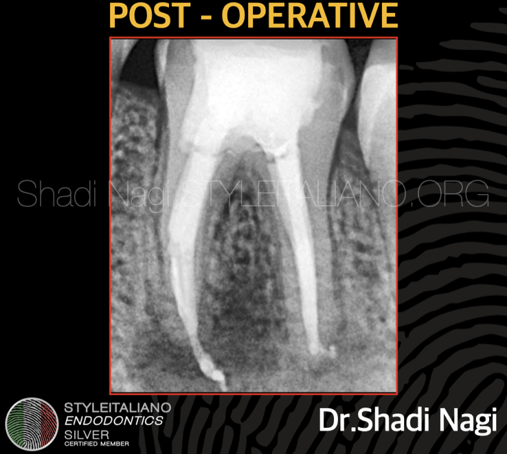
Fig. 12
Post operative radiograph.
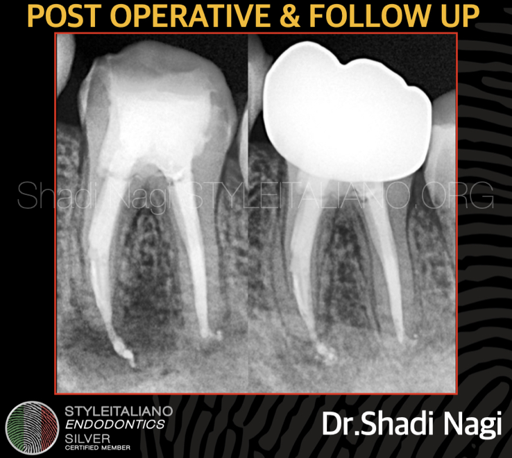
Fig. 13
Post operative and 8 month follow up.
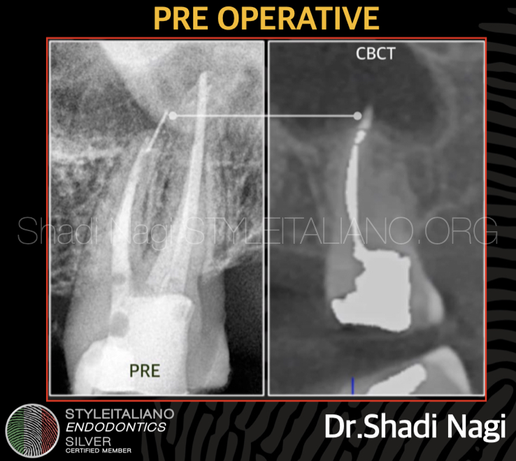
Fig. 14
Case no 6
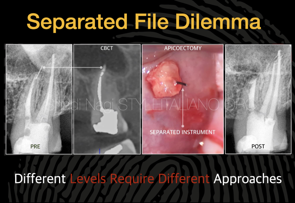
42 years old male patine referred to our clinic for management of separated file at maxillary second molar extended beyond the apex in the maxillary sinus .
The clinical examination showed presence of symptomatic apical periodontitis .
After the CBCT evaluation , the non surgical re-treatment done first followed by the micro surgical approach and removal of the file as a treatment option with very high success rate.
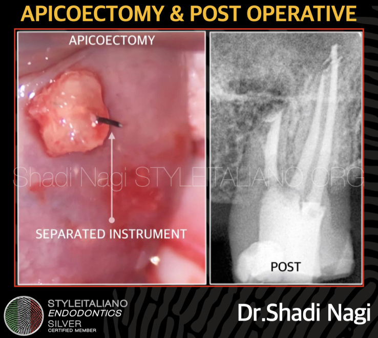
Fig. 15
The separated file removed after root end resection , root end prep followed by root end filling using BC PUTTY .
Post operative radiograph.
Conclusions
The management of the separated instruments from the root canal can be performed under magnification using different techniques such as loop , tube , ultrasonics depending on each case with minimum damage to the tooth and maintaining the original canal shape as much as possible , sometimes micro apical surgery is a backup treatment option .
Bibliography
1-Shen Y, Peng B, Cheung GS: Factors associated with the removal of fractured NiTi instruments from root canal systems. Oral Surg Oral Med Oral Pathol Oral Radiol Endod. 2004, 98:605-10. 10.1016/j.tripleo.2004.04.011
2-AL-ATTAS MH, AL-NAZHAN SA, AL-HARBI MS: Retrieval of separated instruments from root canal space: a case. J Clin Diagnostic Res. 2019, 13:121. 10.7860/JCDR/2019/40512.13111
3- Benoy J., Anjaneyulu K., Aishwarya Ranganath, Riluwan S. Management of Intracanal Separated File Fragment in a Four-Rooted Mandibular Third Molar https://doi.org/10.1155/2021/5547062
4- Alrahabi M, Ghabbani HM. Removal of a separated endodontic instrument by using the modified hollow tube–based extractor system: A case report https://doi.org/10.1177/2050313X20907822