Root fractures: clinical scenarios
19/12/2024
Fellow
Warning: Undefined variable $post in /home/styleendo/htdocs/styleitaliano-endodontics.org/wp-content/plugins/oxygen/component-framework/components/classes/code-block.class.php(133) : eval()'d code on line 2
Warning: Attempt to read property "ID" on null in /home/styleendo/htdocs/styleitaliano-endodontics.org/wp-content/plugins/oxygen/component-framework/components/classes/code-block.class.php(133) : eval()'d code on line 2
Root fracture is a situation that may happen and is generally associated to these symptoms:
- deep probing
- edema/sinus tract in the buccal or lingual aspect
- pain on chewing
Depending on the conditions, the symptoms referred by the patient may vary and, in some cases, painkillers are not able to help managing the pain.
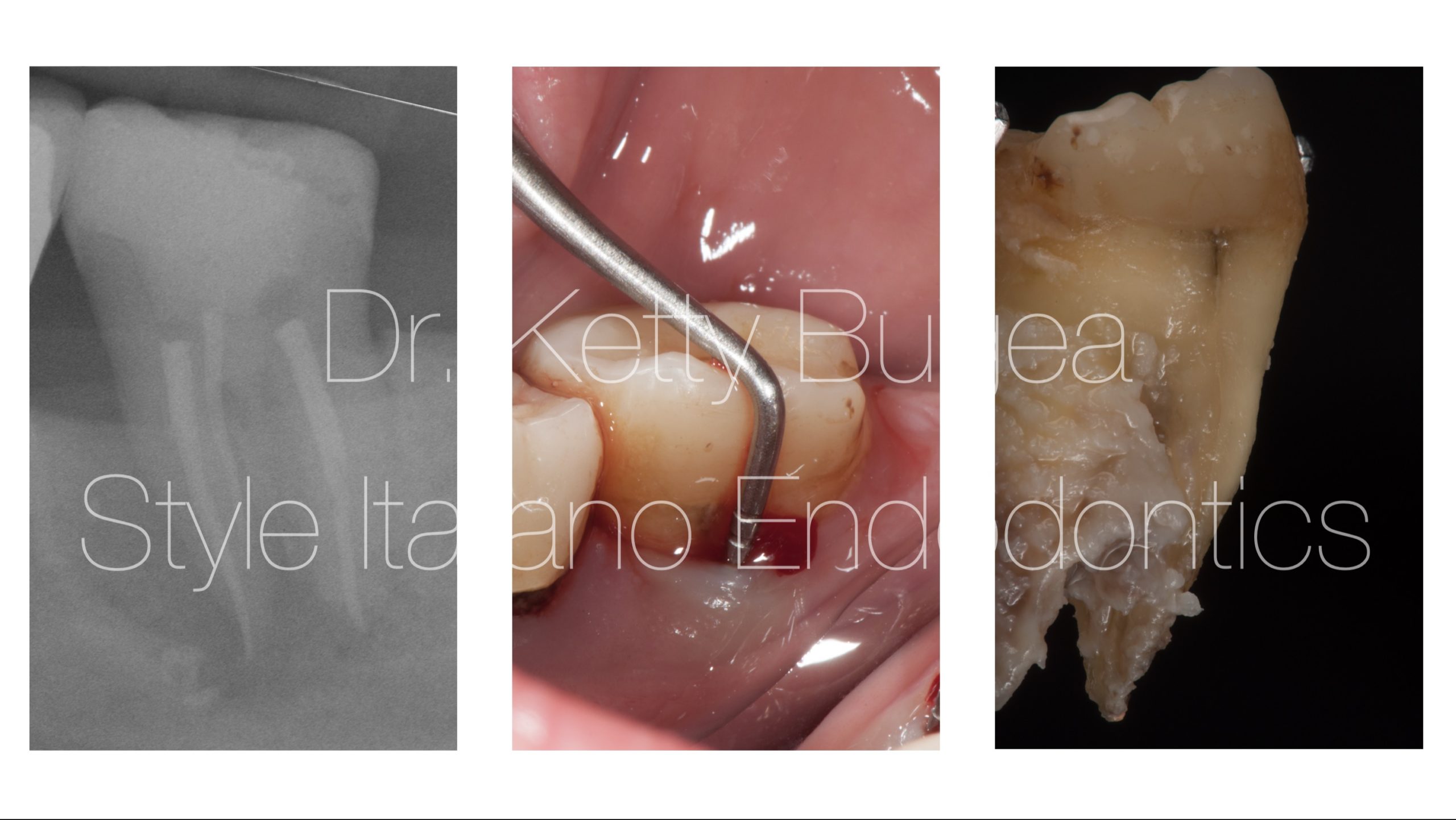
Fig. 1
Case 1
This patient came to my attention because of swelling. The periapical x-ray showed a large J-shaped lesion. typical of root fractures.
In the middle image the probe indicates the fracture line, evidencing a deep tubular defect
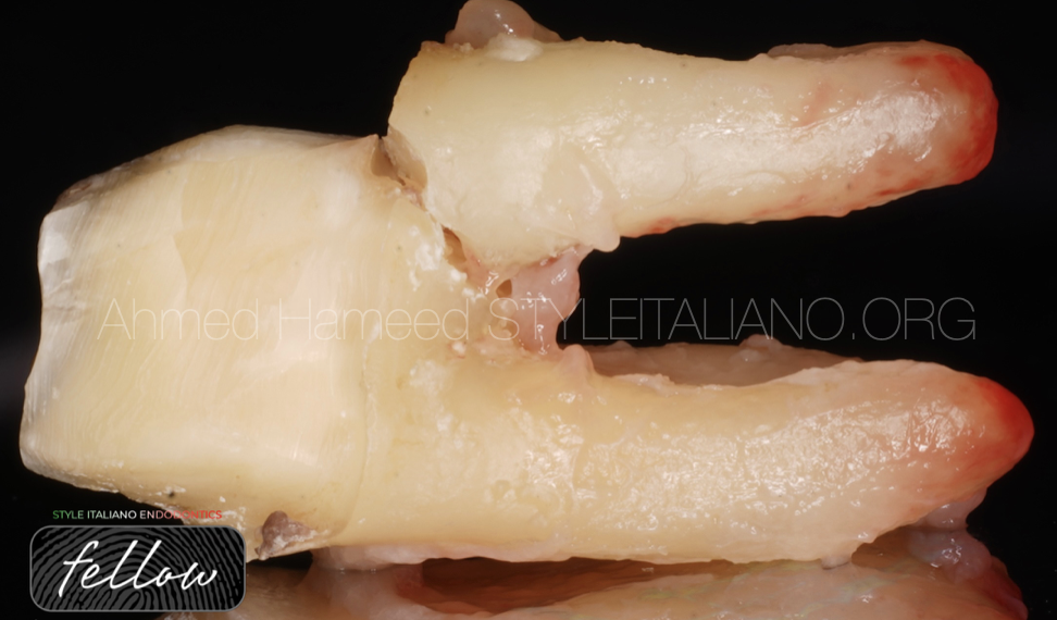
In the third picture, after the extraction we can notice the presence of a fracture in the mesial root.
The tooth was the last in the mandibular arch and it was mesialized, so probably the fracture was due to occlusal load, with the forces distributing along a vector that was not correct.
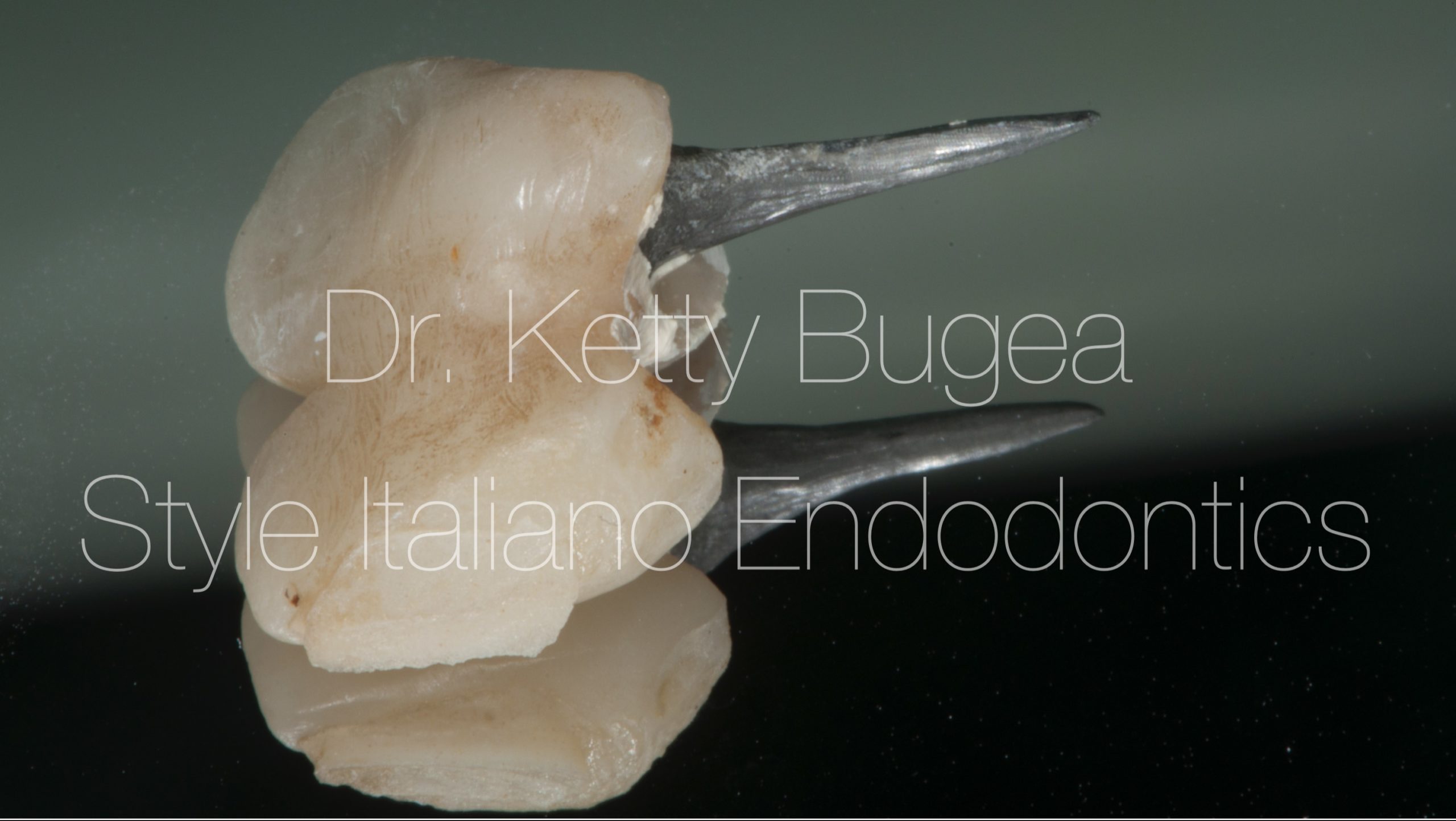
Fig. 2
Metal post
Metal posts are commonly associated with vertical root fracture due to the lateral action forces and to their size and inclination.
The removal of sound dentin to put a bigger post is an "old style" dentistry habitude that might be the cause of fracture.
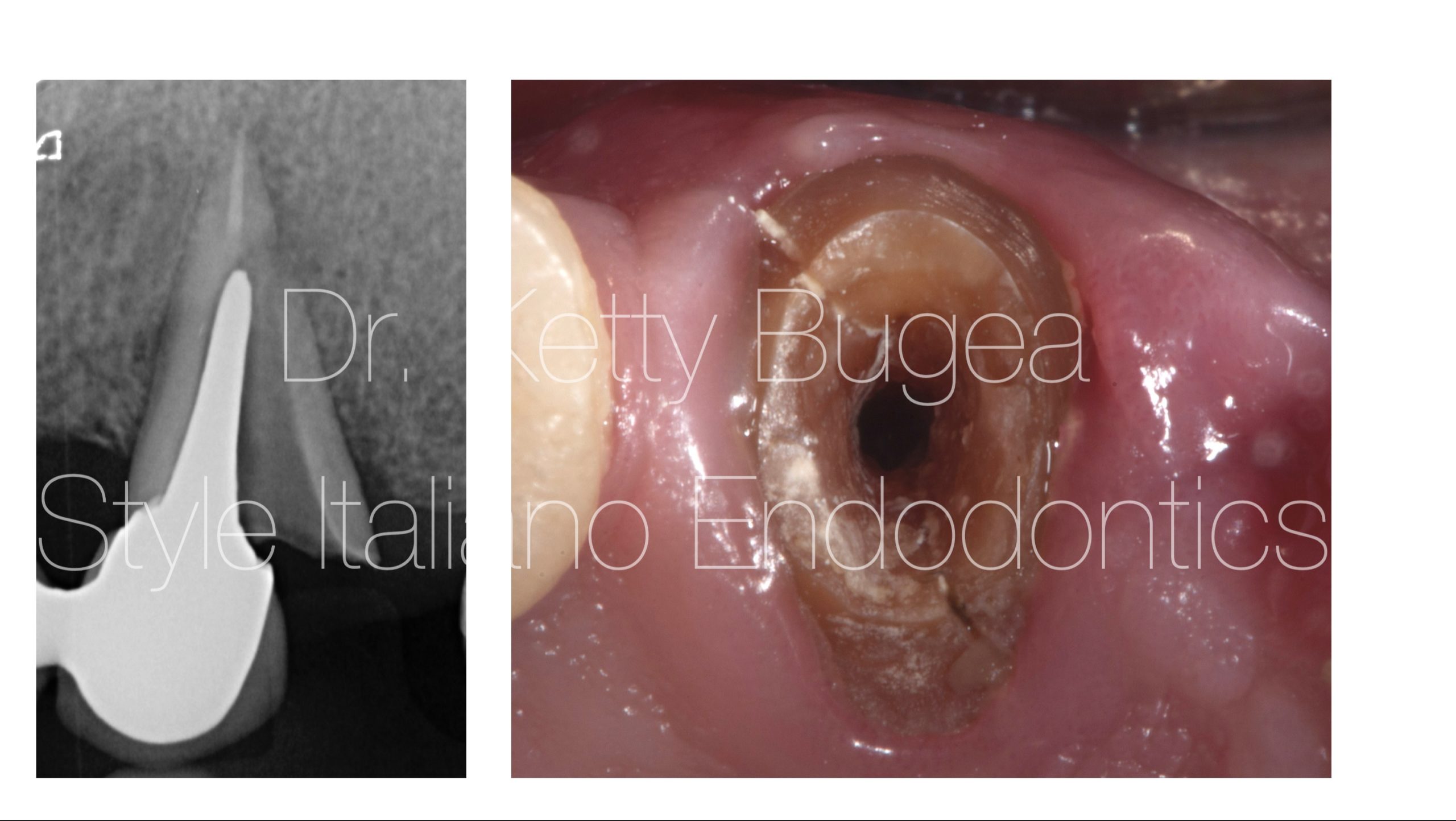
Fig. 3
In the first image the catastrophic fracture happened for the lack of ferule effect, lack of posterior tooth.
In the second image a fracture of an upper canine with a metal post without ferrule effect. The tooth fractured 3 months after the post cementation.
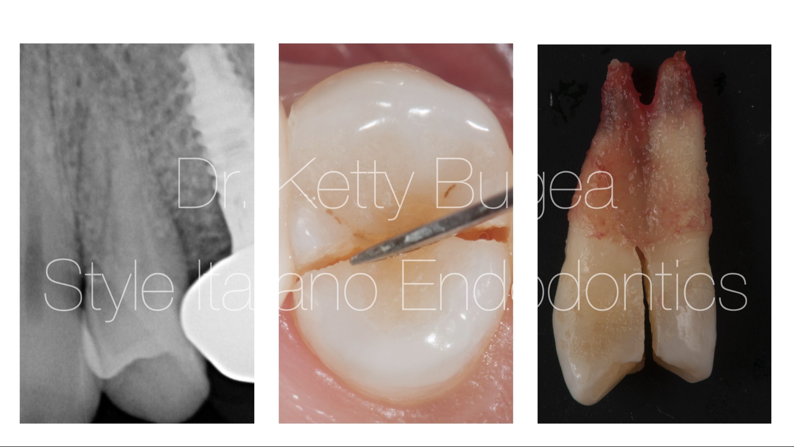
Fig. 4
Sometimes it happens that an healthy tooth with absence of decay, fillings and without root canal treatment begins to be symptomatic while chewing. The pain increases in intensity and the symptoms are similar to an irreversible pulpitis.
In this case the appearance of the tooth on the X-ray was normal, without signs of radiolucency in the apical area.
Probing depth was normal, but the patient had pain on percussion.
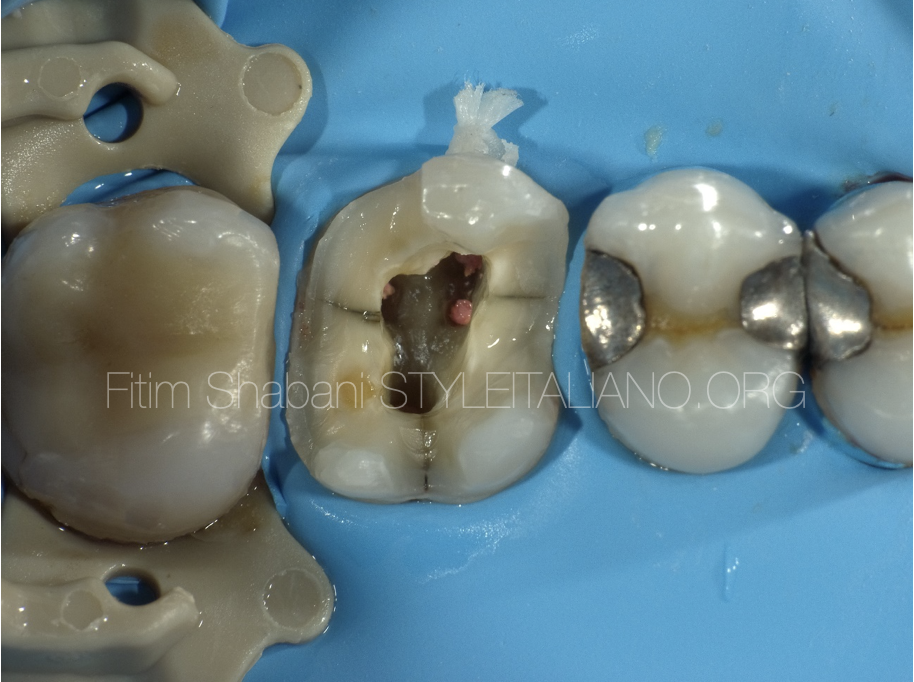
The diagnosis was possible only by inserting a spatula in the middle of the tooth, revealing a complete fracture. The tooth was extracted.

Fig. 5
Fractures can develop alzo horizontally.
In this case it's possible to see a radiolucency only in the middle of the root, with a thin line dividing the apical area from the medium third of the root. After extraction an horizontal fracture was observed. The apical portion was then extracted.
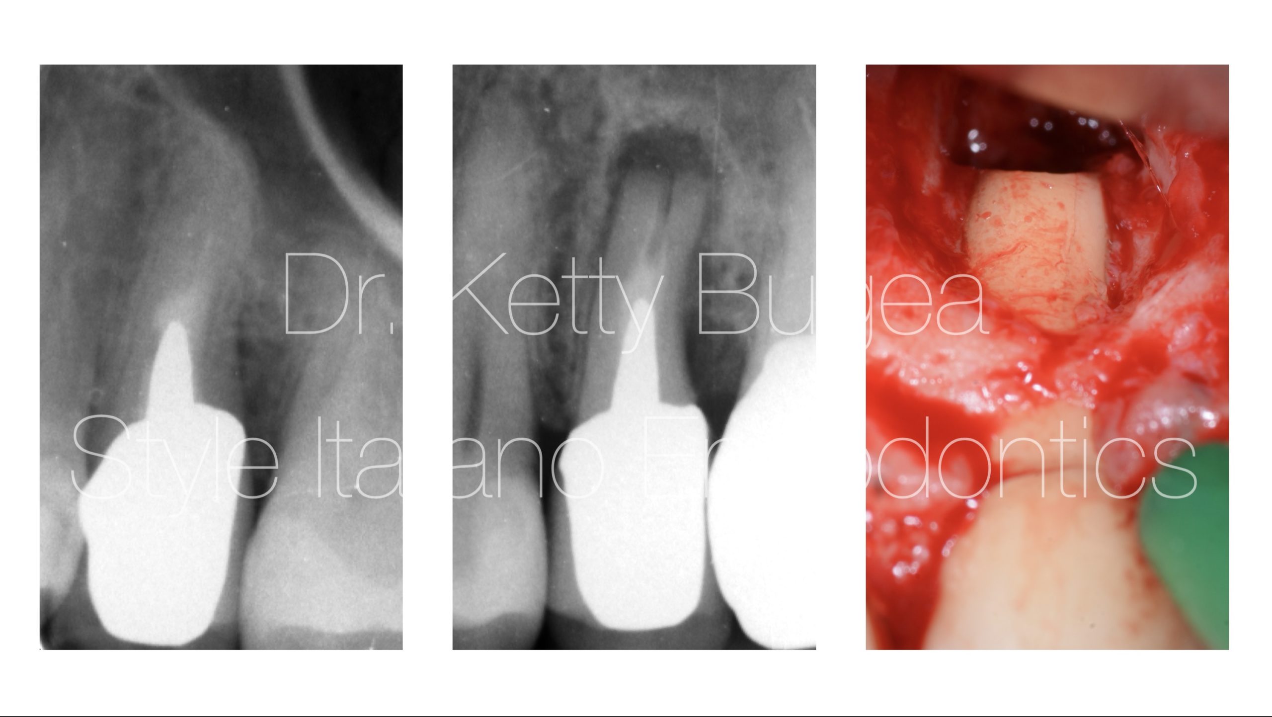
Fig. 6
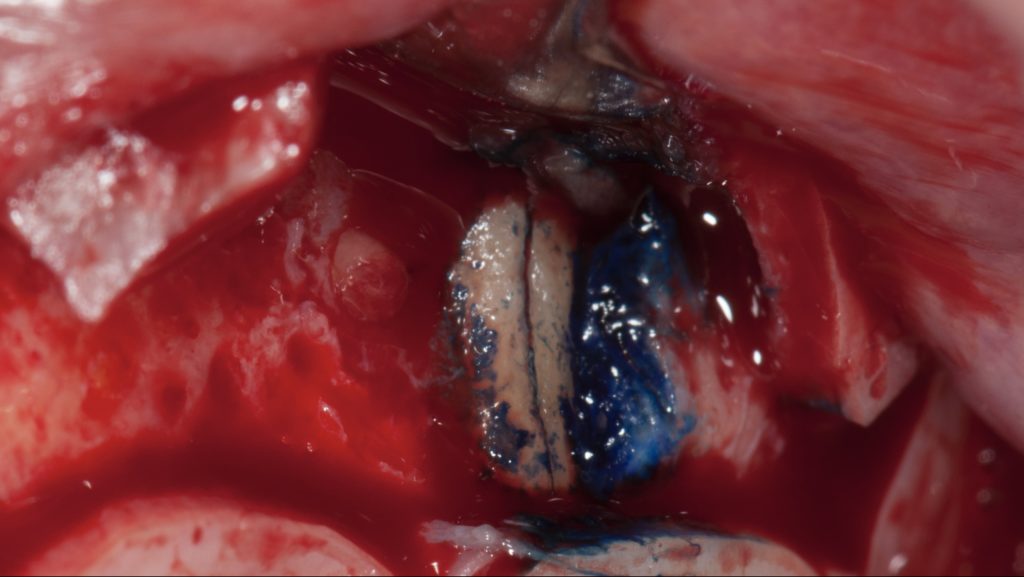
Sometimes the diagnosis of root fracture is very difficult, we can suspect it but in case of trouble an explorative flap may confirm the presence of a fracture.
The patient has undergone an apical surgery from a maxillo facial surgeon, without a microscope, without ultrasonic retropreparation and obturation. Two months later he came to my clinic complaining about the persistence of pain and swelling. After a periapical xray a fracture was suspected. An explorative flap was done and it was possible to evidence a fracture. The the tooth was extracted in the same session

Fig. 7
Born in Agrigento on 22/04/1985
Graduated with honours in dentistry and dental prosthesis from the "Magna Graecia" University of Catanzaro. She mainly dedicates herself to conservative dentistry and endodontics. She works as a consultant in Tolmezzo (UD). Fellow of Style Italiano Endodontics.
Conclusions
Root fractures are sometimes very difficult to diagnose, an appropriate anamnesis, clinical evaluation associated by bimensional and tridimensional X-rays can help the clinicians to formulate a correct diagnosis.
Bibliography
Patel S, Bhuva B, Bose R. Present status and future directions: Vertical root fractures in root filled teeth. Int Endod J. 2022;55(Suppl. 3):804-26
Haueisen H, Gärtner K, Kaiser L, et al. Vertical root fracture: Prevalence, etiology, and diagnosis. Quintessence Int. 2013;44(7):467-74
Haupt F, Wiegand A, Kanzow P. Risk factors for and clinical presentations indicative of vertical root fracture in endodontically treated teeth: A systematic review and meta-analysis. J Endod. 2023;49(8):940-52


