Root canal treatment of an immature permanent mandibular first molar
03/06/2023
Fellow
Warning: Undefined variable $post in /home/styleendo/htdocs/styleitaliano-endodontics.org/wp-content/plugins/oxygen/component-framework/components/classes/code-block.class.php(133) : eval()'d code on line 2
Warning: Attempt to read property "ID" on null in /home/styleendo/htdocs/styleitaliano-endodontics.org/wp-content/plugins/oxygen/component-framework/components/classes/code-block.class.php(133) : eval()'d code on line 2
The root canal treatment of immature permanent teeth poses many challenges, one of which is the obturation technique. These teeth come with wide apices and sometimes even a divergent configuration (or reverse anatomy) in the apical third. The difficulty in such cases is to avoid overextending the filling material into the periapical tissues. (1,2)
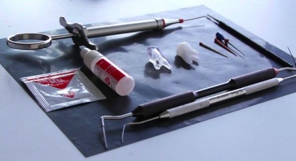
In these cases, apexification is indicated: it consists of creating an apical plug using MTA. Proper delivery and condensation of the MTA to the apical third of the canal is an essential requirement for the success of this procedure (3). However, MTA has difficult handling, especially in apexification where it should be placed in the apical 3-4mm (4). Traditional placement of MTA is difficult because the product was placed in the coronal third then pushed to the apical part. Therefore, to make apexification easier, it appears beneficial to use devices that allow direct delivery of the MTA to the apical third.
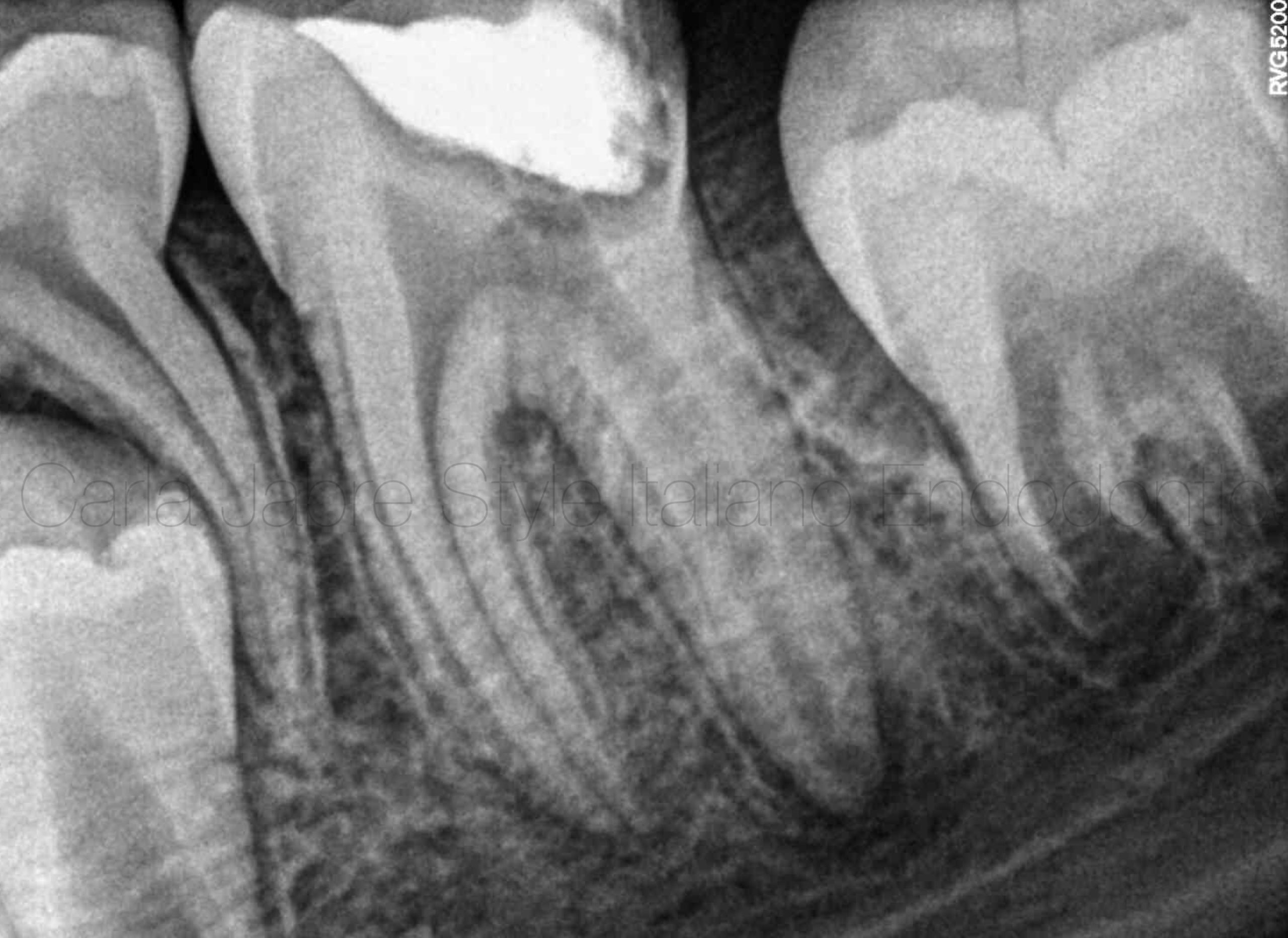
Fig. 1
A 10-year-old patient was referred for a root canal treatment on his mandibular first molar. The diagnosis was pulp necrosis and asymptomatic apical periodontitis.
The initial radiograph shows an immature distal root (reverse anatomy) with an apical radiolucency. However, the mesial root appears fully developed, therefore Regenerative Endodontic Treatment was not an option (5).
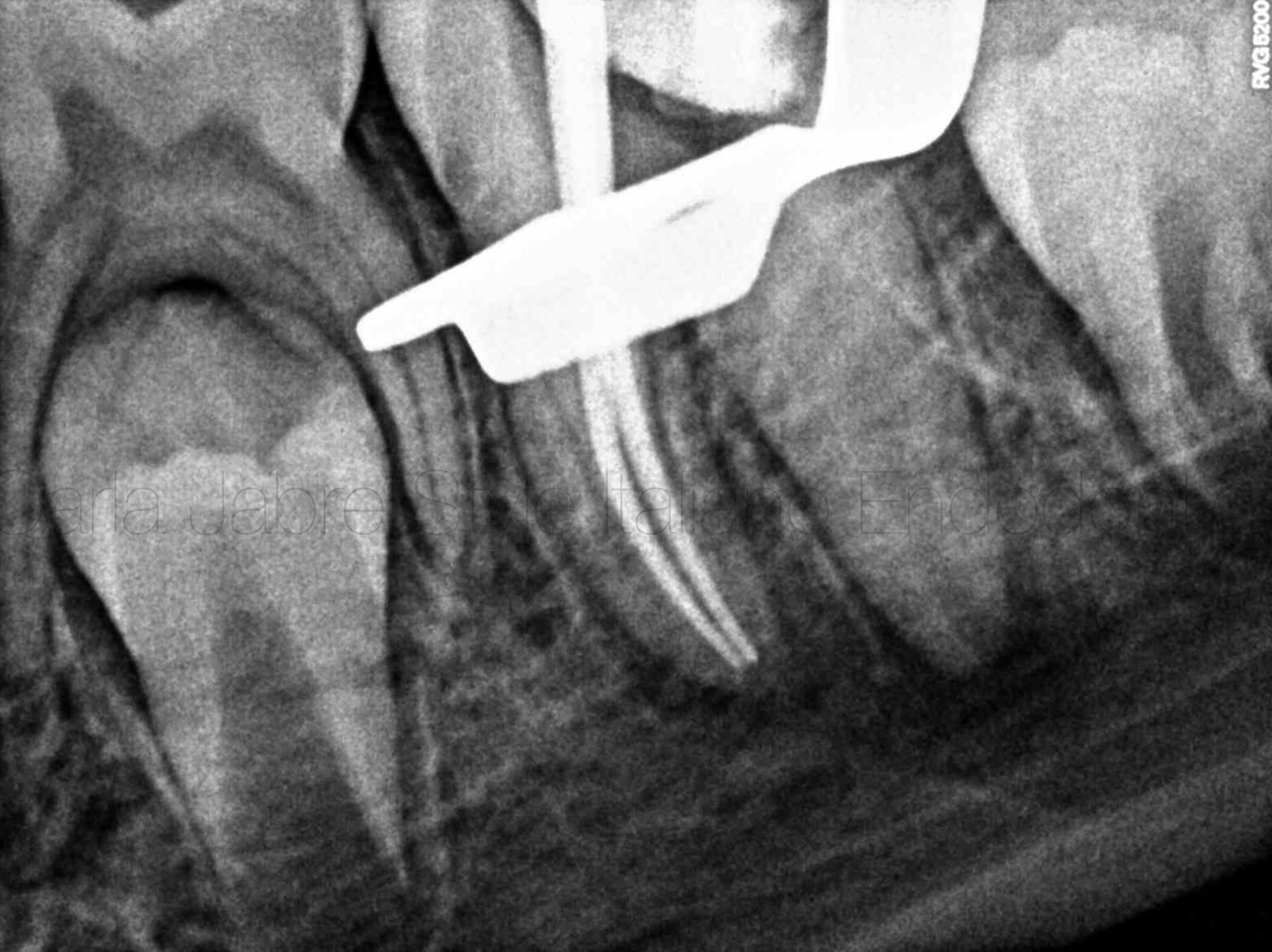
Fig. 2
Once shaping was done, gutta-percha cones were fitted in the mesial canals. A tug-back could be achieved. These canals were filled using the warm condensation technique.
As for the distal canals, the main focus was put on irrigation and sonic activation while avoiding aggressive instrumentation on the already thin canal walls (3).
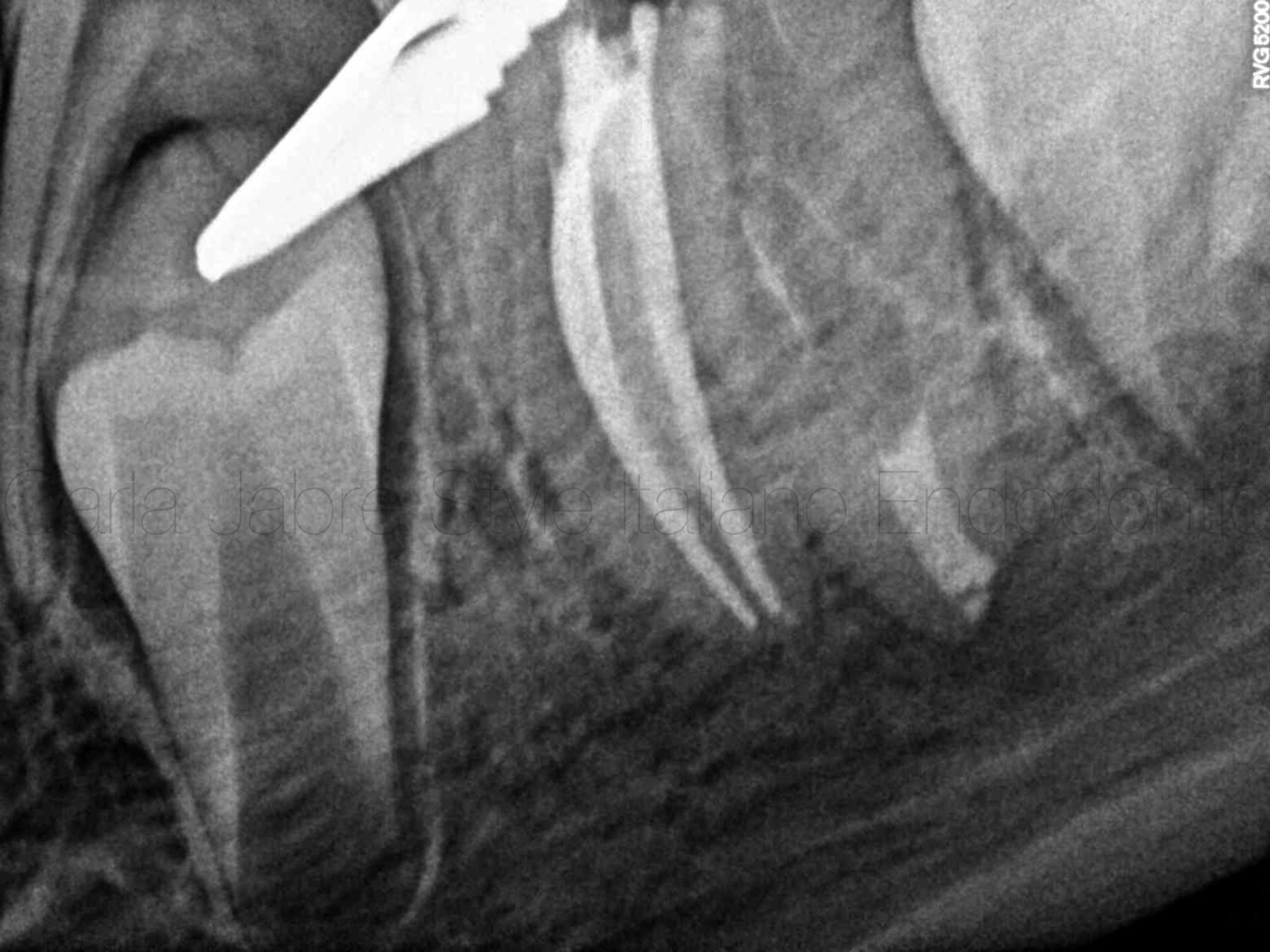
Fig. 3
Obturation of the mesial canals. Note the sealer in the isthmus between the 2 canals.
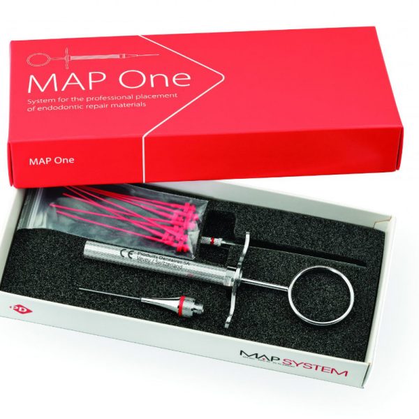
A 4mm apical plug of MTA was done in the DL canal, using the MAP One (Produits Dentaires, Switzerland).
A moist cotton pellet was put under the temporary cement. A second appointment was scheduled 24 hours later for backfill in the distal canals.
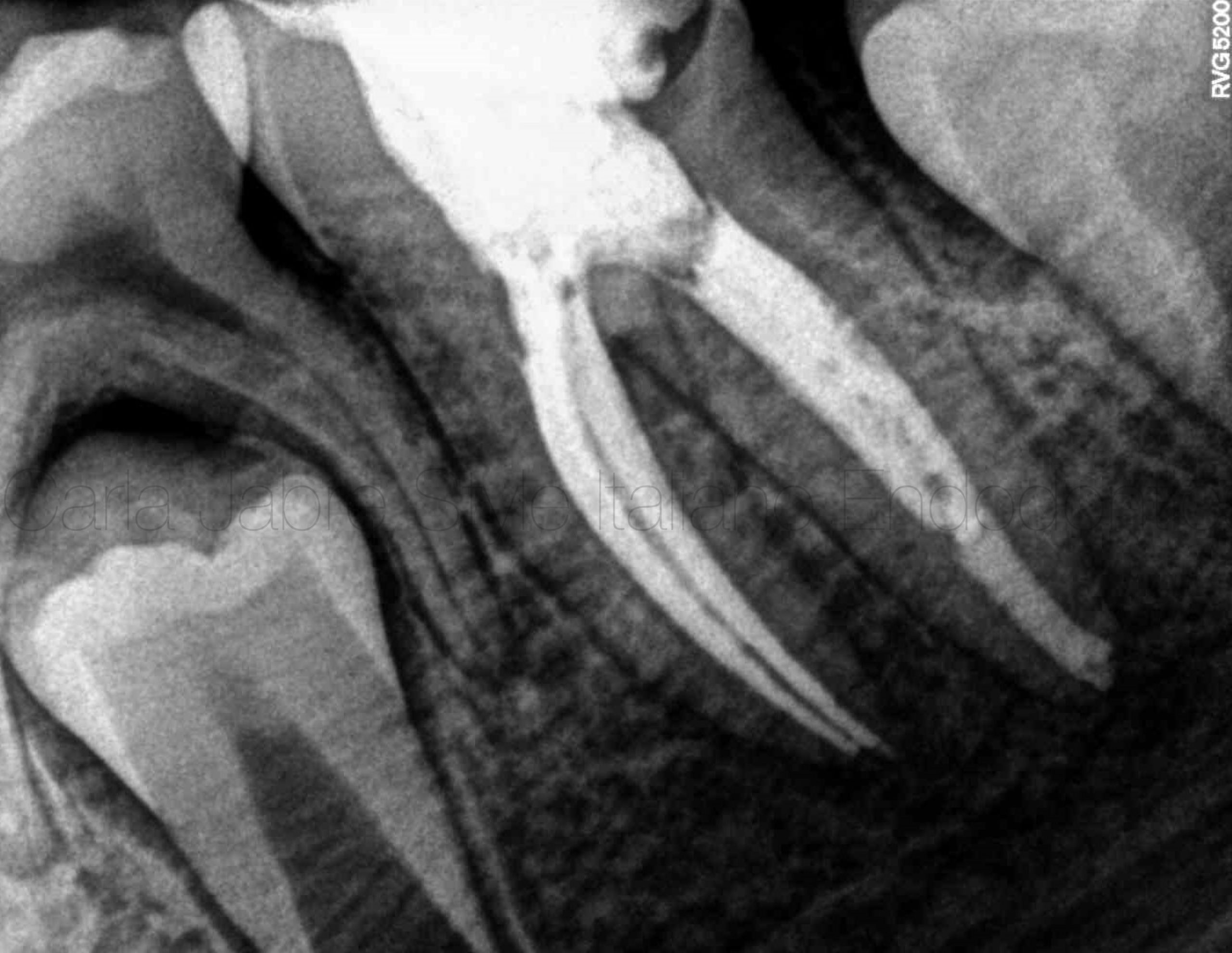
Fig. 4
Final radiograph after backfill in the joining distal canals.
The patient was referred back to the pediatric dentist for the final coronal restoration.
Step by step video
Conclusions
In immature necrotic permanent teeth where conventional canal obturation is impossible, apexification is a predictable alternative for creating an apical seal, especially when using devices that allow the delivery of MTA into the apical area directly.
Bibliography
1: Wigler R, Kaufman A, Lin S, et al. Revascularization: A Treatment for Permanent Teeth with Necrotic Pulp and Incomplete Root Development. J Endod 2013;39:319–326.
2: Harlam SC. Management of incompletely developed teeth requiring root canal treatment. Australian Dental Journal 2016; 61:(1): 95–106.
3: Tolibah, Y.A.; Droubi, L.; Alkurdi, S.; Abbara, M.T.; Bshara, N.; Lazkani, T.; Kouchaji, C.; Ahmad, I.A.; Baghdadi, Z.D. Evaluation of a Novel Tool for Apical Plug Formation during Apexification of Immature Teeth. Int. J. Environ. Res. Public Health 2022, 19, 5304. https://doi.org/10.3390/ ijerph19095304.
4: Nicoloso GF, Goldenfum GM, Dal Pizzol T, et al. Pulp Revascularization or Apexification for the Treatment of Immature Necrotic Permanent Teeth: Systematic Review and Meta Analysis. JOCPD 2019; 43 (5): 305-313.
5: Panda, P.; Mishra, L.; Govind, S.; Panda, S.; Lapinska, B. Clinical Outcome and Comparison of Regenerative and Apexification Intervention in Young Immature Necrotic Teeth—A Systematic Review and Meta-Analysis. J. Clin. Med. 2022, 11, 3909. https://doi.org/10.3390/ jcm11133909.


