Fab Lab: video tutorial of MTA Sandwich Technique
10/03/2017
Riccardo Tonini
Warning: Undefined variable $post in /home/styleendo/htdocs/styleitaliano-endodontics.org/wp-content/plugins/oxygen/component-framework/components/classes/code-block.class.php(133) : eval()'d code on line 2
Warning: Attempt to read property "ID" on null in /home/styleendo/htdocs/styleitaliano-endodontics.org/wp-content/plugins/oxygen/component-framework/components/classes/code-block.class.php(133) : eval()'d code on line 2
Root perforation repair has historically been a treatment with a low success rate; however, recent techniques and materials utilized in root perforation repair, have dramatically improved the prognosis of both surgical and nonsurgical procedures. Root perforation is defined as an artificial communication between the root canal system to the supporting tissues of teeth often caused by using rotary burs inside root canals. In my practice, I have found lot of perforations caused by an inappropriate post space preparation for permanent restoration of endodontically treated teeth.They are located in the middle part of the canal and, according to my personal statistic, 80% of the cases involve the first lower molar: considering this tooth, 60% of perforations are in the mesial root and 40% in the distal root and they are always generated by an over-preparation of post space that has not taken into consideration the geometry of the cross-sectional anatomy of the lower first molar. Another consideration is that large-sized perforations may not respond to repair as well as smaller ones.
DIAGNOSIS: Bacterial infection emanating either from the root canal or the periodontal tissues, or both, prevents healing and brings about inflammatory sequels where exposure of the supporting tissues is inflicted. Thus, painful conditions, suppurations resulting in tender teeth, abscesses, and fistulae including bone resorptive processes may follow (1). A narrow isolated periodontal defect is a possible sign of root perforation. To determine locally isolated vertical bone losses, periodontal probing should be carried out by walking the probe around the tooth while pressing gently on the floor of the sulcus (2).
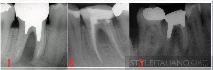
Fig. 1
In the first lower molar an X ray can often show a bone loss between roots and diagnosis is easier than other teeth.
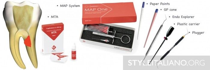
Fig. 2
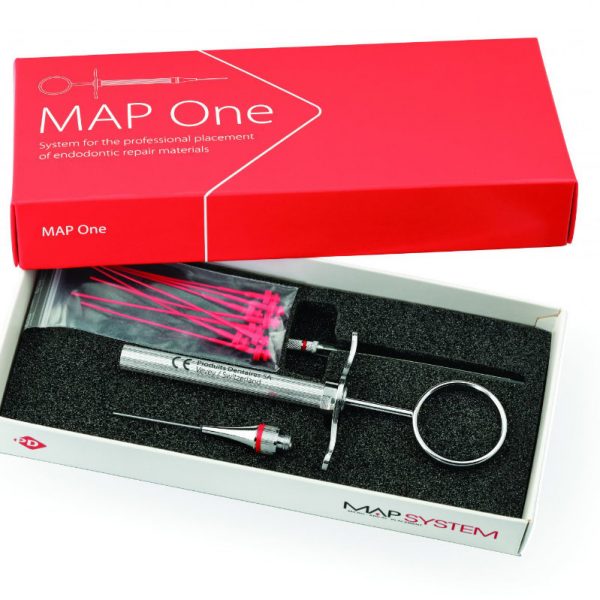
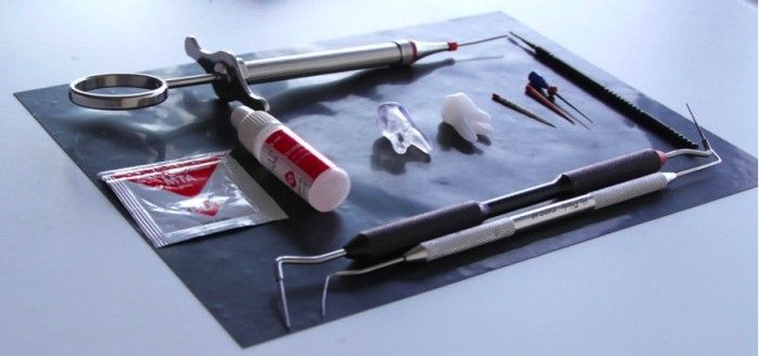
The MTA sandwich technique is the one that i personally reccomand for this kind of perforation and it has been well described by Fabio Gorni in this article But what's the equipment? 1) MTA is my choice material for perforation repair and numerous case reports exist in the literature showing excellent healing results with MTA. 2) The MAP (Micro-Apical Placement) System, provides an efficient method for placing repair materials and is the only one device that lasts over time. It has lot of tips,steel and NiTi, that can satisfy all the requirements. I love the NiTi tip because i can bend it as i want. 3) Paper points 4) One gutta cone 5) One plugger 6) One plastic carrier (usually 40 or 60) 7) One Endo Explorer (Dg16)
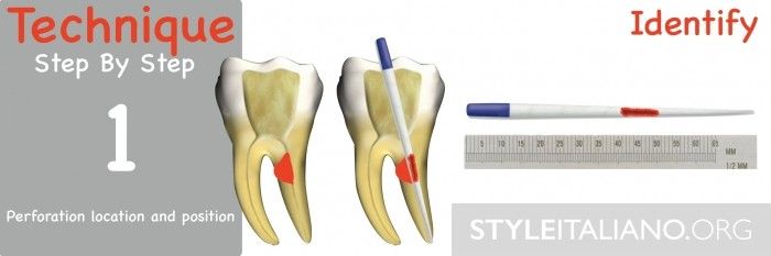
Fig. 3
As accurate detection of root perforations and determination of location are crucial to the treatment outcome. A paper point is enough and the appearance of blood in the middle part of it is the perfect sign for a right diagnosis, detection and location of a perforation.
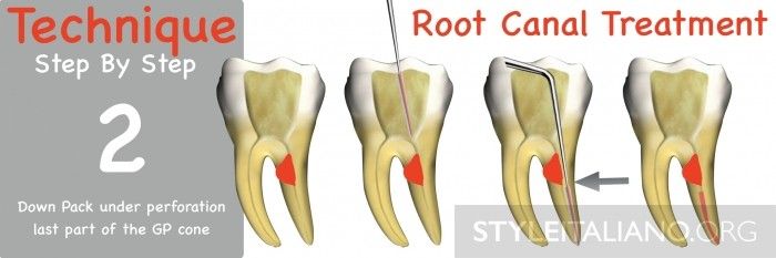
Fig. 4
The second step is represented by a conventional RCT, but obturation is done with last part of a guttapecha cone, starting with warm gauutapercha condensation deeper than the perforation level, avoiding any contamination of perforation area with sealer and GP. A plugger is used for GP condensation
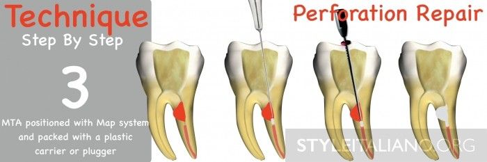
Fig. 5
During the third step MTA is positioned with MAP system. The system consists of a stainless-steel or NiTi applicator with a bayonet catch for several exchangeable applicator cannulas (needles). Inside the cannula there is a plunger made in polymer that is longer than cannula providing a complete extrusion of internal material. The MTA can be taken from a dispenser thrusting the tip into the repair material and placed inside the canal in a sharp way pressing syringe piston to expel the material. An endo Micro Brush can be used to gently pack the MTA or a plastic carrier can be used for a stronger condensation. MTA must be placed to all the extension of perforation.
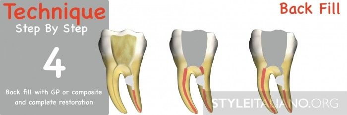
Fig. 6
The last step is rappresented by backfilling the coronal part with GP or composite material in a second visit.
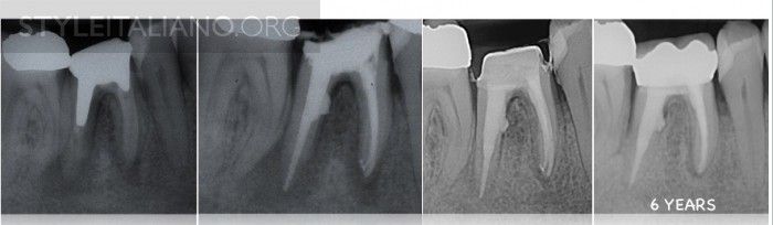
Fig. 7
The complete case with 6 years follow-up
Conclusions
Right techniques and right materials and devices ensure good results and success over time. The MTA sandwich technique is a safe and repeatable procedure that must be considered for root canal perforations repair.
Bibliography
IGOR TSESIS & ZVI FUSS Diagnosis and treatment of accidental root perforations ,Endodontic Topics 2006, 13, 95107
Cohen S, Liewehr F. Diagnostic procedures. In: Cohen S, Burns RC, eds. Pathways of the Pulp, 8th edn. St Louis, MO: CV Mosby, 2002: 12.
Kvinnsland I, Oswald RJ, Halse A, Gronningsaeter AG. A clinical and roentgenological study of 55 cases of root perforation. Int Endod J 1989: 22: 7584.
Nicholls E. Treatment of traumatic perforations of the pulp cavity. Oral Surg Oral Med Oral Pathol 1962: 15: 603612.
Abou-Rass M, Frank AL, Glick DH. The anticurvature filing method to prepare the curved root canal. J Am Dent Assoc 1980: 101: 792794.
Eleftheriadis GI, Lambrianidis TP. Technical quality of root canal treatment and detection of iatrogenic errors in an undergraduate dental clinic. Int Endod J 2005: 38: 725734.
Sinai IH. Endodontic perforations: their prognosis and treatment. J Am Dent Assoc 1977: 95: 9095.
Gutmann JL, Harrison JW. Surgical Endodontics. Boston, MA: Blackwell Scientific Publications, 1991, p. 409.
Fuss Z, Tsesis I, Lin S. Root resorption diagnosis, classification and treatment choices based on stimulation factors. Dent Traumatol 2003: 19: 175182.
Ingle JL. A standardized endodontic technique utiliz- ing newly designed instruments and filling materials. Oral Surg Oral Med Oral Pathol 1961: 14: 8391.