Retreatment of a Mandibular Second Molar with a Periapical Lesion in a C-Shaped Canal
28/08/2025
Mohamed Milad Ganawo
Warning: Undefined variable $post in /home/styleendo/htdocs/styleitaliano-endodontics.org/wp-content/plugins/oxygen/component-framework/components/classes/code-block.class.php(133) : eval()'d code on line 2
Warning: Attempt to read property "ID" on null in /home/styleendo/htdocs/styleitaliano-endodontics.org/wp-content/plugins/oxygen/component-framework/components/classes/code-block.class.php(133) : eval()'d code on line 2
Endodontic retreatment of a mandibular second molar with a C-shaped canal configuration presents unique clinical challenges due to its complex and variable anatomy. C-shaped canals, are commonly found in mandibular second molars, retreatment becomes complicated by the difficulty in completely cleaning, disinfecting, and obturating the root canal .
Reasons related to treatment failure include missed canals, inadequate debridement, and poor sealing of the C-shaped anatomy. Successful retreatment requires advanced diagnostic tools, such as CBCT imaging, enhanced visualization with dental microscopes, and specialized techniques for canal negotiation and disinfection.
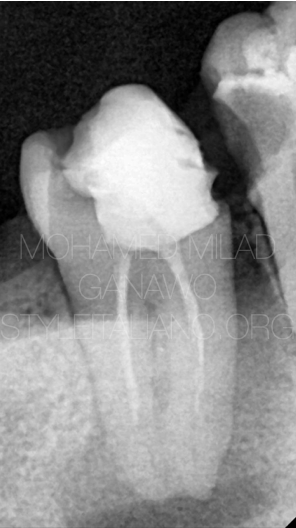
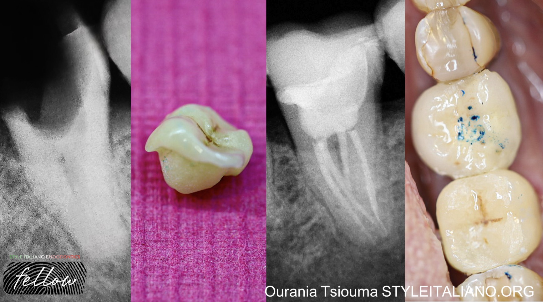
Fig. 1
Initial situation shows poor permanent restoration, poor and under extended fill, and apical radiolucency.
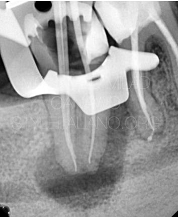
Fig. 2
After old fill was entirely removed canals cleaned, now cone fit as shown on radiograph.
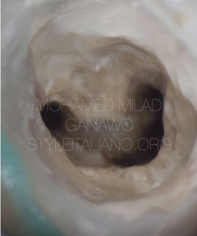
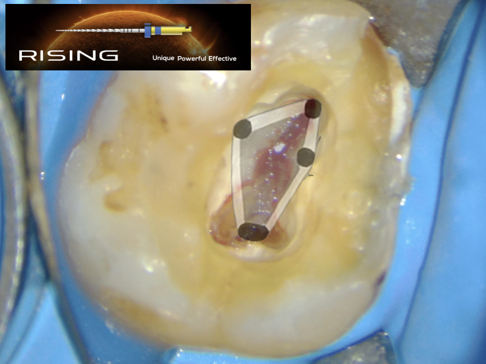
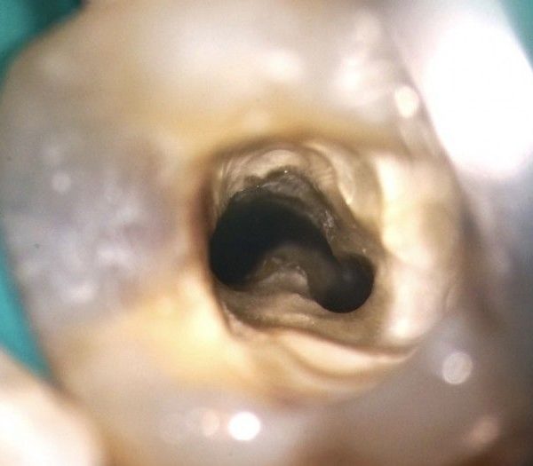
Fig. 3
Microscopic shot shows root canal map in C shaped way.
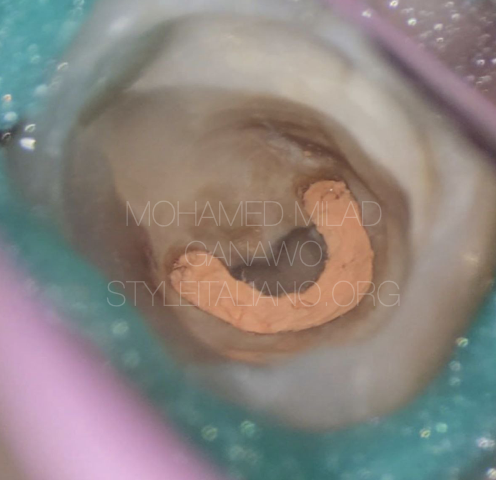
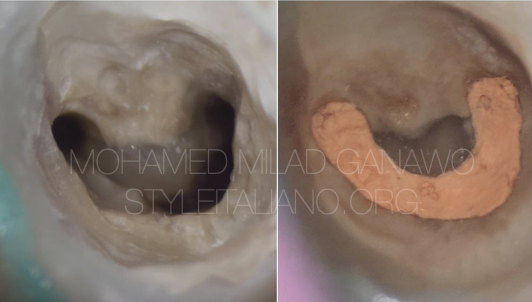
Fig. 4
Immediate microscopic shot right after.obturation shows C shaped obturation
Video of the procedure
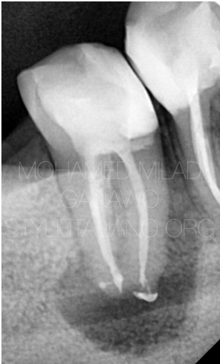
Fig. 5
Obturation done , and reveals lateral canals
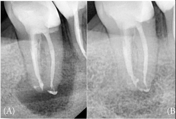
Fig. 6
- pre-operative.
- One year follow up.
Conclusions
The retreatment of a mandibular second molar with a C-shaped canal and associated periapical radiolucency demands Proper diagnosis using CBCT, and use of magnification and ultrasonic instruments.
Effective disinfection with sodium hypochlorite imagination and calcium hydroxide medication improves the chances of periapical healing. Obturation techniques such as thermoplasticized gutta-percha or bioceramic sealers help achieve a three-dimensional seal.
even highly complex C-shaped canal cases can be successfully retreated, preserving the tooth and restoring function, just keep the patient under regular follow up.
Bibliography
1. Fan, B., Cheung, G. S., Fan, M., Gutmann, J. L., & Bian, Z. (2004). C-shaped canal system in mandibular second molars: Part I—Anatomical features. Journal of Endodontics, 30(12), 899-903.
2. Jin, G. C., Lee, S. J., & Roh, B. D. (2006). Anatomical study of C-shaped canals in mandibular second molars by analysis of computed tomography. Journal of Endodontics, 32(1), 10-13.
3. Mauger, M. J., Schindler, W. G., & Walker, W. A. (1998).An evaluation of canal morphology at different levels of root resection in mandibular first molars. Journal of Endodontics, 24(9), 607-609.
4. Zhang, R., Yang, H., Yu, X., Wang, H., Hu, T., & Dummer, P. M. (2011).Use of CBCT to identify the morphology of mandibular permanent molar teeth in a Chinese subpopulation. International Endodontic Journal, 44(2), 162-169.
5. Gutmann, J. L., & Gao, Y. (2012).
Alteration in the inherent metallic and surface properties of nickel-titanium root canal instruments to enhance performance, durability, and safety: A focused review. International Endodontic Journal, 45(2), 113-128.