RCT Of A First Lower Molar with Five Canals And An Additional Distal Root
01/03/2025
Fellow
Warning: Undefined variable $post in /home/styleendo/htdocs/styleitaliano-endodontics.org/wp-content/plugins/oxygen/component-framework/components/classes/code-block.class.php(133) : eval()'d code on line 2
Warning: Attempt to read property "ID" on null in /home/styleendo/htdocs/styleitaliano-endodontics.org/wp-content/plugins/oxygen/component-framework/components/classes/code-block.class.php(133) : eval()'d code on line 2
The chemomechanical preparation of the canal system is one of the most important phases of endodontic treatment and can be compromised in certain cases when performing a minimally invasive approach if several factors are not taken into account.
By reducing the size of the instruments we will deform the canal less, creating a faster and more anatomical mechanical instrumentation.
The main problem will be the chemical part of the irrigation, which due to the reduction in size of the mechanical preparation, will make it more difficult to reach the apical third with our irrigation needles.
Anatomy will also play a very important role in this aspect, as due to its great variability and complexity, it can compromise the success of our chemomechanical preparation.
In this case, we are treating a lower molar with five canals and an additional distolingual root (Radix Entomolaris).
The anatomy of the additional root often causes problems during instrumentation due to its severe curvature. The use of CBCT is essential in order to fully analyse the anatomy and position of the canals as well as the degree of curvature and the convergence points between the canals.
Although they are not frequent, we must be aware of this anatomical variation in order not to make mistakes during the preparation of the canal system.
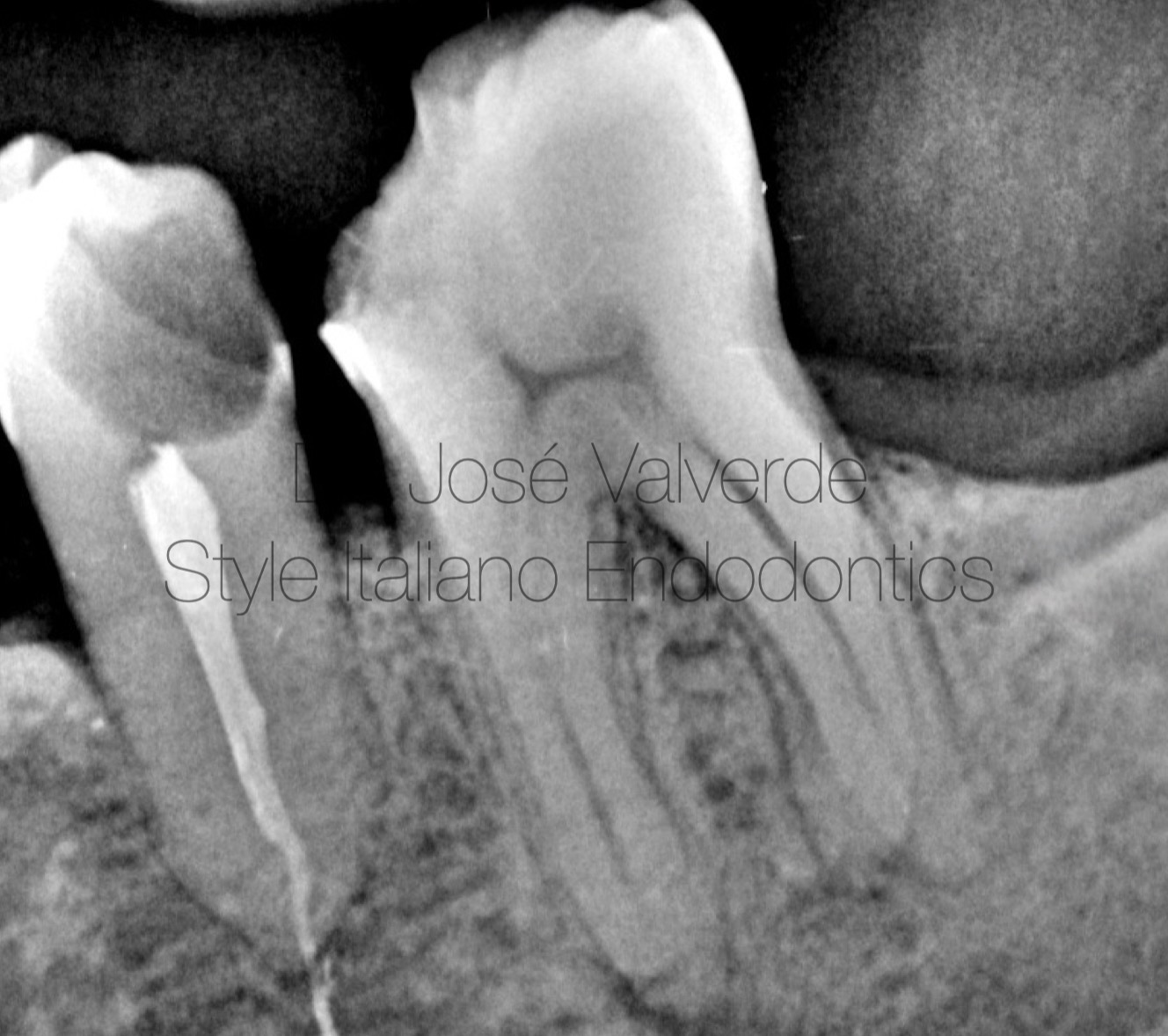
Fig. 1
A 35-year-old patient comes to the office with severe pain when drinking cold drinks and while chewing.
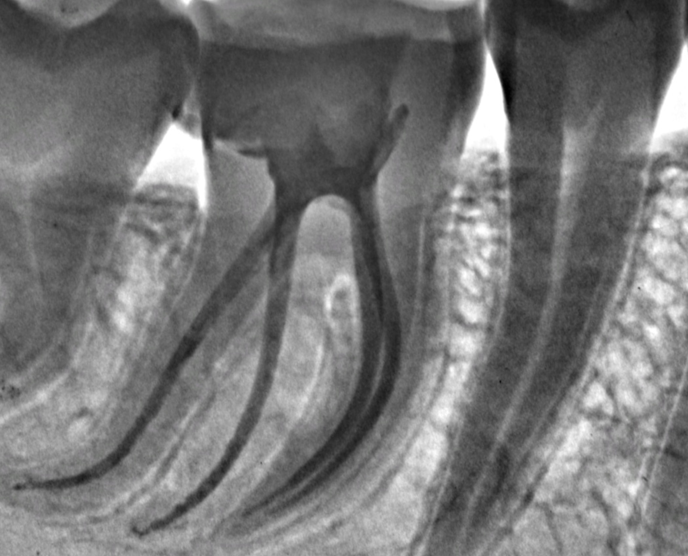
In the preoperative X-ray we can suspect an additional distal root. Due to the complexity of the anatomy, we decided to take a CBCT to analyse the position and curvature of the canals.
The diagnosis was irreversible pulpitis without periodontal tissue involvement.

Fig. 2
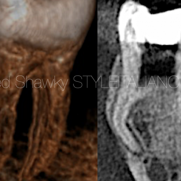
Axial view showing the crowded shape of the distal canal and the presence of at least 3 canals in the distal root.
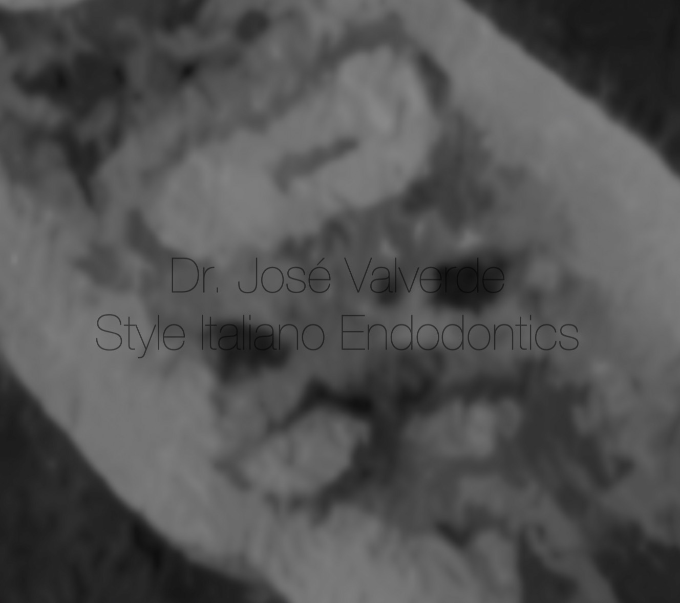
Fig. 3
We can see how in the apical zone they separate into two roots.
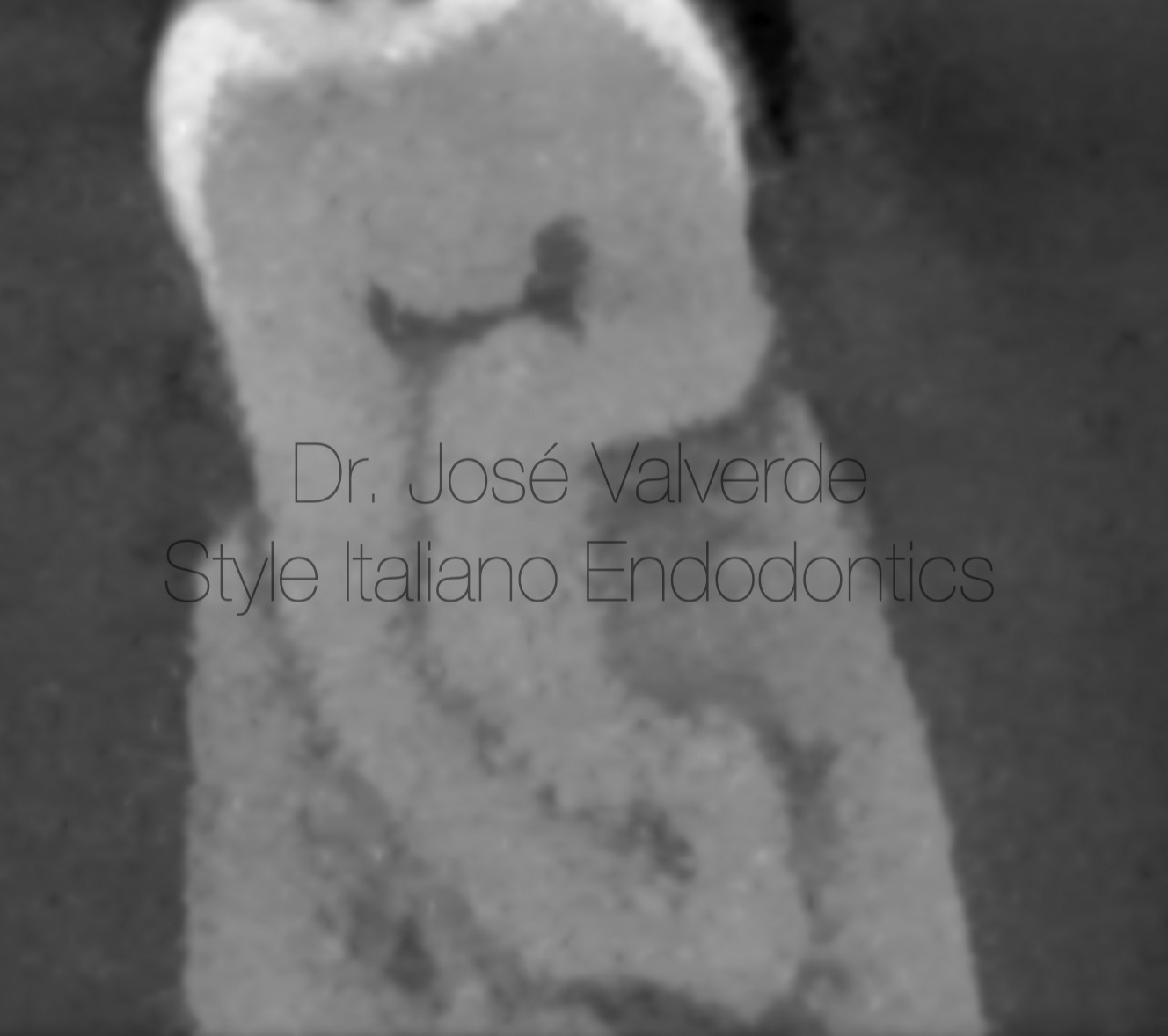
Fig. 4
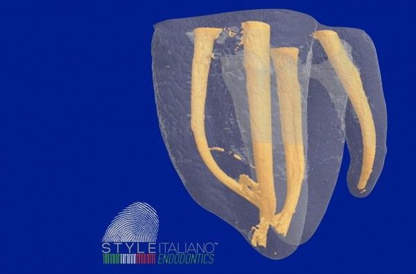
Double curvature of the mesial root.
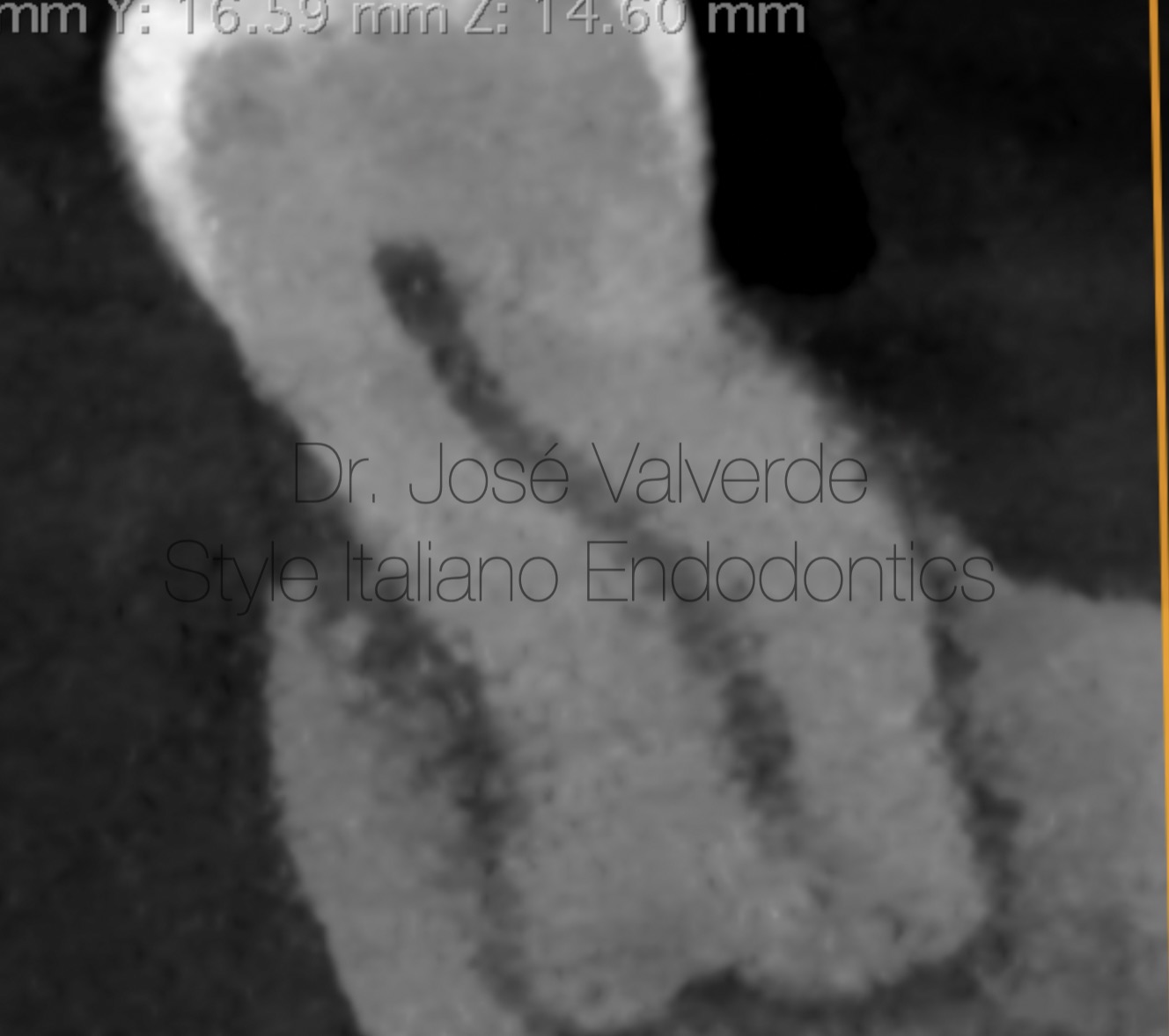
Fig. 5
We can see the complexity of the distal root and the bifurcation in the apical third.
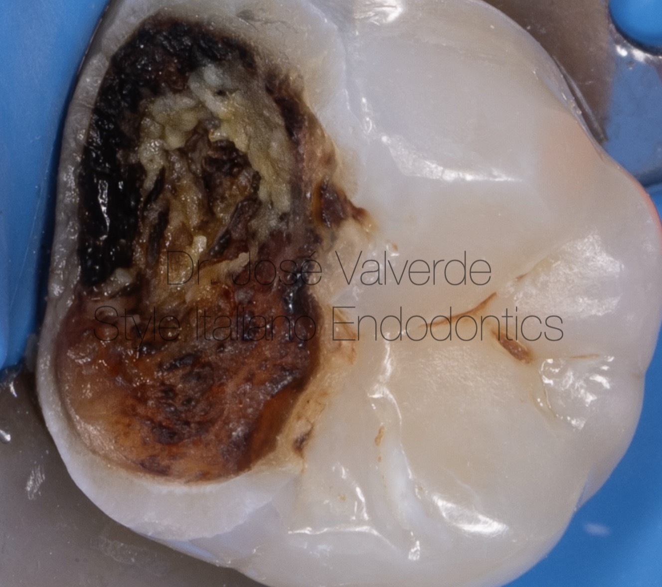
Fig. 6
Initial condition of the patient's tooth, which had deep caries in contact with the pulp chamber.
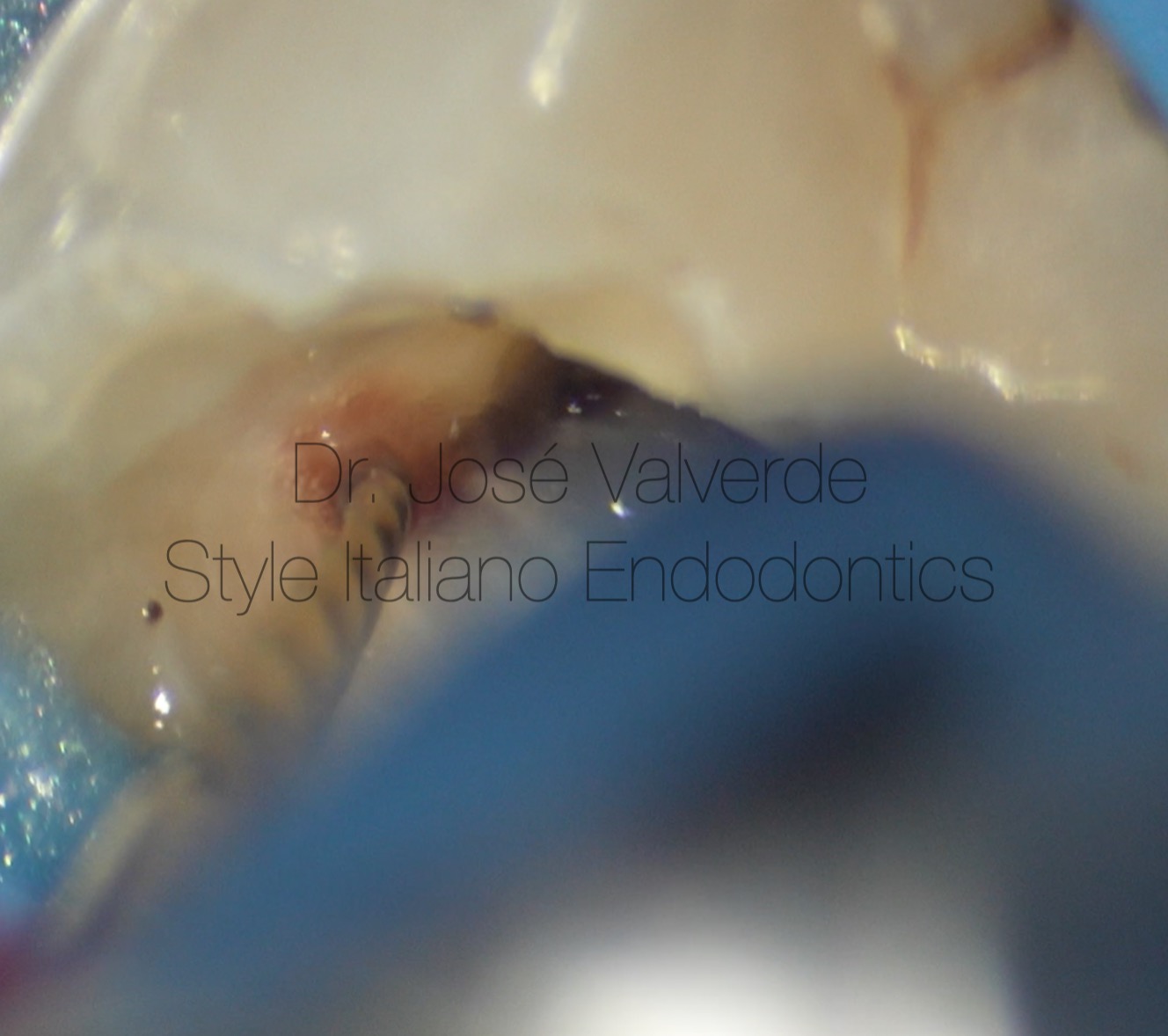
Fig. 7
Preflaring of the coronal third of the canal with SlimShaper Pro ZS1 (15/02).
We shape the coronal third of the canal up to the first resistance.
Fig. 8
Before reaching the apical third with the rotary files, we perform a minimal glidepath with K06, 08 and 10 files. When we have managed to get the K10 file completely loose in the canal, we start working in thirds of each of the canals, using a minimally invasive instrumentation system (SlimShaper Pro).
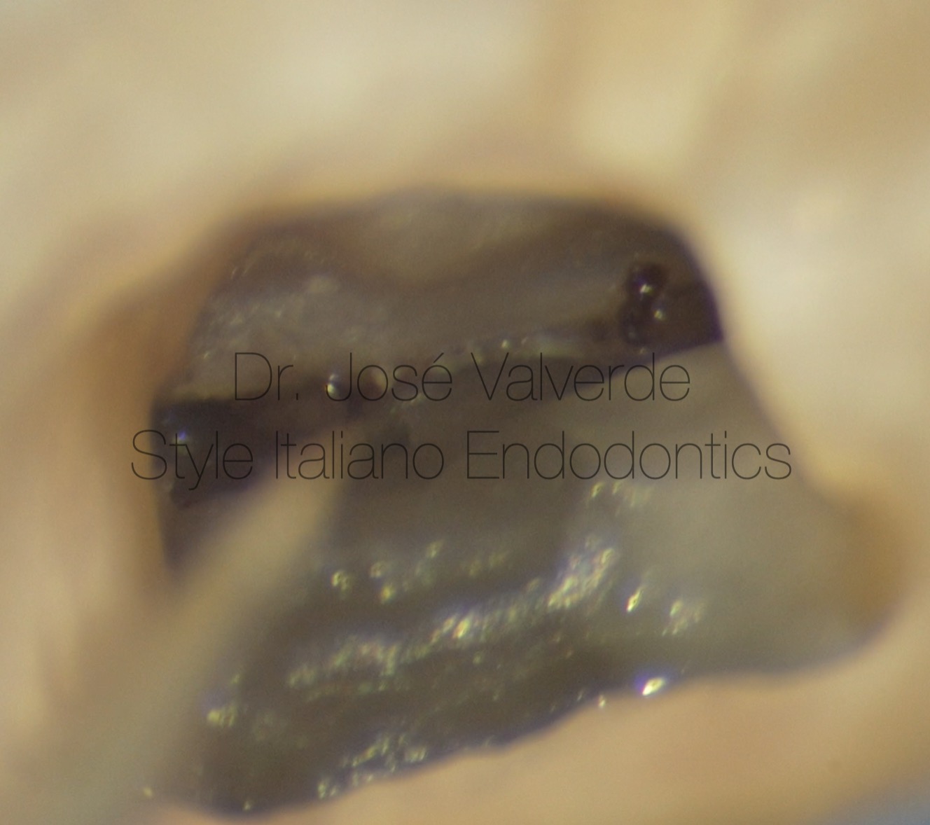
Fig. 9
To help us locate additional distal root anatomy, we use a Micro-opener, which allows us to remove the coronal dentine from the distocentral canal and clean the isthmus by indirect ultrasonic vibration.
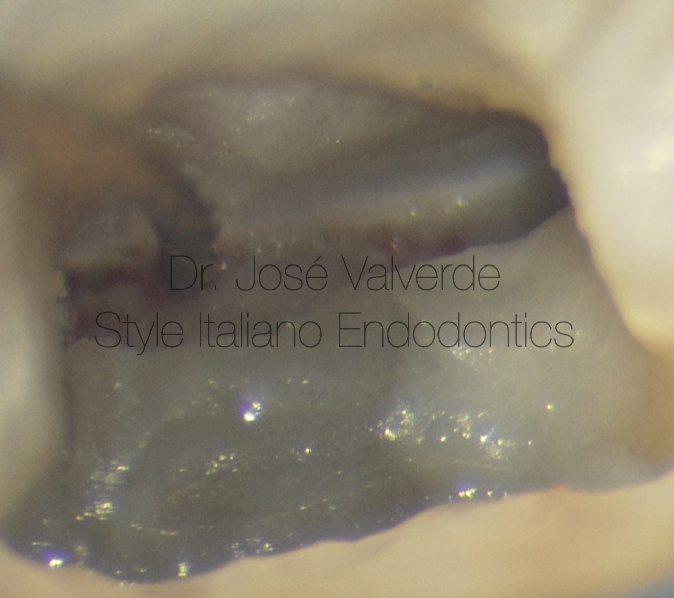
Fig. 10
After the canals have been located, we proceed to instrument each of them in thirds, using in this case ZS1 (15/02).
Patency was performed after each rotary file and irrigation with Z-Rinse during mechanical shaping.
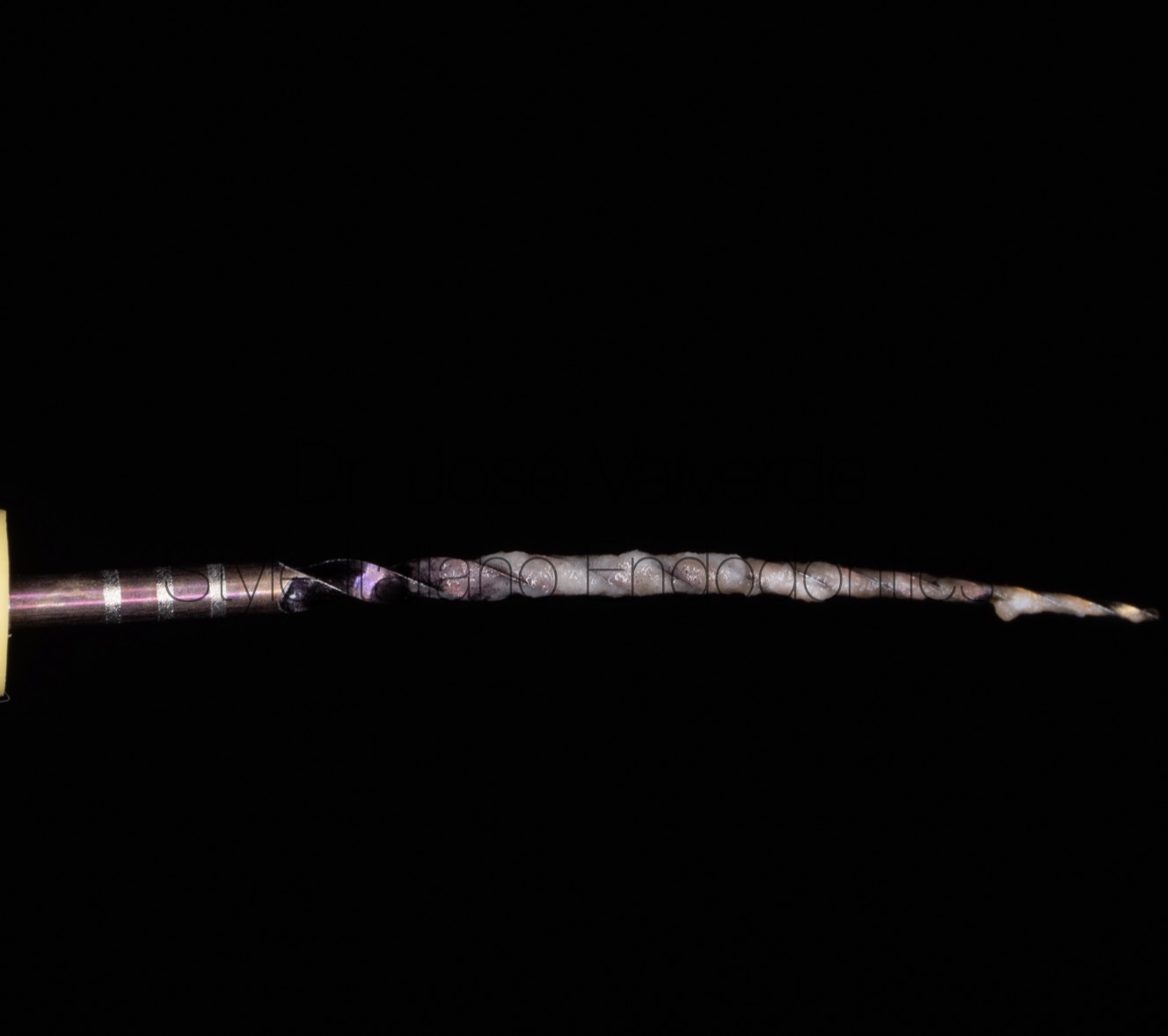
Fig. 11
Once we have instrumented the five canals with ZS1 and ZS2, we observe that the 20/04 file is working in the apical third and given that we are dealing with a vital tooth, we decide to finish the mechanical preparation at this point.
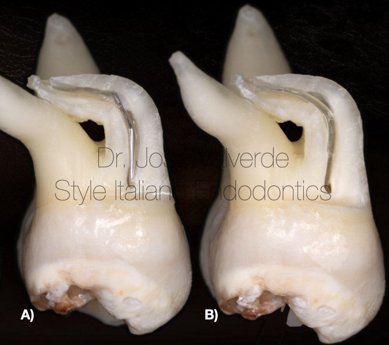
Fig. 12
One of the main problems we find in complex anatomies and minimally invasive preparations is the depth of the irrigation needle.
In this case, during treatment we use a 30G metal needle (A) and for the final irrigation protocol we use an Irriflex needle (B).
This combination allows us to irrigate safely during treatment and allows the NaOCl and EDTA 17% to reach the apical third, creating a constant renewal of irrigant in the critical area of the canal.
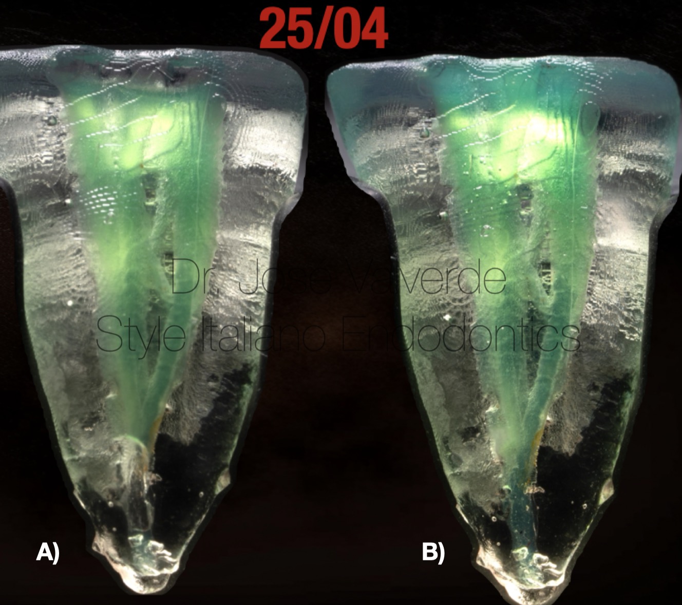
Fig. 13
We can see how in minimally invasive preparations the irrigant will reach the apical third with the Irriflex needle (B), while in the case of the metal needle, the irrigant will remain in the middle third (A).
Although both needles have the same diameter at the tip, the flexibility of Irriflex (B) allows it to adapt better to the original anatomy of the canal, and to carry the irrigant to the apical third.
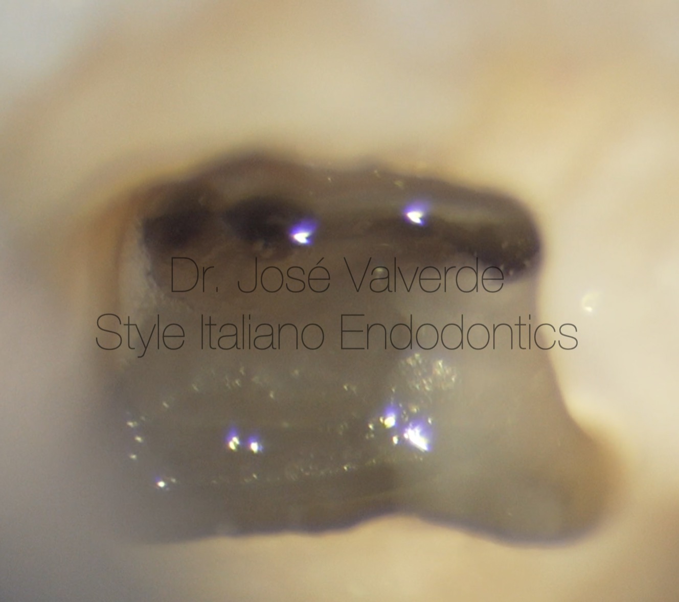
Fig. 14
View of the three distal canals after 20/04 preparation with SlimShaper Pro ZS2.
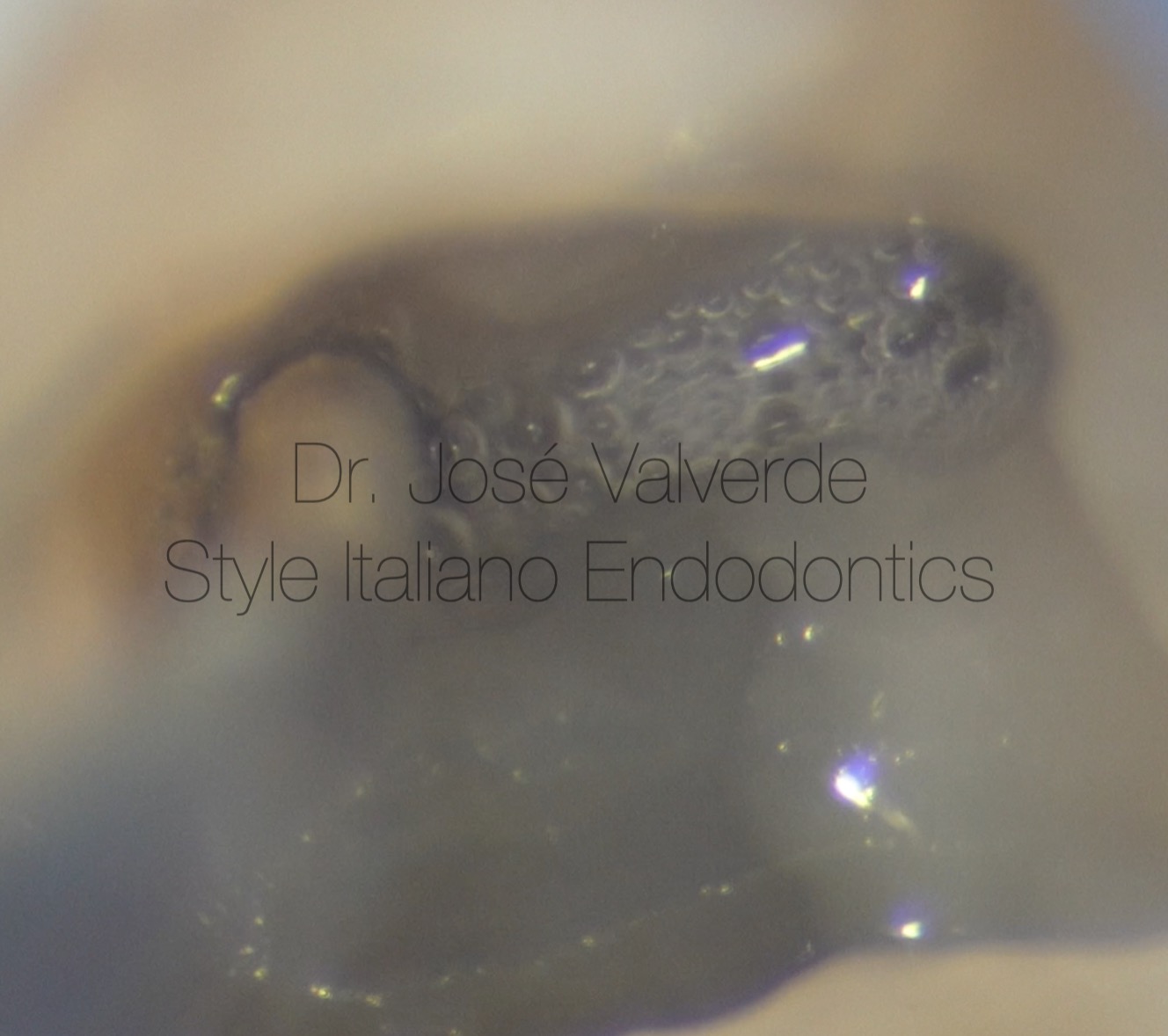
Fig. 15
Intracanal heating of NaOCl 5.25% was performed with the 30/04 plugger of the Elements Free system.
We placed the plugger at - 3mm from the WL and heated the irrigant at 180º for 4 seconds. After heating, we sonically activate the irrigant.
Repeat this step on all canals.
After washing and drying the canals, we irrigate the five canals with EDTA 17% for 60’, activating the irrigant manually with a calibrated 2% gutta percha tip.
Finally, NaOCl 5.25% is used again in the 5 canals, this time without activation.
In the final irrigation protocol, we used Irriflex instead of Z-Rinse.
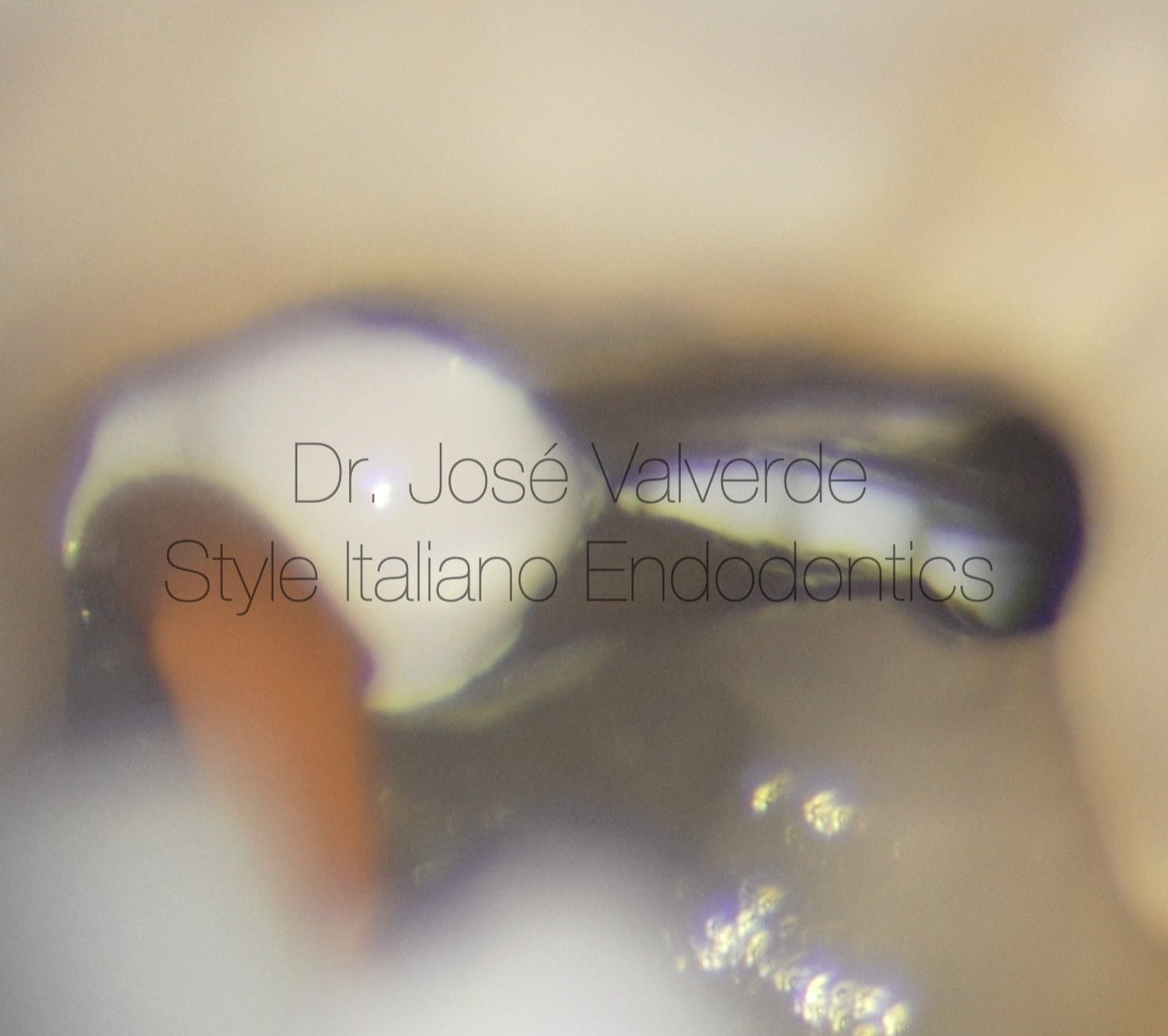
Fig. 16
After rinsing the canals and drying them by keeping some moisture, we used NeoSealer Flo in all five canals.
During the injection of the sealer, we observed how it spreads throughout the anatomy of the distal root.
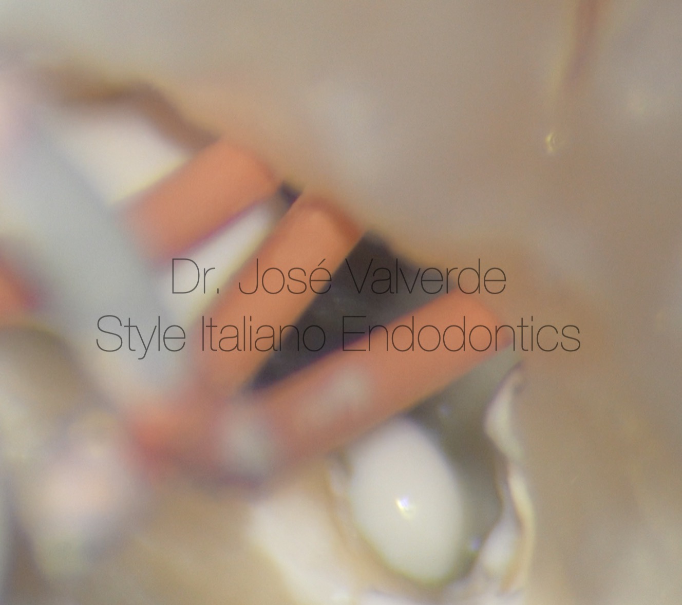
Fig. 17
We repeat the procedure on the mesial canals, also observing that there is a connection between them.
After injection of the bioceramic sealer, we calibrate 2% gutta percha cones and perform vertical compaction with the 30/04 plugger of the Elements Free system.
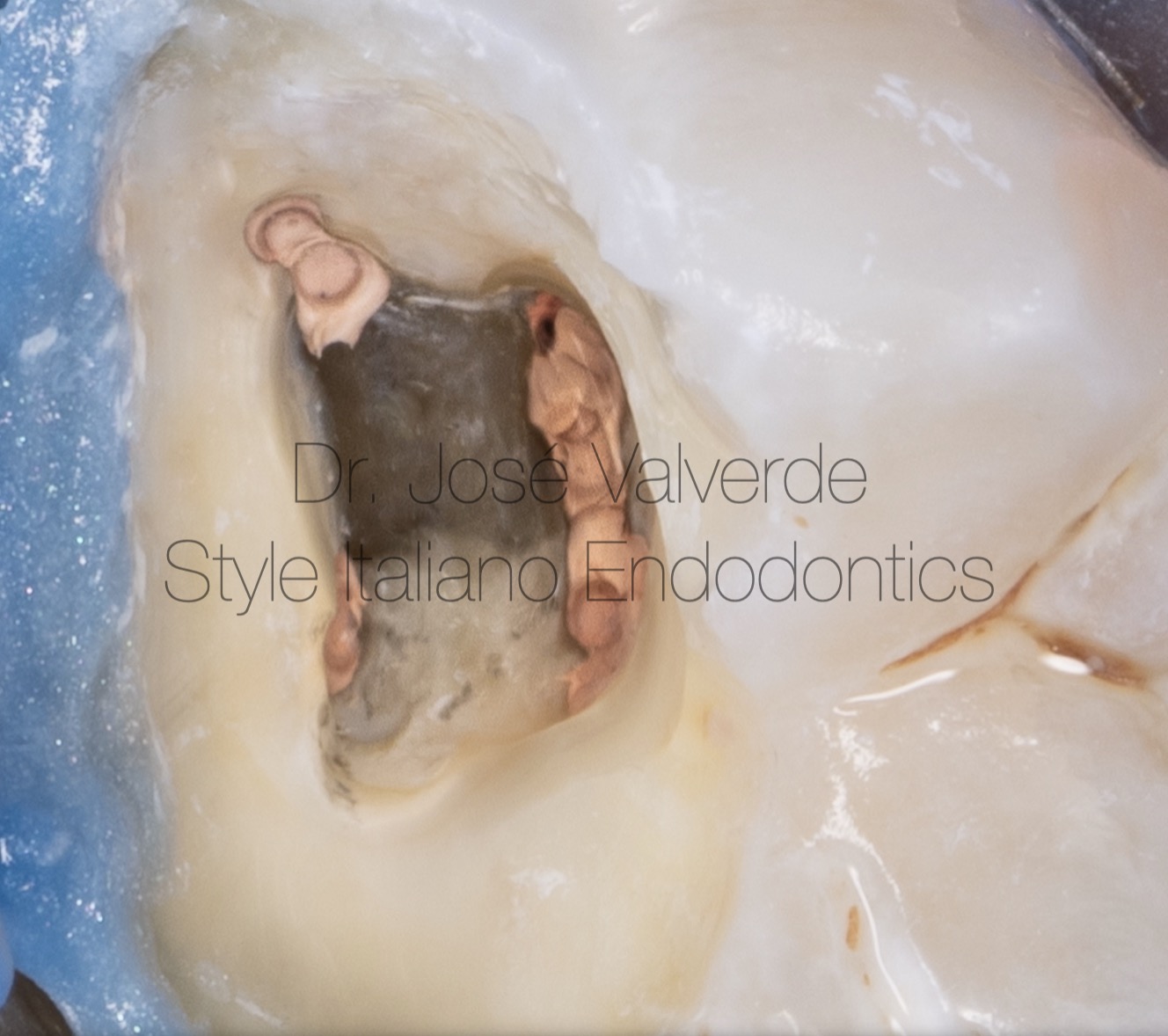
Fig. 18
Photo of the pulp chamber after obturation of the five canals.
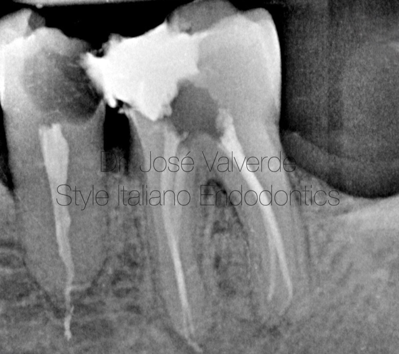
Fig. 19
Orthorradial angulation. We can observe the minimally invasive shaping of the canals.
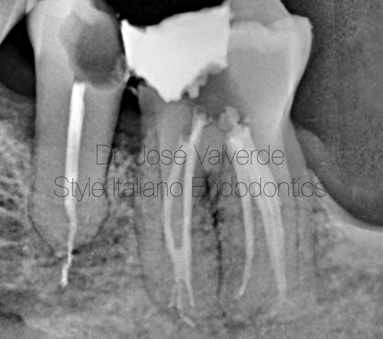
Fig. 20
Mesiorradial angulation showing the complexity of the apical third of the mesial root
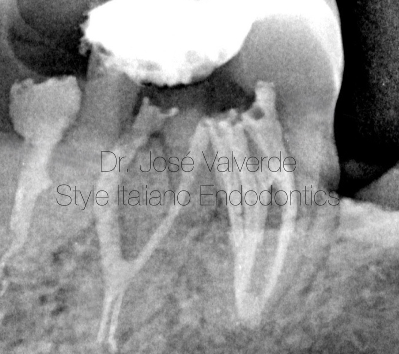
Fig. 21
In the distorradial angulation we can see the isthmus in the middle third and the conection between the main canal and the others in the apical third.

Fig. 22
About the Author
Dr. Jose Valverde raduated in dentistry from the Alfonso X El Sabio University, Madrid, Spain - European Master in Microscopic Endodontics and Apical Surgery from the Rey Juan Carlos University, Madrid, Spain. - PhD candidate in Universidade Grande Rio, Rio de Janeiro, Brasil - Proffesor of the Master in Microscopic Endodontics and Apical Surgery at Rey Juan Carlos University - Proffesor of the Expert in Endodontics and Apical Surgery at Universidad La Salle - Titular Member of the Spanish Association of Endodontics (AEDE) - Member of the American Association of Endodontics (AAE) - Member of the International Association of Dental Traumatology (IADT) - Fellow Member of Style Italiano Endodontics - Opinion Leader for Zarc4Endo - Active participant in national and international endodontic congresses, such as AAE, ESE, AEDE or IFEA. - Works as exclusive endodontist in Gran Canaria, Spain, and is dedicated to research and education
Conclusions
As chemomechanical preparation is one of the fundamental parts of root canal treatment, we must be aware of the limitations of the instruments we use in order to create strategies that allow us to be more effective in cleaning and disinfecting the root canal system.
Using irrigation needles that are adapted to this minimally invasive preparation and that also adapt to large curvatures and complex anatomies will be key to improving our irrigation.
Bibliography
CALBERSON, Filip L.; DE MOOR, Roeland J.; DEROOSE, Christophe A. The radix entomolaris and paramolaris: clinical approach in endodontics. Journal of endodontics, 2007, vol. 33, no 1, p. 58-63.
MARTÍNEZ-BERNÁ, Arturo; RUIZ-BADANELLI, Pedro. Maxillary first molars with six canals. Journal of endodontics, 1983, vol. 9, no 9, p. 375-381.
ABELLA, F., et al. Managing severe curvature of radix entomolaris: three‐dimensional analysis with cone beam computed tomography. International Endodontic Journal, 2011, vol. 44, no 9, p. 876-885.
BAASCH, Alessandra, et al. Effects of the Irrigation Needle Design on Root Canal Disinfection and Cleaning. Journal of Endodontics, 2024.