Precise endodontic treatment of a maxillary canine using the RISING by Fanta Dental
04/04/2026
Warning: Undefined variable $post in /home/styleendo/htdocs/styleitaliano-endodontics.org/wp-content/plugins/oxygen/component-framework/components/classes/code-block.class.php(133) : eval()'d code on line 2
Warning: Attempt to read property "ID" on null in /home/styleendo/htdocs/styleitaliano-endodontics.org/wp-content/plugins/oxygen/component-framework/components/classes/code-block.class.php(133) : eval()'d code on line 2
Endodontic treatment of maxillary canines requires precision due to their long roots and potential anatomical variations. This case demonstrates a structured and efficient approach to root canal therapy performed using the Rising Kit by Fanta Dental.
Pre-operative radiographic evaluation confirmed a single-rooted maxillary canine with a patent canal and no significant curvature complications. The treatment plan focused on achieving thorough cleaning, controlled shaping, and a dense three-dimensional obturation.
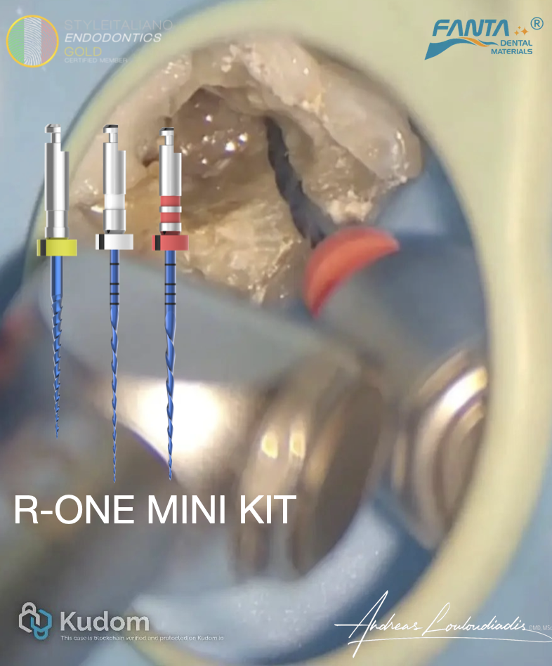
The Rising Kit enabled a streamlined workflow, offering excellent tactile control and consistent canal shaping while preserving the original anatomy. Irrigation and disinfection protocols were carefully implemented throughout the procedure to maximize microbial control.
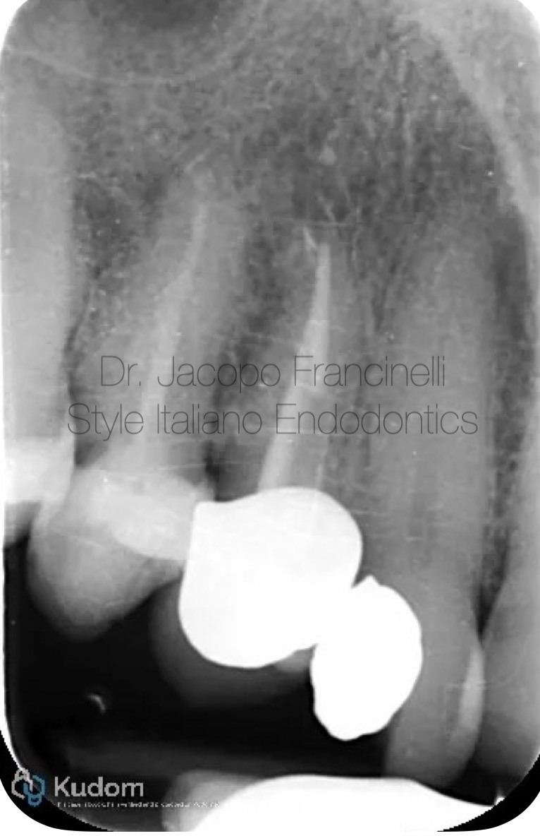
Fig. 1
Pre-operative X-ray:
The initial radiograph shows a maxillary canine with a single, clearly defined canal. The periapical region appears without significant radiolucency, though clinical symptoms indicated pulpal involvement. The canal anatomy appears straight and accessible, making it suitable for efficient mechanical preparation.
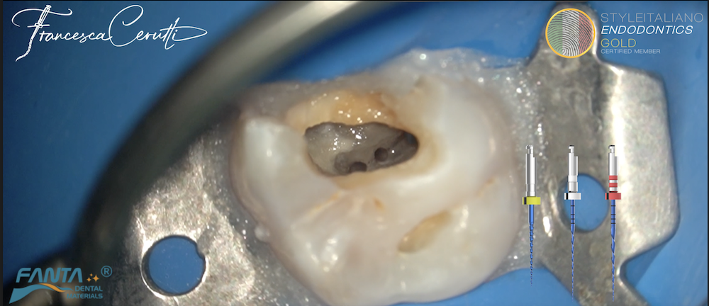
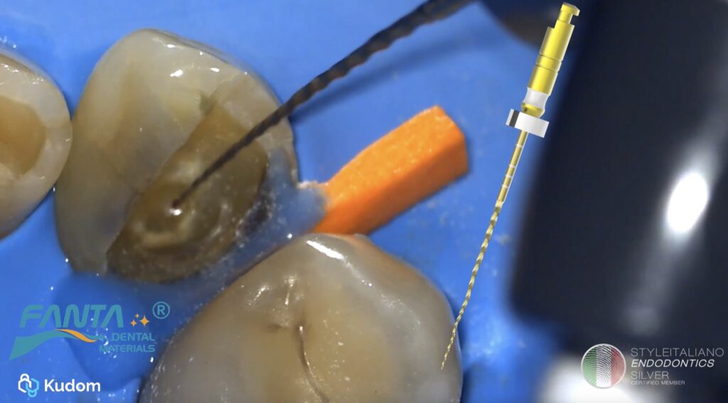
The clinical video illustrates key steps of the procedure, including access preparation, canal negotiation, and shaping using the Rising Kit by Fanta Dental reaching R3 (30/.04). It highlights the smooth progression of instruments, efficient debris removal, and clinician control throughout the procedure.
Close-up sequences emphasize the system’s cutting efficiency and flexibility, demonstrating how it maintains canal integrity while achieving an ideal taper. The video reinforces the predictability and ease of use of the system in daily clinical practice.
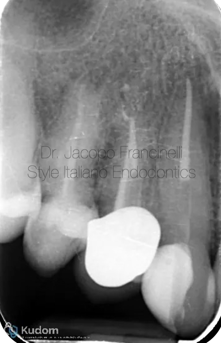
Fig. 2
Post-operative X-ray (Final Obturation and Reconstruction):
The final radiograph shows a dense and homogeneous root canal obturation extending to the correct working length, with excellent adaptation to canal walls and no visible voids.
Additionally, the image demonstrates the placement of an intracanal endodontic post within the prepared canal space. The post is well-centered and aligned with the root axis, contributing to optimal retention and structural reinforcement of the remaining tooth. This reconstruction ensures long-term stability and supports the coronal restoration, completing the endodontic treatment successfully.
Conclusions
This case underscores the importance of integrating proper endodontic protocols with reliable instrumentation and restorative planning. The Rising Kit by Fanta Dental facilitated efficient canal preparation while maintaining anatomical integrity, leading to a high-quality obturation.
The addition of a well-positioned intracanal post further enhances the structural durability of the tooth, ensuring long-term clinical success. This system represents a dependable solution for clinicians seeking precision, efficiency, and consistency in modern endodontic practice.
Bibliography
- Kakehashi S, Stanley HR, Fitzgerald RJ. The effects of surgical exposures of dental pulps in germ-free and conventional laboratory rats. Oral Surg Oral Med Oral Pathol 1965 20(3):340-49.
- Bergenholtz G. Micro-organisms from necrotic pulp of traumatized teeth. Odontol Revy. 1974;25(4):347-58.
- Sjögren U, Figdor D, Persson S, Sundqvist G. Influence of infection at the time of root filling on the outcome of endodontic treatment of teeth with apical periodontitis. Int Endod J. 1997;30:297, 6
- Marshall FJ, Pappin J. A crown-down pressureless preparation of root canal enlargement: technic manu- al. Dept of Endodontics, Oregon health Sciences University, Portland, Oregon 1980.
- Goerig AC, Michelich RJ, Schultz HH. Instrumentation of root canals in molar using the step-down technique. J Endod 1982; 8: 550-4.
- Morgan LF, Montgomery S. An evaluation of the crown-down pressureless technique. J Endod 1984;10: 491-8.