Open apex management using bioceramics
20/03/2023
Mohamad Zaafrany
Warning: Undefined variable $post in /home/styleendo/htdocs/styleitaliano-endodontics.org/wp-content/plugins/oxygen/component-framework/components/classes/code-block.class.php(133) : eval()'d code on line 2
Warning: Attempt to read property "ID" on null in /home/styleendo/htdocs/styleitaliano-endodontics.org/wp-content/plugins/oxygen/component-framework/components/classes/code-block.class.php(133) : eval()'d code on line 2
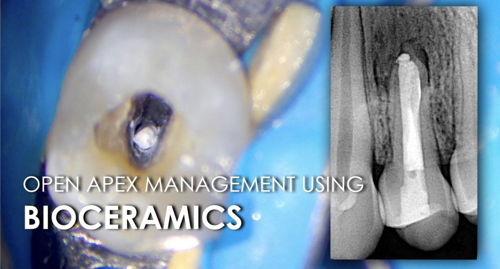
In the presented case we’ll describe a management of a maxillary premolar with open apex and large periapical lesion.
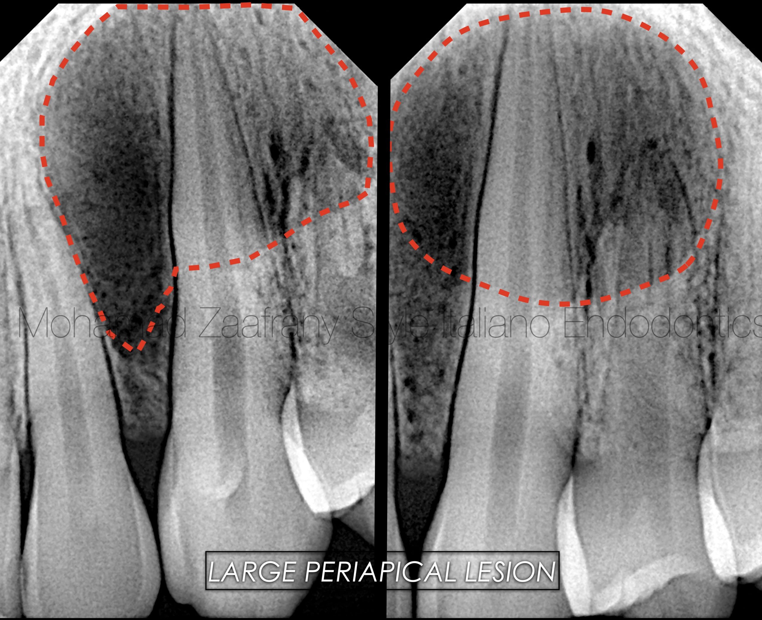
Fig. 1
Figure 1: Preoperative radiographic analysis
-16 years old male patient presented with complain of discomfort on chewing on the upper left side for 2 weeks, with old history of dental trauma.
-On clinical examination teeth are sound with tenderness to percussion on teeth no. 23,24.
-Preoperative radiograph showing large radiolucency related to maxillary canine and premolar region with resorption like lesion to the apex of tooth no. 24
-Clinical tests reveals a non responsive tooth no. 24 to cold and hot test with positive response of neighboring teeth.
-Case diagnosed as necrotic tooth no. 24 with Chronic Apical Periodontitis.
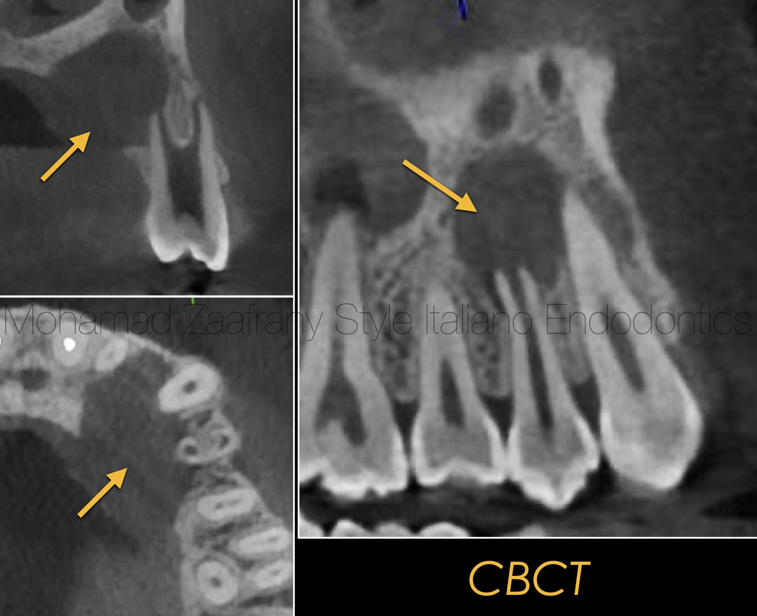
Fig. 2
Figure 2:
CBCT cuts showing the lesion is more related to palatal root
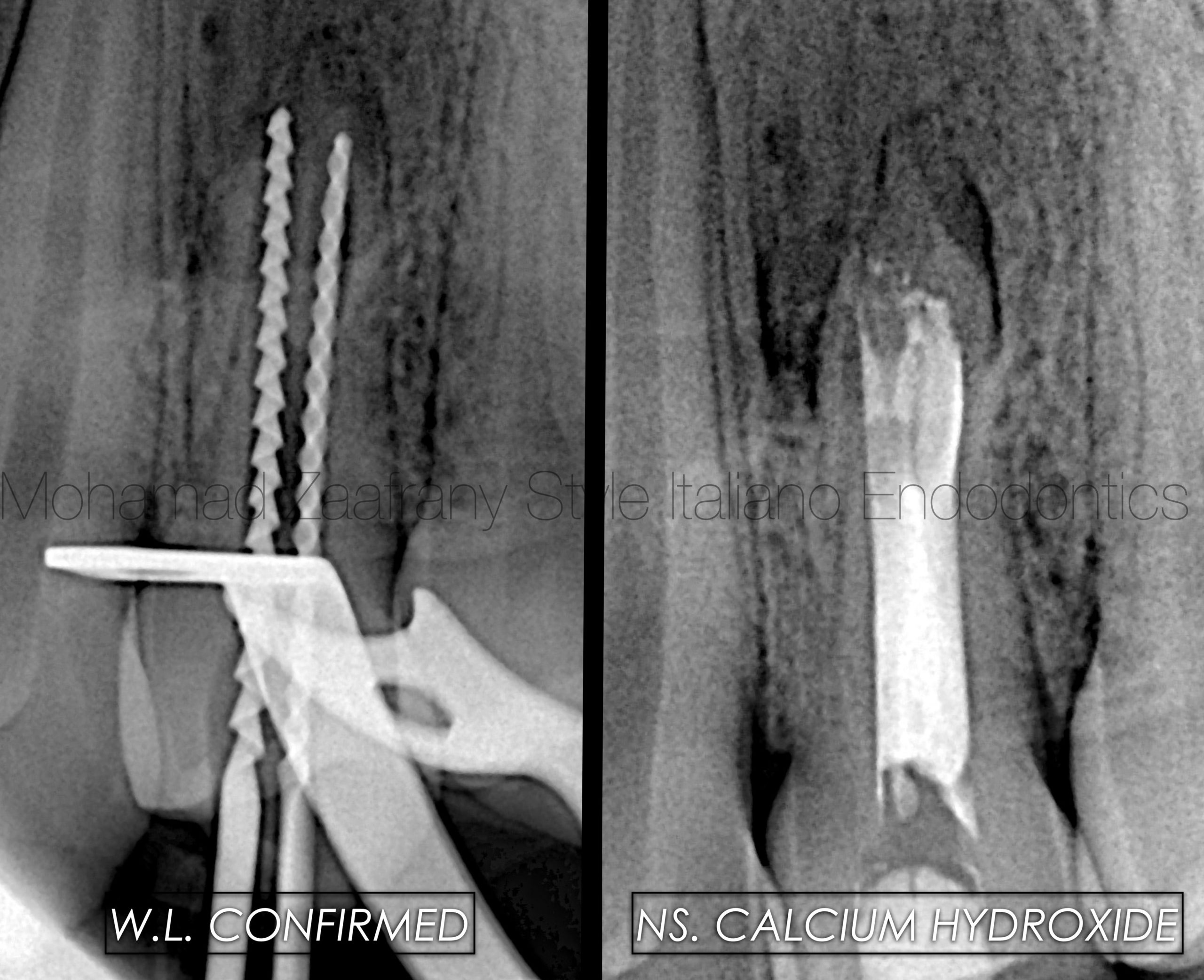
Fig. 3
Figure 2:
After local anesthesia administration, Access opened.
W L confirmed, root canal system disinfected using 5.25% Sodium Hypochlorite , EDTA solution 17% with sonic and ultrasonic activation.
Non setting Calcium Hydroxide kept inside the canal for 2 weeks as intracanal medication as to raise the PH of the environment to be more suitable to receive the plug material.
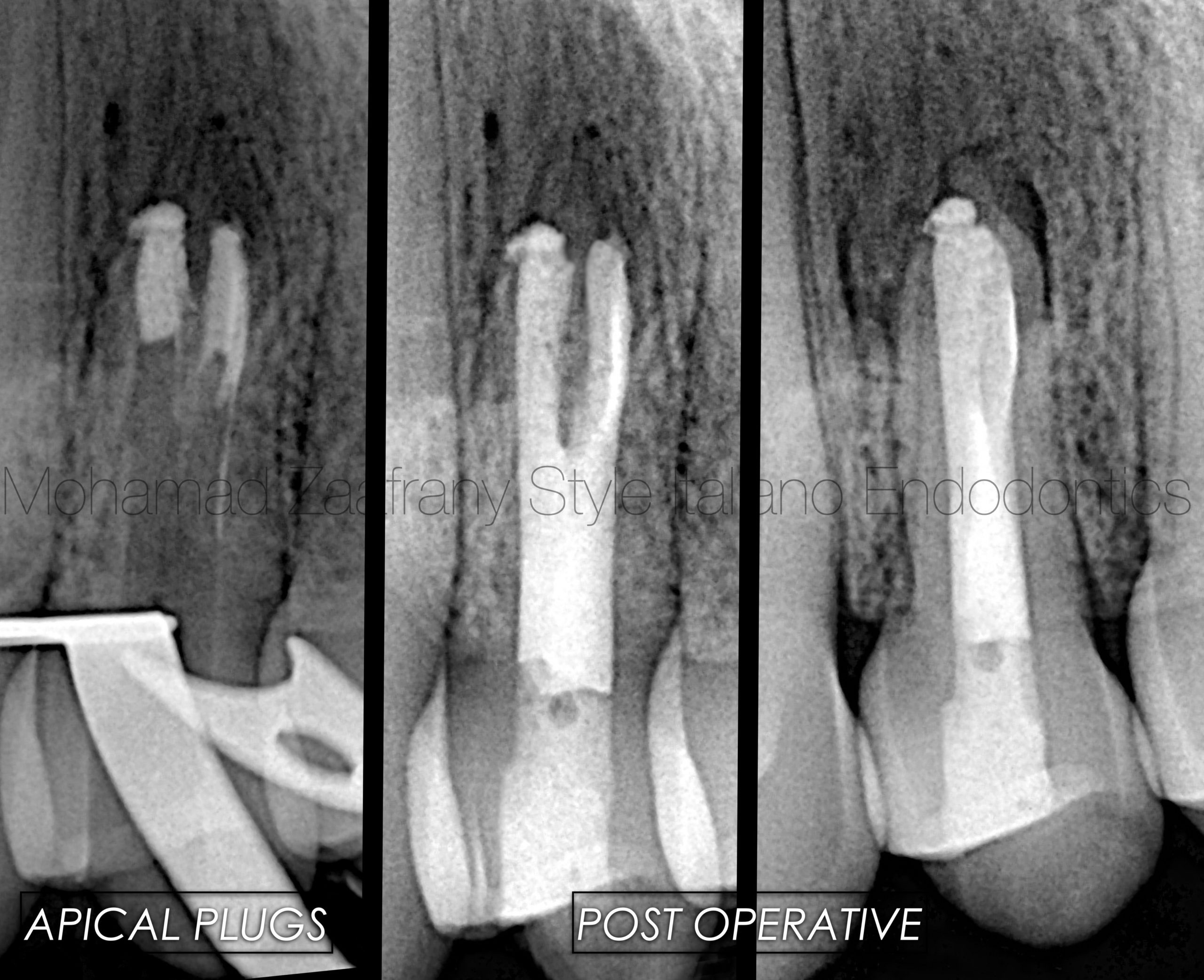
Fig. 4
Figure 3:
Bioceramic Putty material applied to the apex for a more controlled apical seal followed by backfilling the canal using warm Gutta Percha.
Post operative radiograph showing a well adapted apical plugs of putty Bioceramics material with a well filled pulp space.
In the video a step by step application of the Putty Bioceramic material to the apex and backfilling the root space using warm Gutta Percha.
Conclusions
Diagnosis should be performed in a very systematic method applying all needed clinical tests to avoid over treatment or treatment to teeth that are not involved as a reason for disease.
Managing immature roots providing 3D sealing to the root canal system might represent a challenge to the operator.
Using biocompatible filling materials like MTA or Bioceramics that provide superior sealing properties over conventional Gutta percha in this kind of cases is considered a solution for more predictable results.
The use of magnification and right tools would facilitate the job for the clinician in such challenging procedures.
Bibliography
MTA: A clinical review Peter Z Tawil, DMD, MS, FRCD, Dip. ABE, Derek J. Duggan, BDentSc, MS, Dip. ABE, and Johnah C. Galicia, DMD, MS, PhD. Compend Contin Educ Dent. 2015 Apr; 36(4): 247–264.
Sjögren U, Figdor D, Spanberg L, Sundqvist G. The antimicrobial effect of calcium hydroxide as a short‐term intracanal dressing. Int Endod J 1991;24(3):119–25.
Antimicrobial effect of calcium hydroxide as an intracanal medicament in root canal treatment: a literature review - Part I. In vitro studies Dohyun Kim and Euiseong Kim, Restor Dent Endod. 2014 Nov; 39(4): 241–252.
Wang Z. Bioceramic materials in endodontics. Endod Top 2015;32(1):3–30.
21 Best SM, Porter AE, Thian ES, Huang J. Bioceramics: Past, present and for the future. J Eur Ceram Soc 2008;28:1319–27.
Apexification: A systematic review. Fabricio Guerrero, Asunción Mendoza, David Ribas, and Karla Aspiazu J Conserv Dent. 2018 Sep-Oct; 21(5): 462–465.
Sealing properties of Mineral Trioxide Aggregate orthograde apical plugs and root fillings in an In vitro apexification model. JOE 2007 Mar;33(3):272-5. doi: 10.1016/j.joen.2006.11.002.


