Non-surgical endodontic retreatment of a maxillary central incisor with open apex using MTA Apical Plug Technique
21/10/2021
José Conde Pais
Warning: Undefined variable $post in /home/styleendo/htdocs/styleitaliano-endodontics.org/wp-content/plugins/oxygen/component-framework/components/classes/code-block.class.php(133) : eval()'d code on line 2
Warning: Attempt to read property "ID" on null in /home/styleendo/htdocs/styleitaliano-endodontics.org/wp-content/plugins/oxygen/component-framework/components/classes/code-block.class.php(133) : eval()'d code on line 2
The major goals of root canal treatment are to clean and shape the root canal system and seal it in 3 dimensions to prevent reinfection of the tooth. Although initial root canal therapy has been shown to be a predictable procedure with a high degree of success , failures can occur after treatment. Lack of healing is attributed to persistent intraradicular infection residing in previously uninstrumented canals, dentinal tubules, or in the complex irregularities of the root canal system. The extraradicular causes of endodontic failures include periapical actinomycosis , a foreign body reaction caused by extruded endodontic materials, an accumulation of endogenous cholesterol crystals in the apical tissues , and an unresolved cystic lesion
The orthograde retreatment of dental elements previously treated with the most varied techniques is a fairly common clinical practice, particularly for endodontic specialists . This article presents a case report of failed endodontics treatment in maxillary central incisor with open apex.
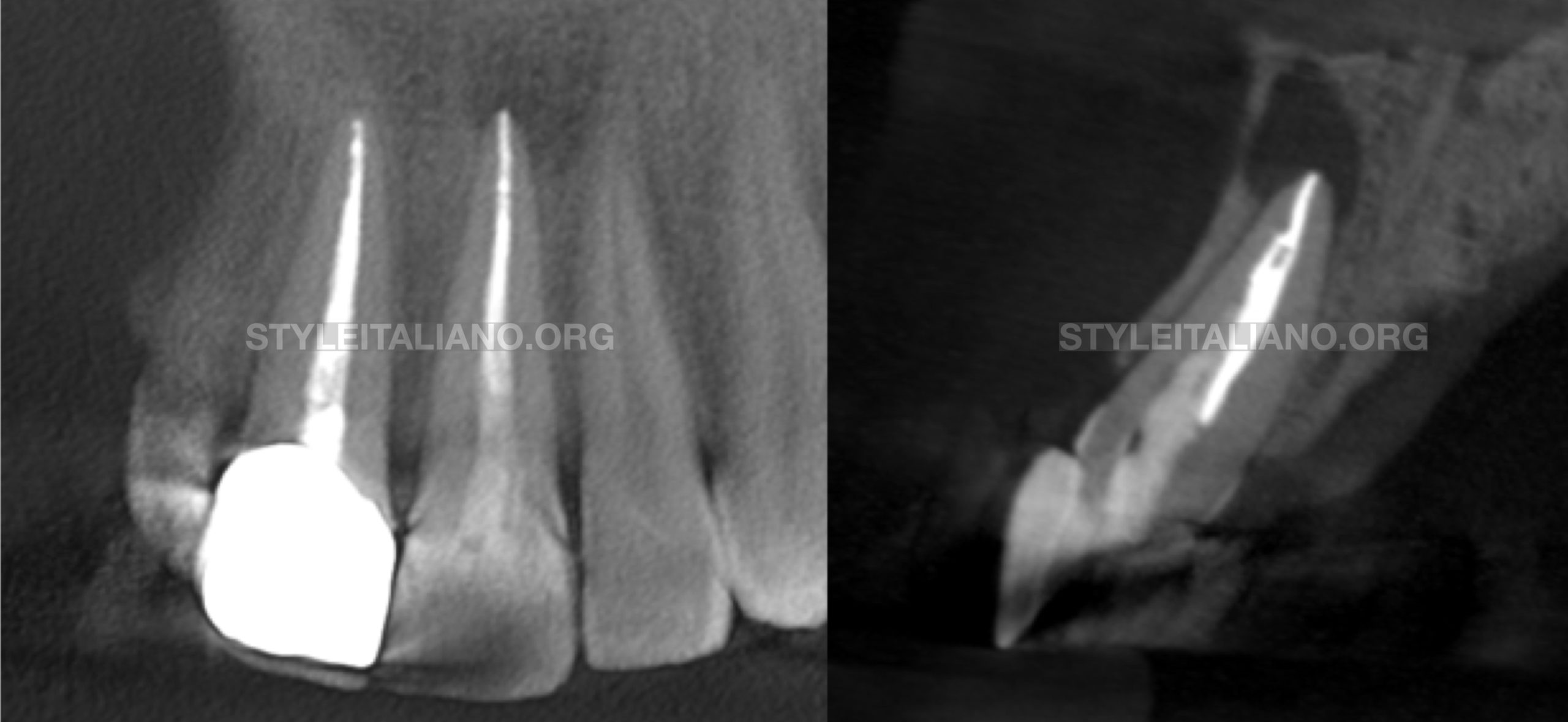
Fig. 1
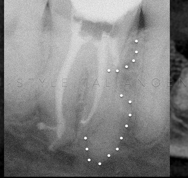
A 34-year-old male patient presented in our private endodontic office for evaluation of tooth 21, which had received nonsurgical endodontic treatment years ago caused by trauma. The presenting symptoms included swelling in the buccal vestibule and pain. His health history was unremarkable and radiographic examination revealed two fiber post into the root, incomplete sealing associated with extensive periapical bone loss. Clinical examination disclosed fluctuant swelling in the vestibule, no mobility and pain on percussion. The diagnosis was acute peri-apical abscess, after discussing treatment options, we decided on an orthograde retreatment. The patient was informed of the pros and cons of the treatment and informed consent was obtained.
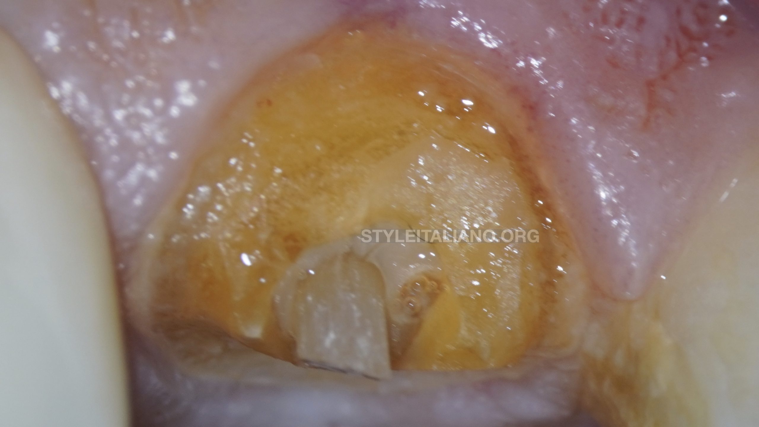
Fig. 2
In this image we can see the preoperative situation and the location of the fiber post.
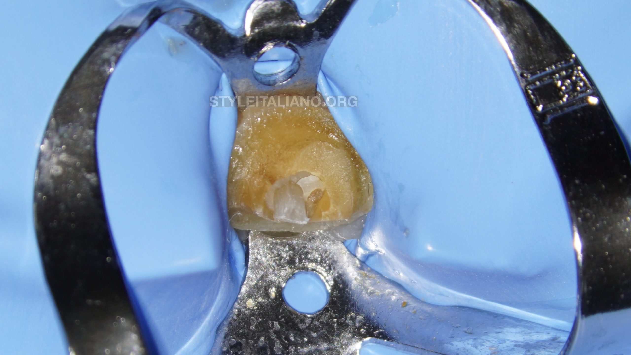
Fig. 3
After anesthesia the tooth was isolated
The Fiber Post was removed using ultrasonic tip.
Old guttapercha was removed using Hedstrom FIles
After removing the old gutta-percha, we discovered a second fiber post in the vestibular wall.
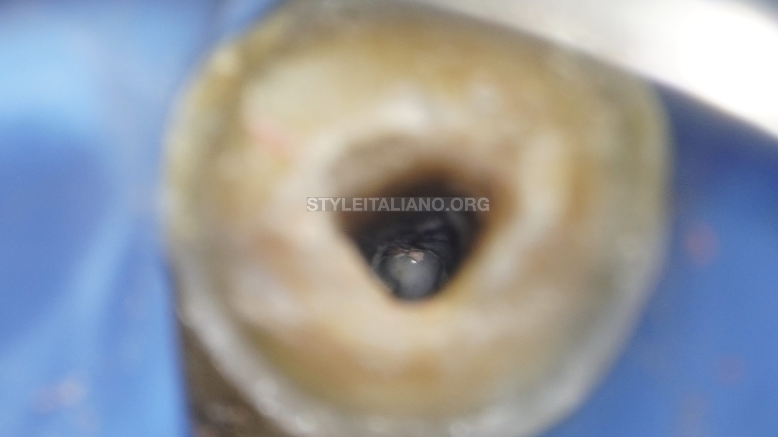
Fig. 4
The canal was chemomechanically shaped and cleaned using the following protocol:
Desobturation Technique : Hedstrom Files
Shaping : Protaper Next X1- X5 , K Files 60/70/80
Irrigation; Sodium hypochlorite 5,25% + Saline Solution + Edta 17% PUI + sodium hypochlorite 5,25% PUI
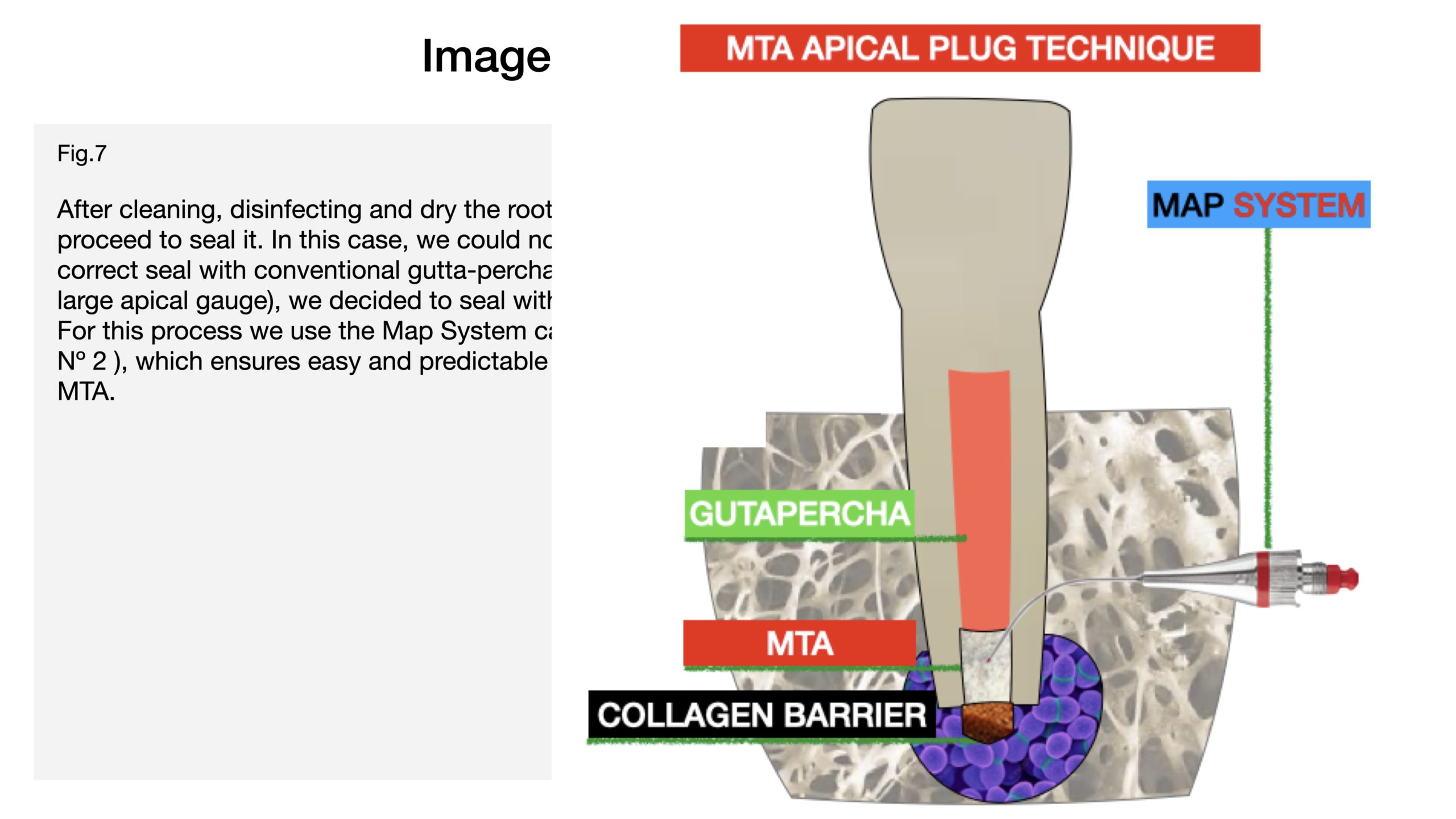
Fig. 5
After cleaning, disinfecting and dry the root canal we proceed to seal it. In this case, we could not ensure a correct seal with conventional gutta-percha (due to the large apical gauge), we decided to seal with MTA plug.
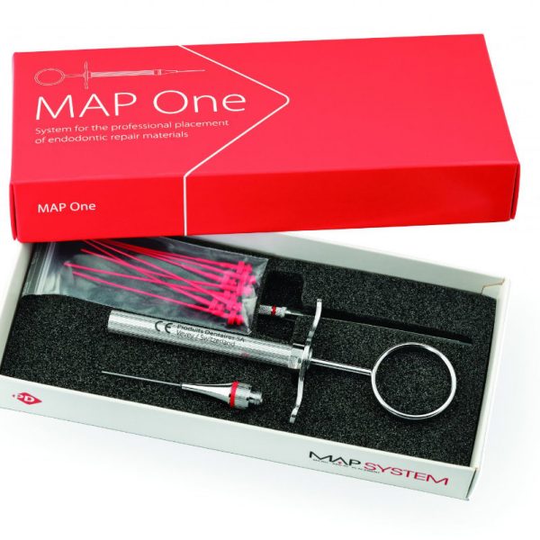
For this process we use the Map System carrier ( Needle Nº 2 ), which ensures easy and predictable handling of the MTA.
This video shows the Apical Plug Technique
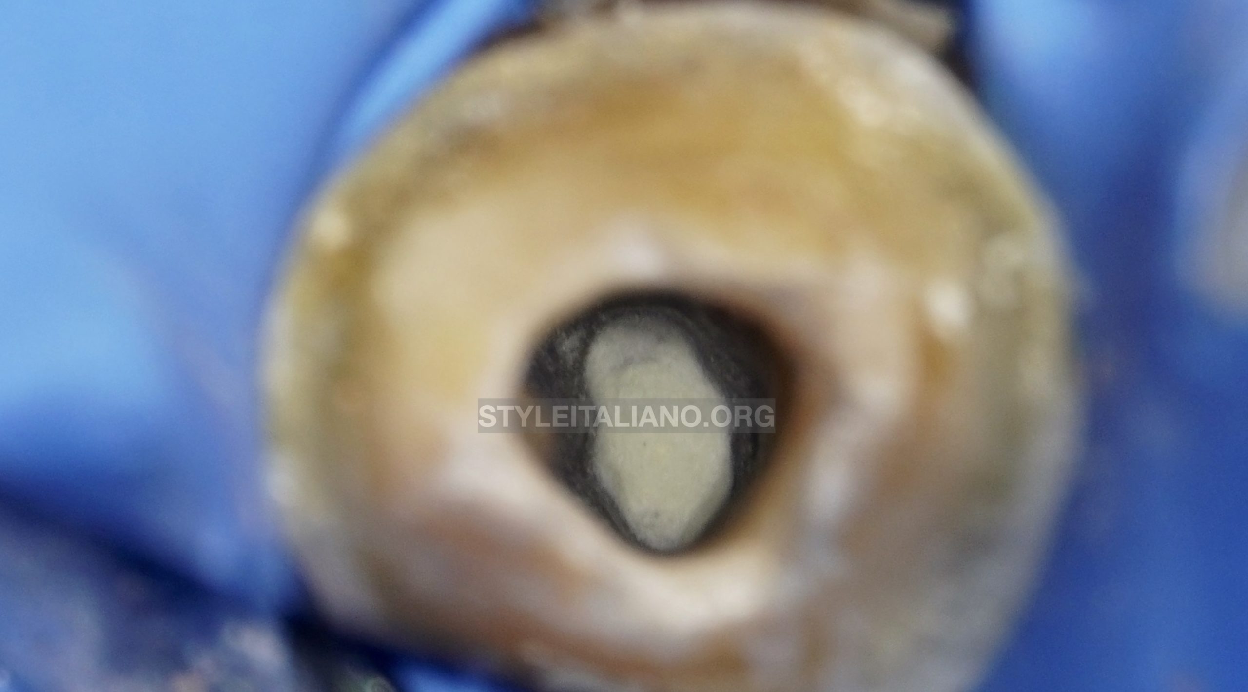
Fig. 6
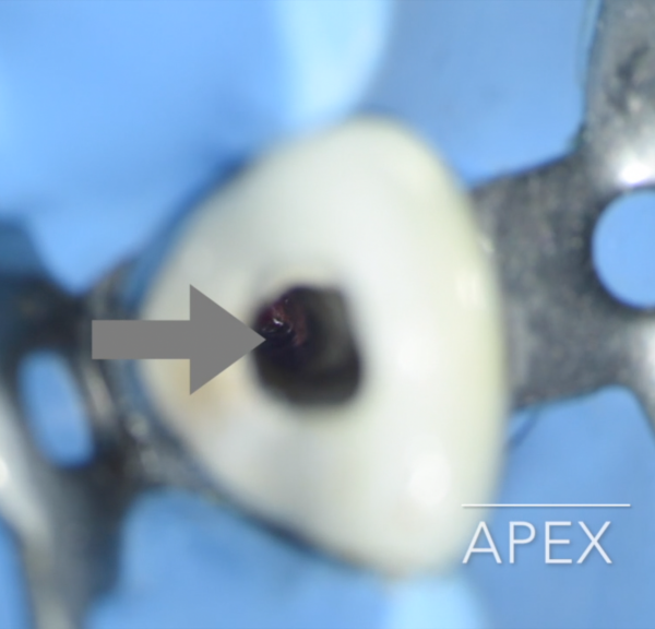
In this picture we can see the apical Plug.
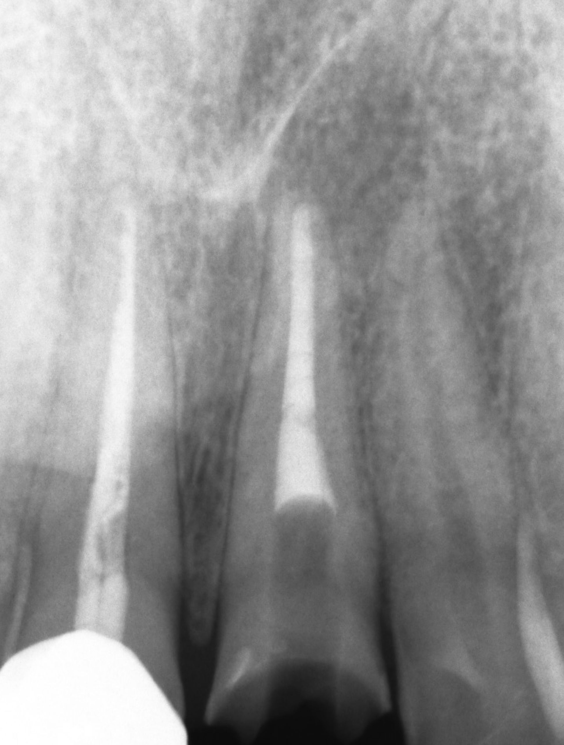
Fig. 7
Final Xray
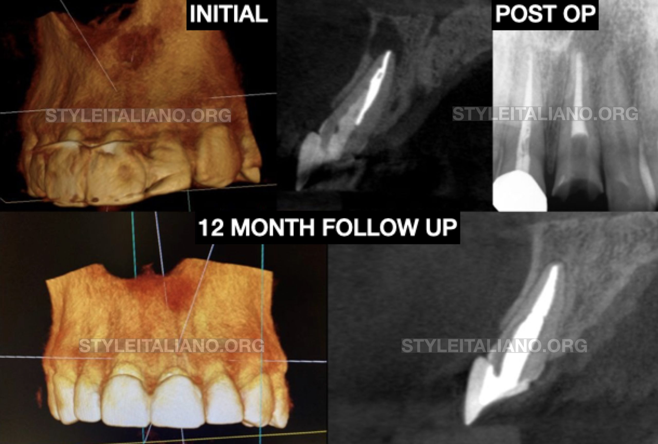
Fig. 8
12 MONTH FOLLOW UP
Conclusions
The MTA apical plug technique is a repeatable, predictable and safe technique in cases with open apex or incomplete root development. We must know and handle this type of technique to ensure a correct apical seal in all cases.
Bibliography
Gorni FG, Gagliani MM. The outcome of endodontic retreatment: a 2-yr follow-up. J Endod. 2004 Jan;30(1):1-4.
Torabinejad M, Corr R, Handysides R, Shabahang S. Outcomes of nonsurgical re- treatment and endodontic surgery: a systematic review. J Endod 2009;35:930–7


