MTA pulp capping in an adult
19/04/2020
Francesca Cerutti
Warning: Undefined variable $post in /home/styleendo/htdocs/styleitaliano-endodontics.org/wp-content/plugins/oxygen/component-framework/components/classes/code-block.class.php(133) : eval()'d code on line 2
Warning: Attempt to read property "ID" on null in /home/styleendo/htdocs/styleitaliano-endodontics.org/wp-content/plugins/oxygen/component-framework/components/classes/code-block.class.php(133) : eval()'d code on line 2
MTA has a broad field of use in endodontics thanks to its biocompatibility and its capability to harden in a moist environment.
Several articles proved it to be effective as a pulp capping material, although most of the literature focuses on the pulp capping in young patients...but what happens when if do this in an adult?
A clinical case follows in which a direct pulp capping with MTA was executed in an adult patient.
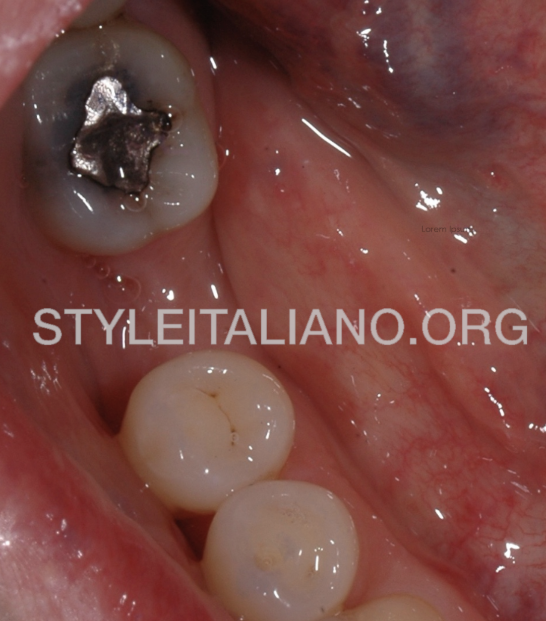
Fig. 1
An adult patient came to me to replace a chipped amalgam restoration. The marginal discoloration suggested the presence of bacterial infiltrate.
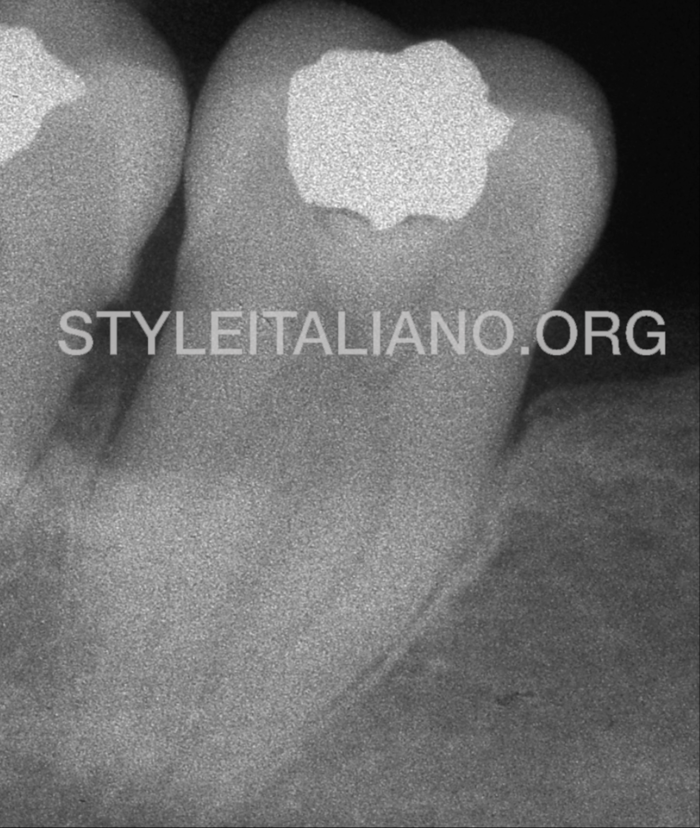
Fig. 2
The pre-operative x-ray showed the presence of a void between the amalgam and the cavity liner.
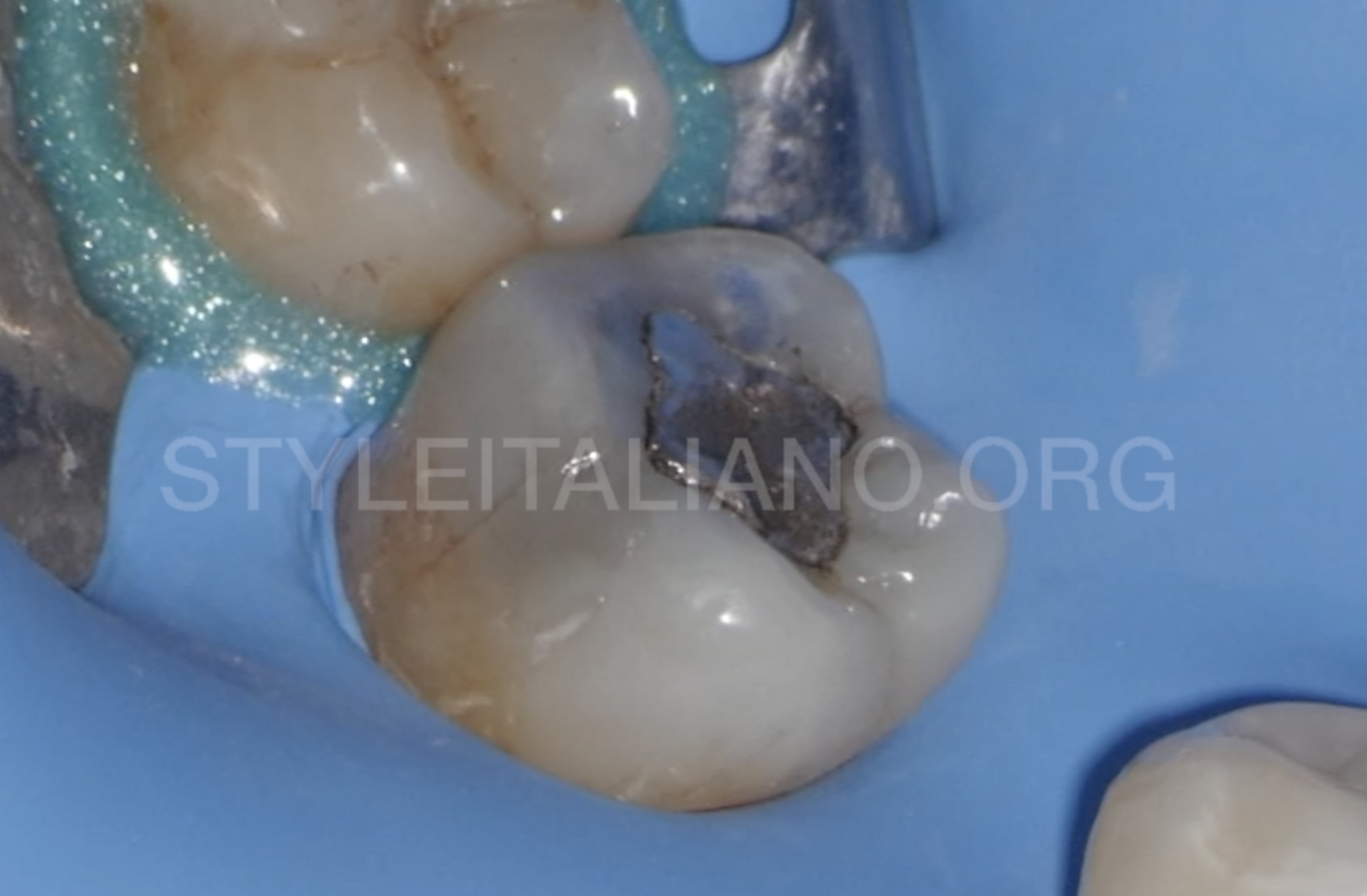
Fig. 4
First of all, the tooth was isolated with the rubber dam by placing a clamp on the tooth 4.8.
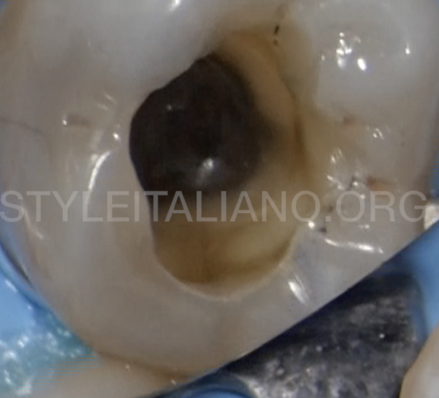
Fig. 5
The amalgam restoration was removed, showing the presence of brown stains. Obviously they have to be removed in order to achieve a good cavity cleaning and to improve the adhesion.
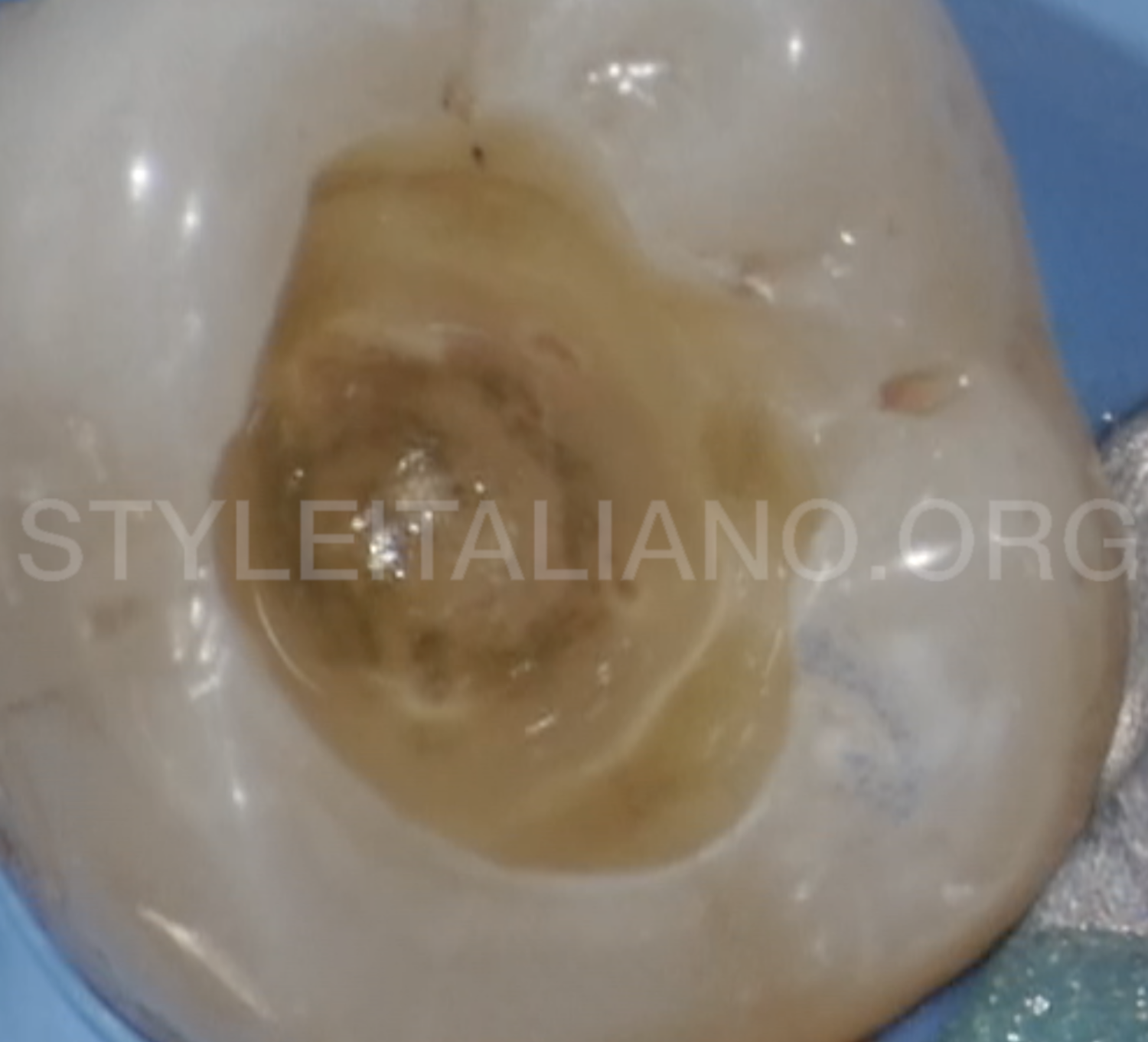
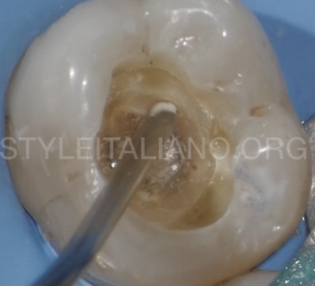
Fig. 6
After removing the decayed tissue, the communication between the pulp chamber and the cavity was visible.
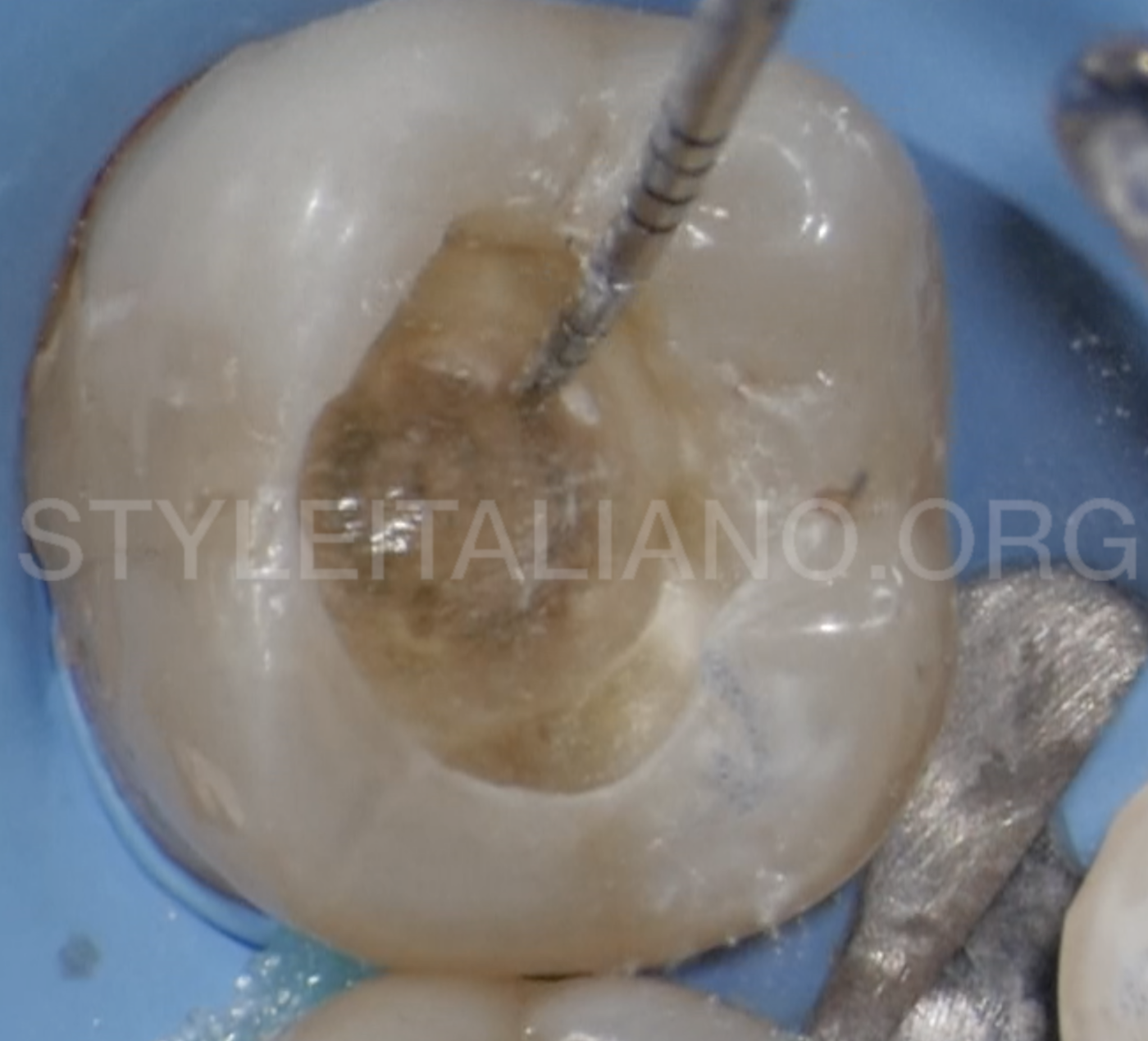
Fig. 8
A probe was used to confirm the setting of the MTA.
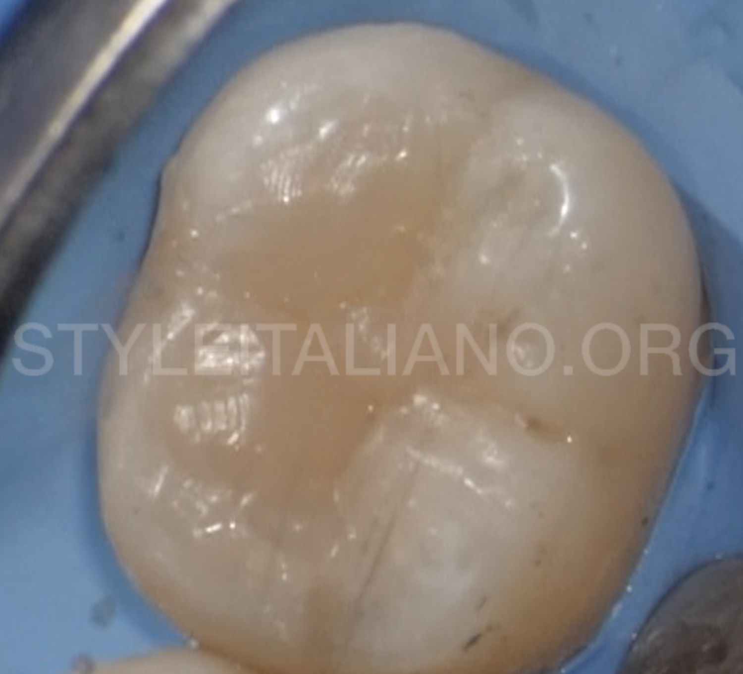
Fig. 9
At this point, a direct composite restoration was executed.
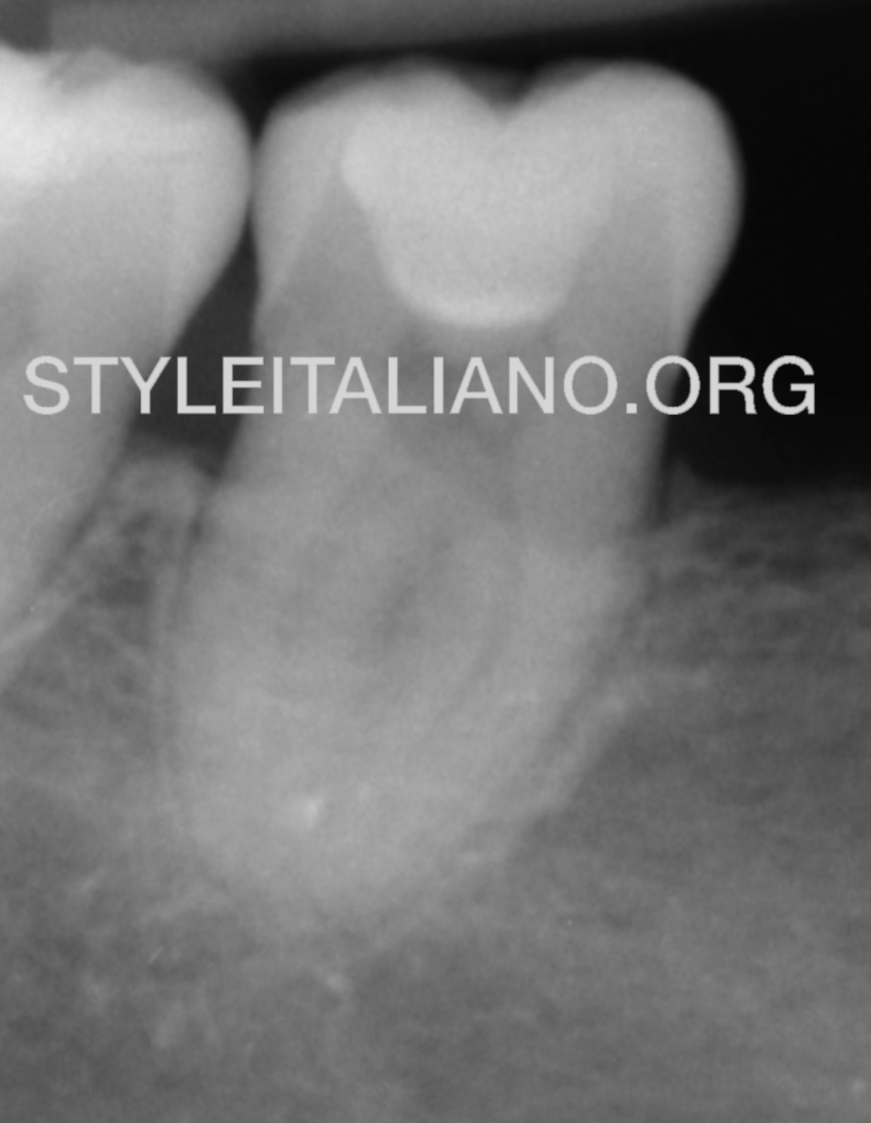
Fig. 10
The 1 year-recall x-ray confirms the tooth is still vital.
Conclusions
Even if the direct pulp capping is not so diffused in adult teeth, MTA offers us the possibility to keep the tooth vital for a longer period of time, according to a minimally invasive approach. The use of a bismuth-free material does not expose the tooth to the risk of staining.
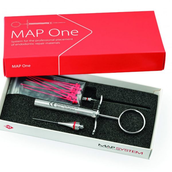
A dedicated carrier makes the positioning easy and predictable.
Bibliography
Aguilar P, Linsuwanont P. Vital pulp therapy in vital permanent teeth with cariously exposed pulp: a systematic review. Journal of endodontics. 2011;37(5):581-7.
Brizuela C, Ormeno A, Cabrera C, Cabezas R, Silva CI, Ramirez V, et al. Direct Pulp Capping with Calcium Hydroxide, Mineral Trioxide Aggregate, and Biodentine in Permanent Young Teeth with Caries: A Randomized Clinical Trial. J Endod. 2017;43(11):1776-80.
Dammaschke T, Stratmann U, Wolff P, Sagheri D, Schafer E. Direct pulp capping with mineral trioxide aggregate: an immunohistologic comparison with calcium hydroxide in rodents. J Endod. 2010;36(5):814-9.