MTA apical plug in second lower molar
09/04/2020
Fabio Gorni
Warning: Undefined variable $post in /home/styleendo/htdocs/styleitaliano-endodontics.org/wp-content/plugins/oxygen/component-framework/components/classes/code-block.class.php(133) : eval()'d code on line 2
Warning: Attempt to read property "ID" on null in /home/styleendo/htdocs/styleitaliano-endodontics.org/wp-content/plugins/oxygen/component-framework/components/classes/code-block.class.php(133) : eval()'d code on line 2
In case of large foramen the traditional techniques with gutta-percha and cement are less efficient in term of sealing , for this reason the use of different techniques are nowadays the gold standard when we have to face resorbed apexes , zipped apexes or big foramen
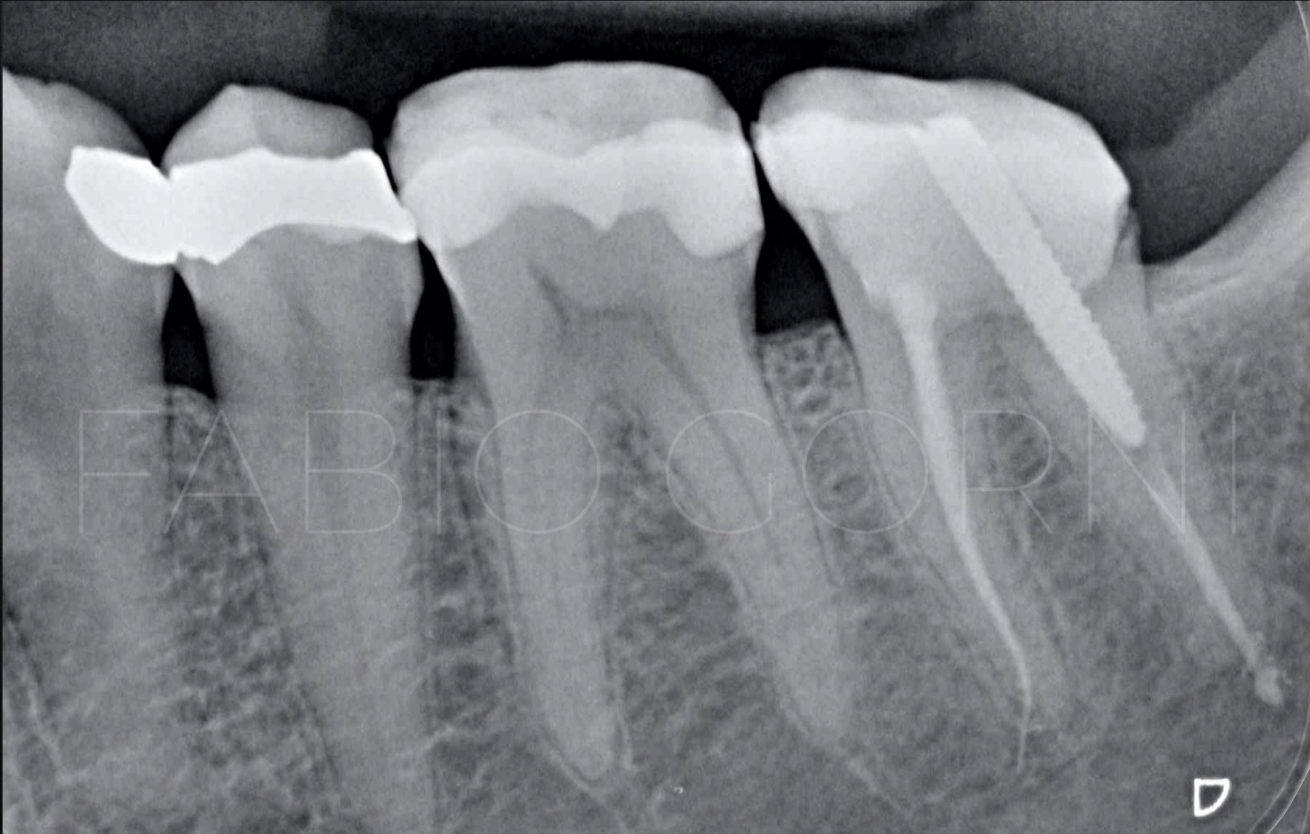
Fig. 1
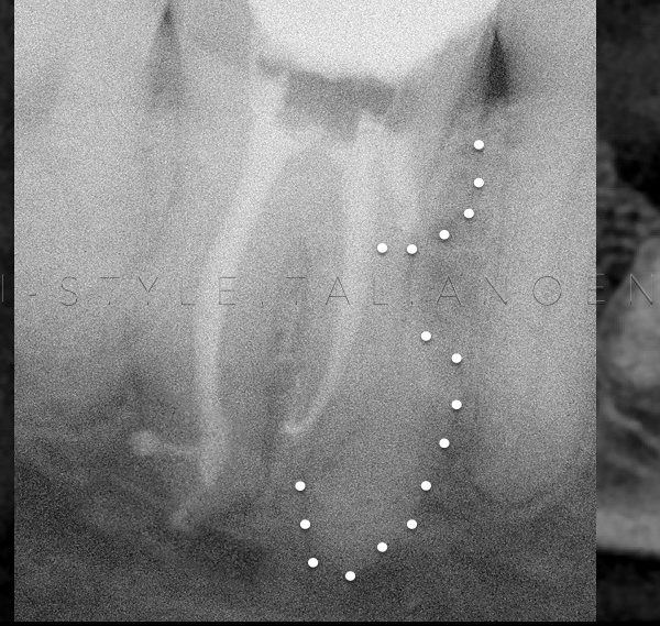
Female 63 yrs old , the patient has been referred to the office for a problem on distal root in second lower molar , the patient has pain during the chewing and radiographically is evident an overfilling due to a wrong obturation technique .
Two different operative options , endodontic surgery or non surgical retreatment , but in this case we preferred the retreatment option for the unfavorable anatomy and the difficulty in the surgical access even in presence of materials beyond the apex .
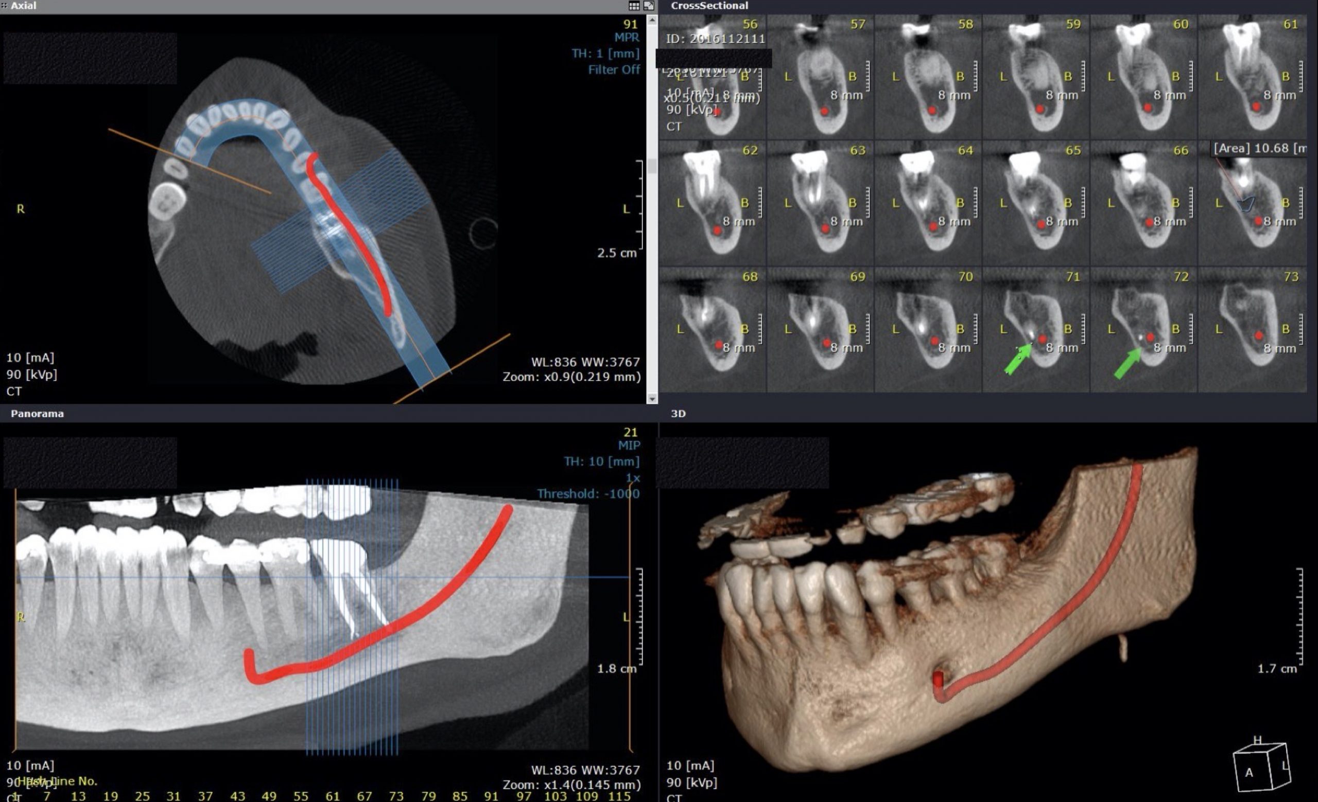
Fig. 2
In the CBCT the anatomy situation is clear , it’s possible to see the relationships between the apex and the mandibular nerve as well as the materials over the apex.
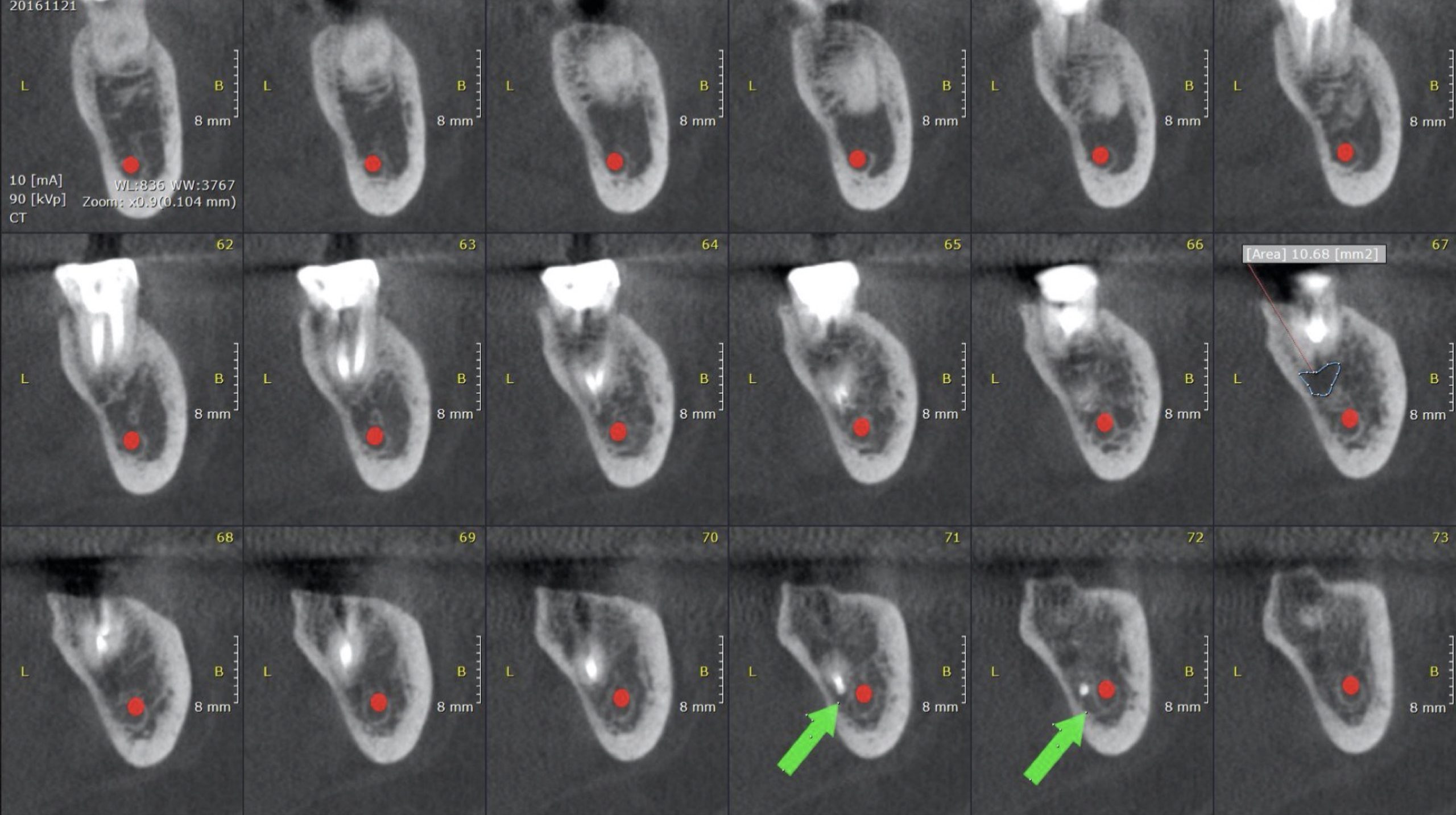
Fig. 3
CBCT cross sections
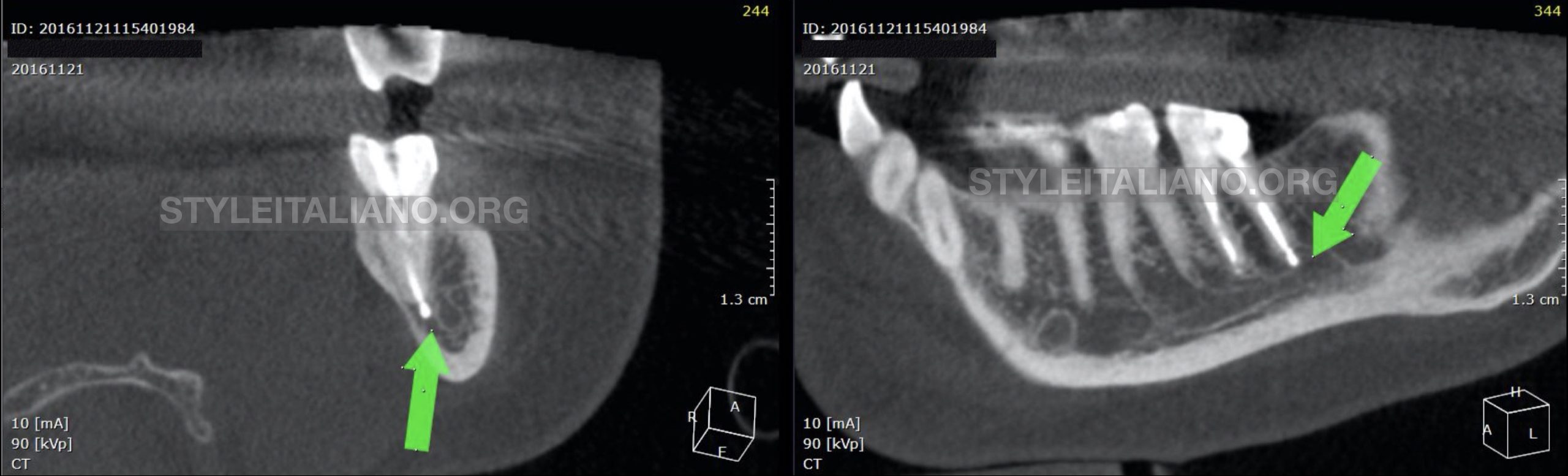
Fig. 4
Green Arrows show the overextended material in two different CBCT views
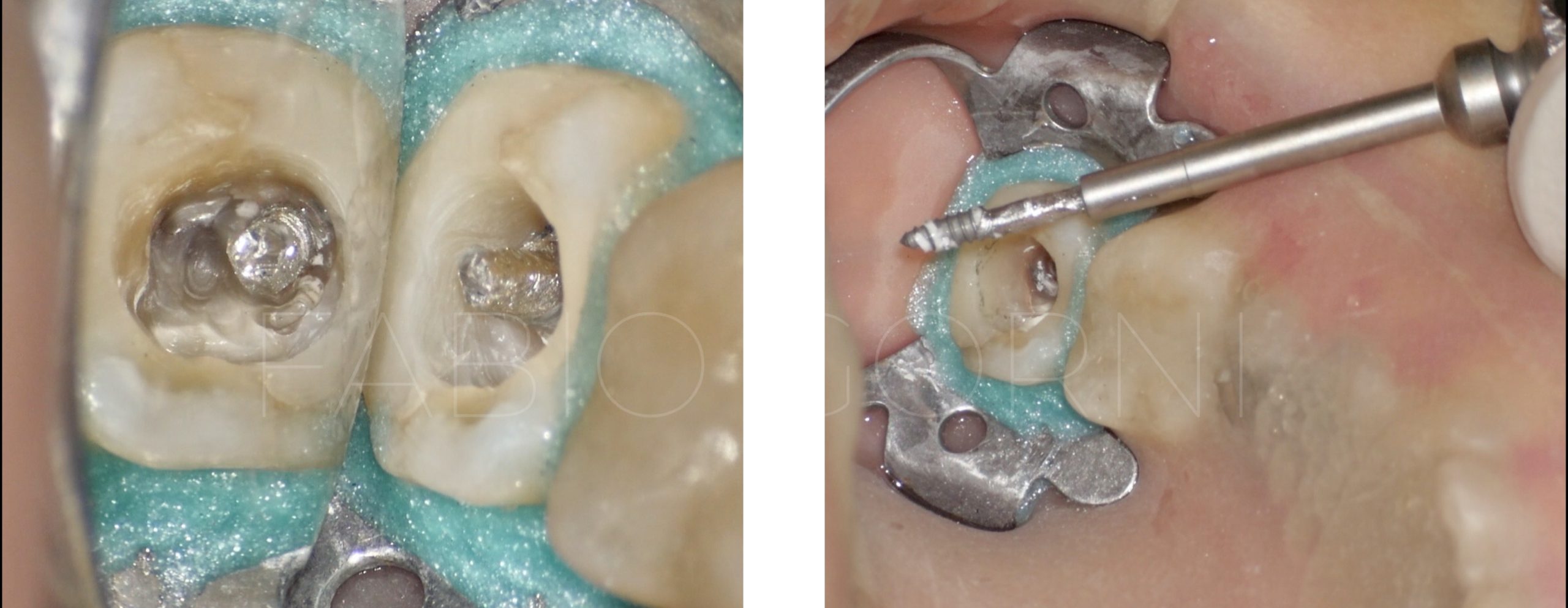
Fig. 5
We removed the big screw post thanks to the a special screw driver and after that all the cement using ultrasonic tips , in the previous treatment the colleague used thermafil technique and part of the core is out of the apex .
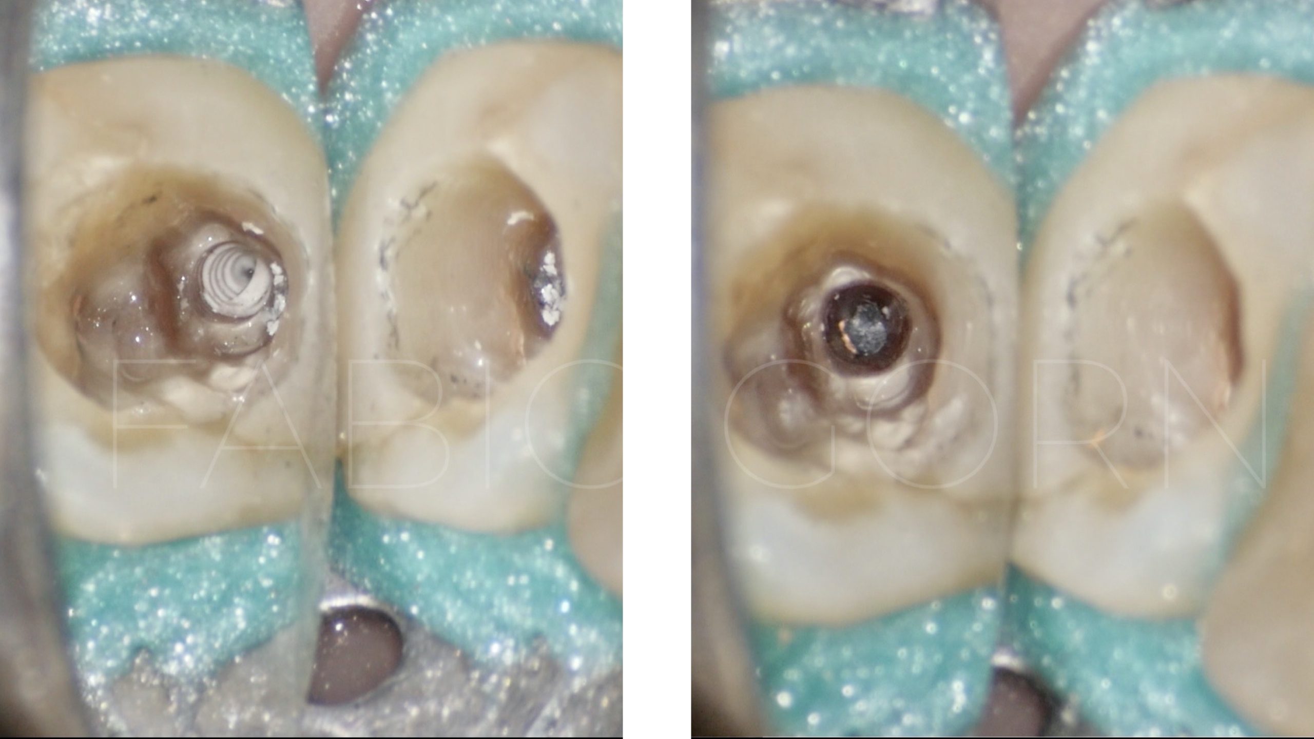
Fig. 6
After the disassembling we can see the cement in the canal and the plastic core that occlude the space
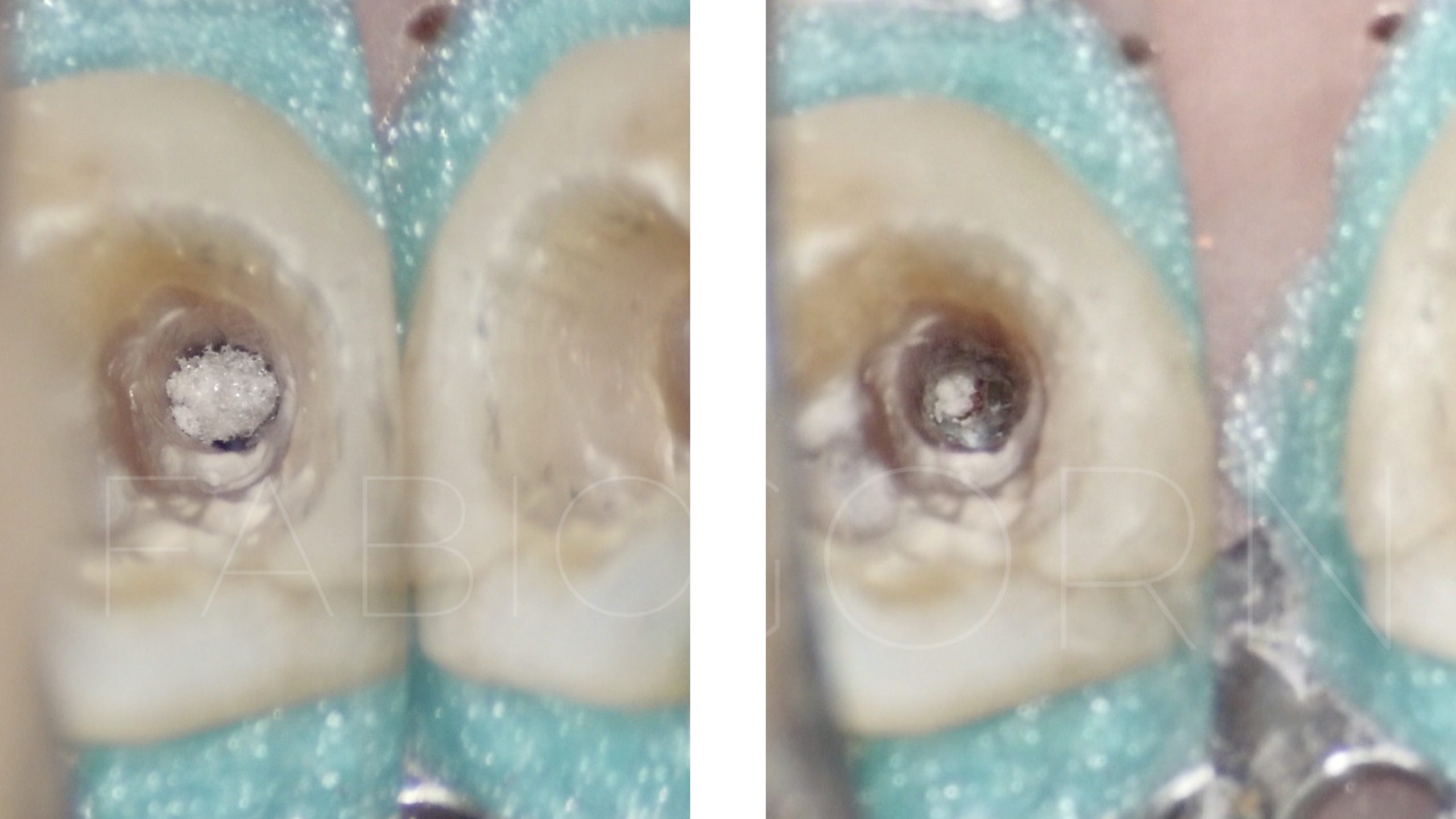
Fig. 7
When the disassembling has been completed , the canal shaped and cleaned , we positioned a collagen matrix beyond the foramen to obtain a better control during the apical plug .
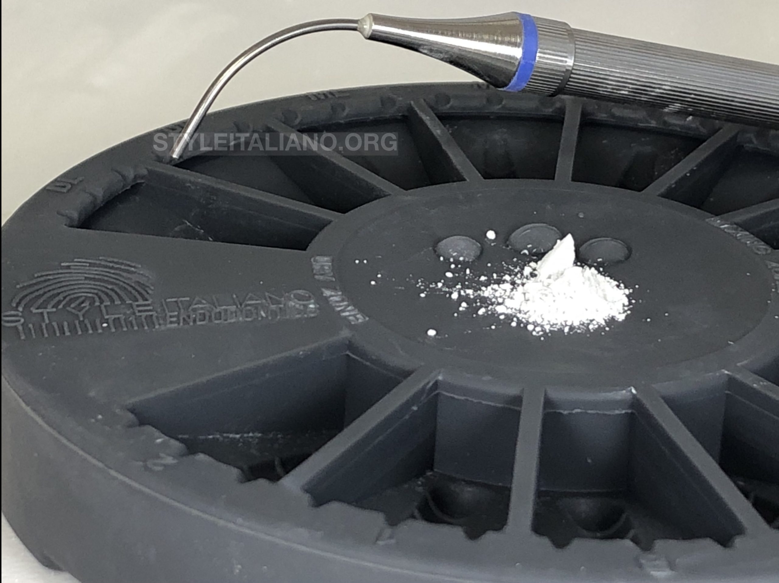
Fig. 8
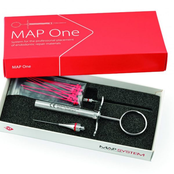
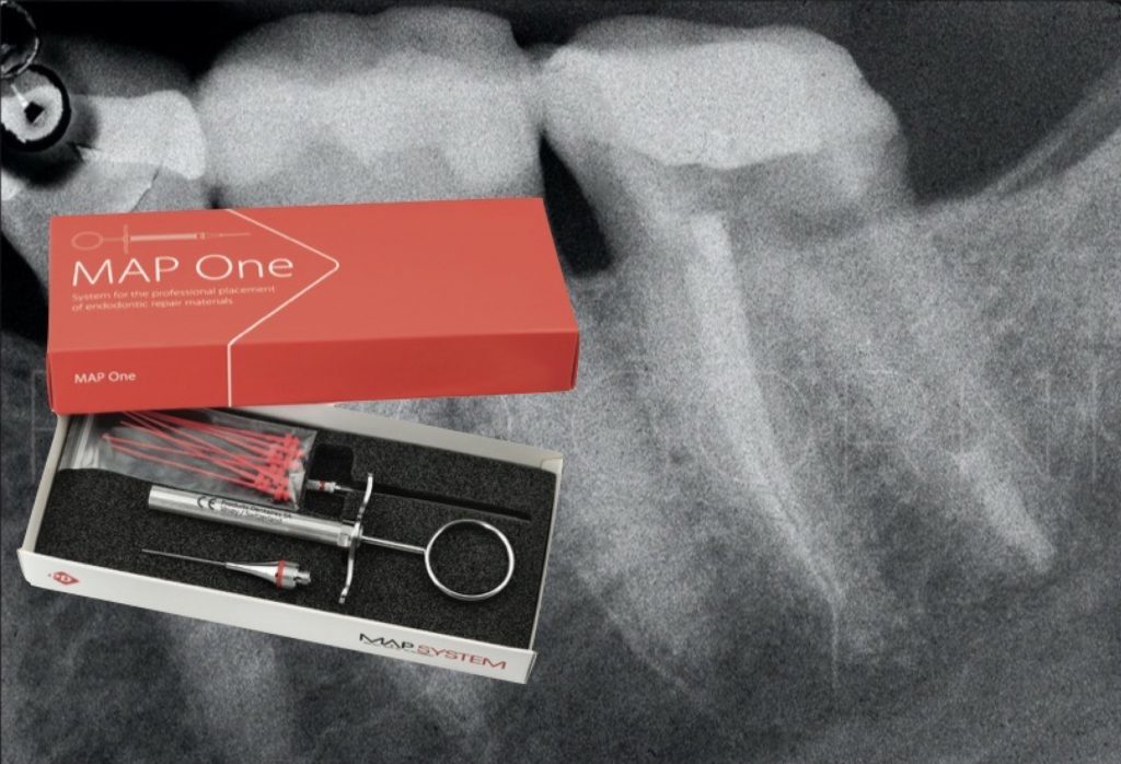
MAP SYSTEM , MTA and PLANO
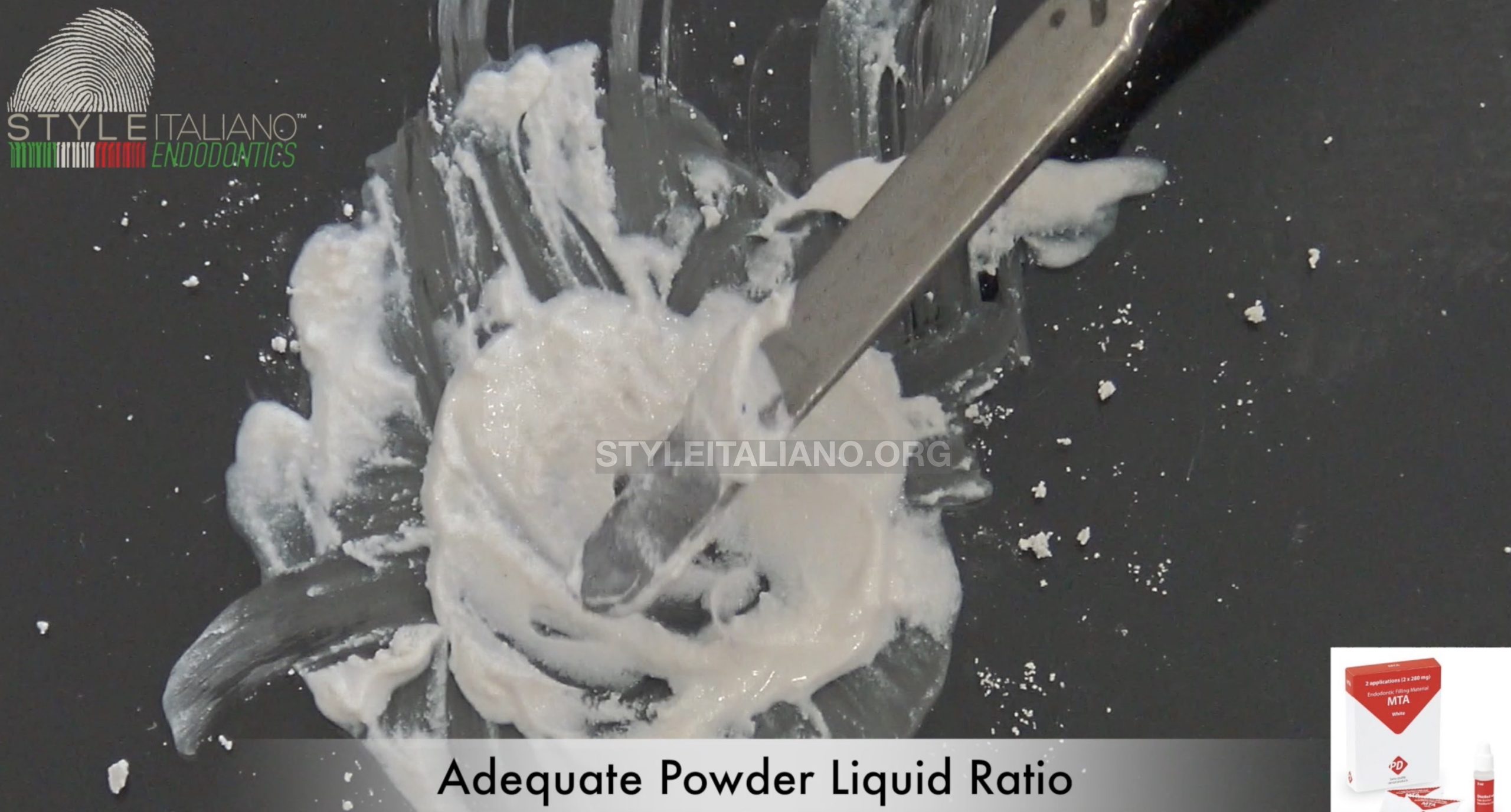
Fig. 9
the cement must be mixed correctly with the right powder liquid ratio
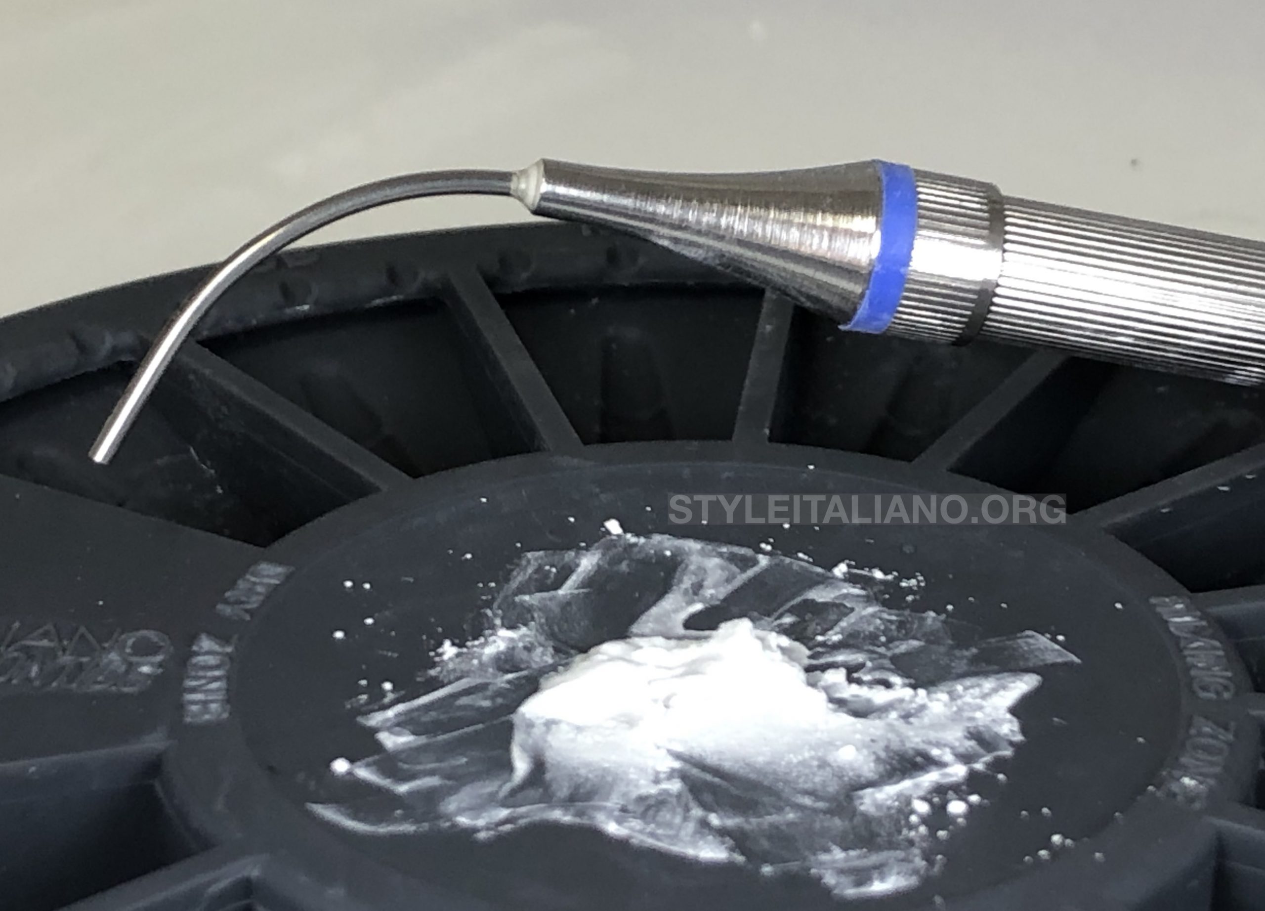
Fig. 10
MTA mixed and ready to use
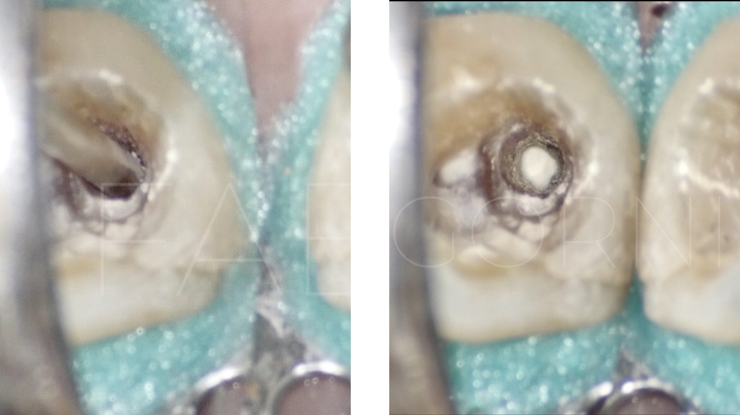
Fig. 11
The MAP-ONE bring the MTA in the apical one third
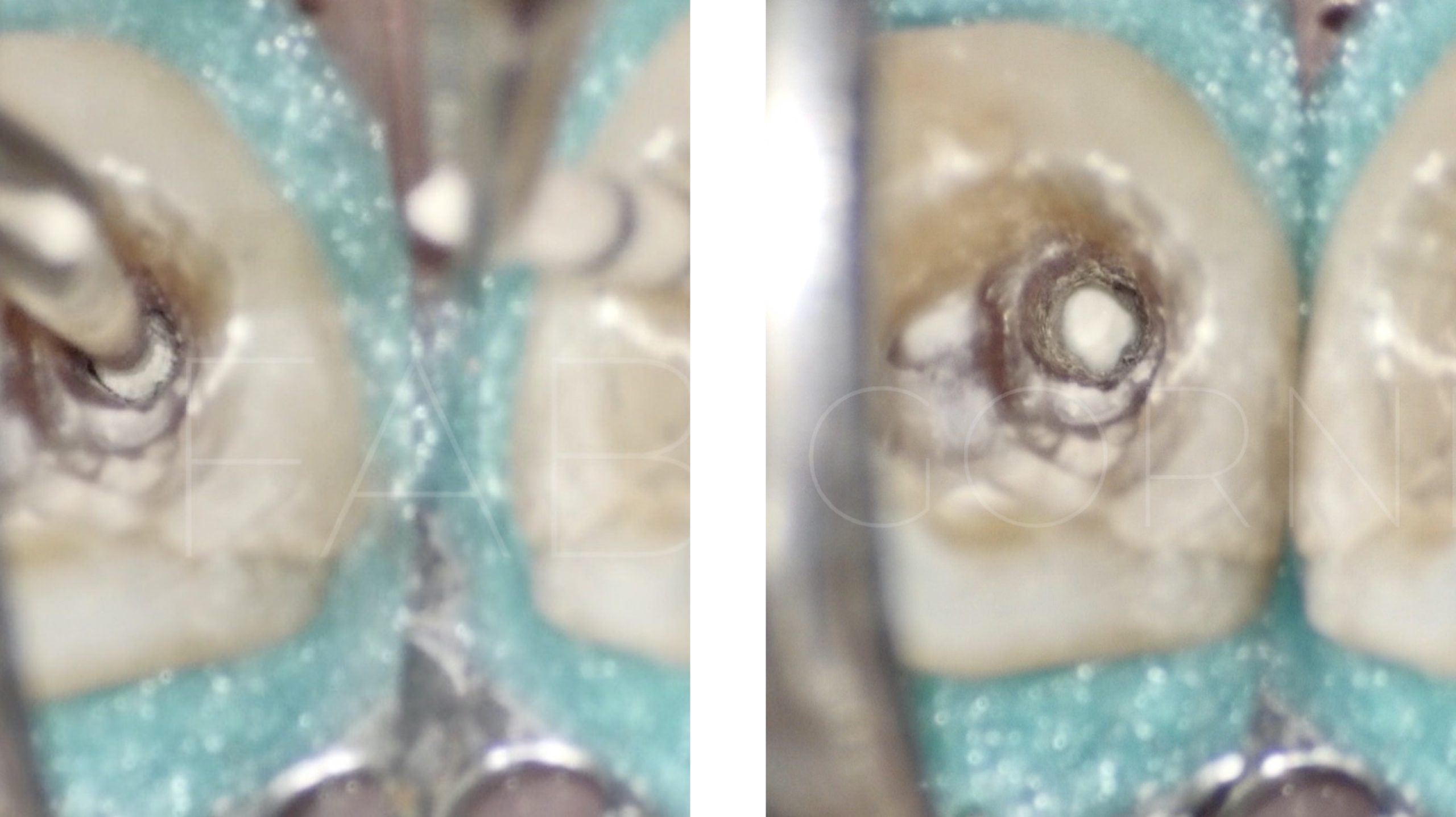
Fig. 12
Using the MAP-ONE it’s easy to bring the MTA in the apical one third , subsequently the material is pushed and adapted gently to the foramen, using a paper point .
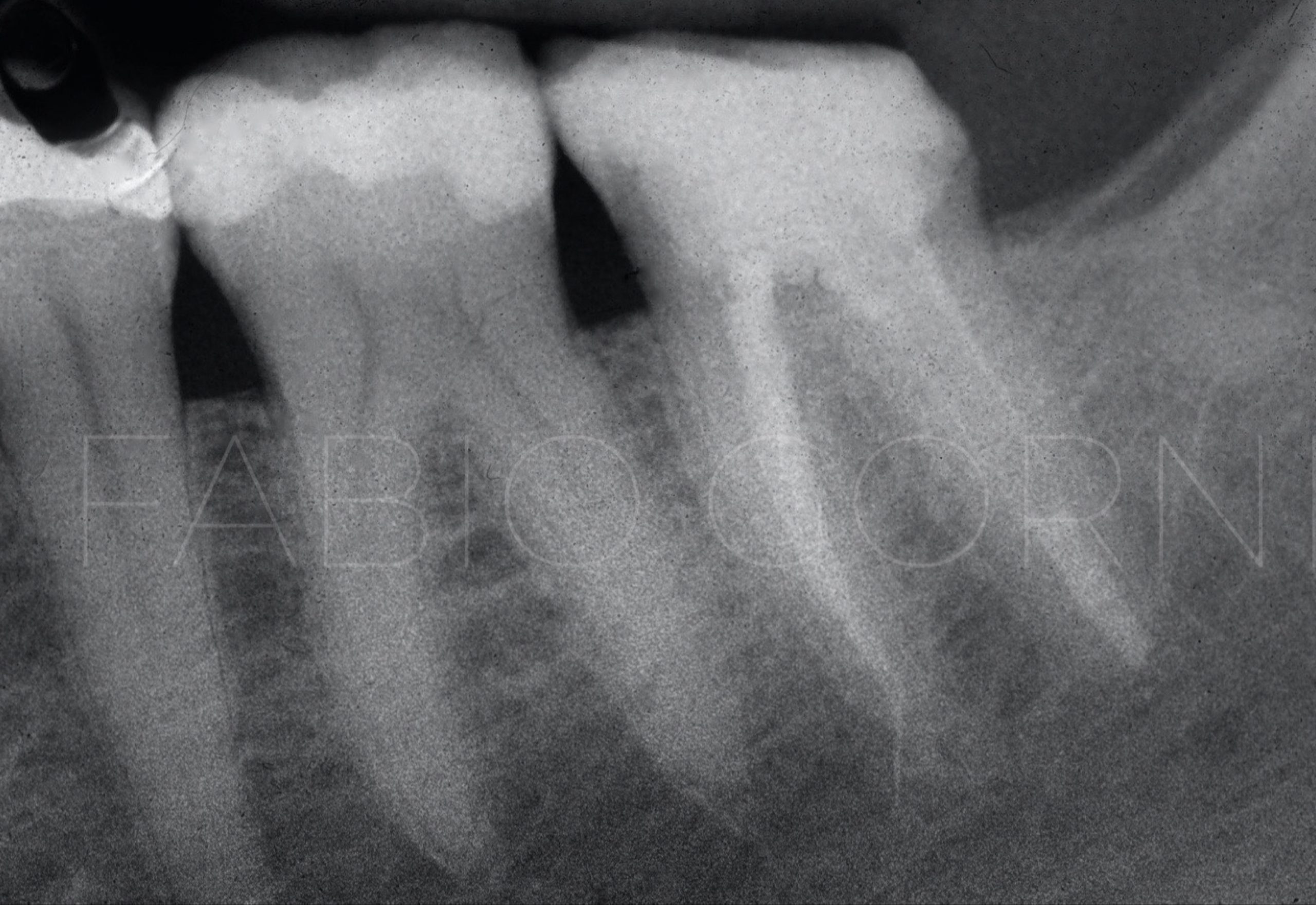
Fig. 13
Post-op xray , the plug is perfectly positioned , no overfilling and the obturation is tridimensional
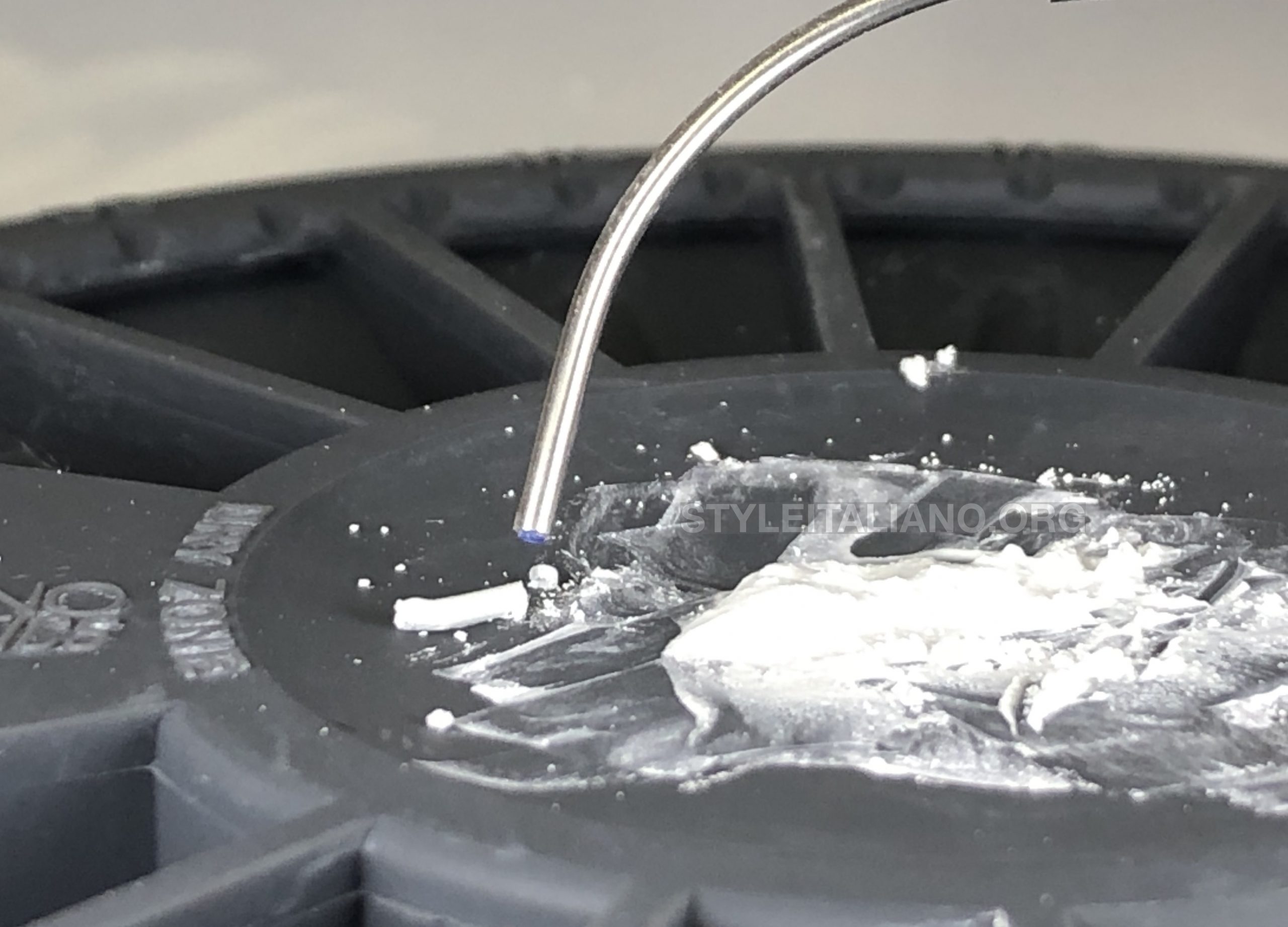
Fig. 14
With MAP system it's possible to obtain a MTA block, 5 mm leght ideal to do apical plug
Conclusions
To obtain a perfect adaptation of the cement in the apical one third is necessary bring the right amount of material in the apex and at the right level , that’s why the use of MAP-ONE is mandatory
Bibliography
Torabinejad M, Parirokh M, Dummer PMH. Mineral trioxide aggregate and other bioactive endodontic cements: an updated overview - part II: other clinical applications and complications. Int Endod J. 2018 Mar;51(3):284-317.
Parirokh M, Torabinejad M, Dummer PMH. Mineral trioxide aggregate and other bioactive endodontic cements: an updated overview - part I: vital pulp therapy. Int Endod J. 2018 Feb;51(2):177-205.