Molars anatomy Pt.2
13/03/2025
Gabriele Ragucci
Warning: Undefined variable $post in /home/styleendo/htdocs/styleitaliano-endodontics.org/wp-content/plugins/oxygen/component-framework/components/classes/code-block.class.php(133) : eval()'d code on line 2
Warning: Attempt to read property "ID" on null in /home/styleendo/htdocs/styleitaliano-endodontics.org/wp-content/plugins/oxygen/component-framework/components/classes/code-block.class.php(133) : eval()'d code on line 2
Endodontic success relies on thorough debridement, disinfection, and obturation of the root canal system.
The maxillary first molar, which frequently presents anatomical variations, is a major challenge in clinical endodontics.
Understanding its complex morphology is critical in reducing treatment failure caused by untreated or undetected canals (Cleghorn et al., 2006; Estrela et al., 2015).
This review aims to provide a comprehensive synthesis of the external and internal root canal anatomy of the maxillary first molar, supported by anatomical studies utilizing CBCT and micro-CT.

Fig. 1
External Root Morphology
The maxillary first molar typically exhibits a triradicular structure with three well-differentiated roots:
•Mesiobuccal root (MBR) – The most complex and variable root, frequently containing a second canal (MB2).
•Distobuccal root (DBR) – Generally shorter and more tapered than the mesiobuccal root, with a single canal.
•Palatal root (PR) – The longest and most robust root, usually containing a single wide canal.
Variations in Root Form
Although the classical three-rooted form is predominant, anatomical variations exist:
• Fusion of the distobuccal and palatal roots (7–15%) results in a C-shaped canal morphology (Kim et al., 2018).
• Radix mesiolingualis, a supernumerary root, is rarely reported (Bolger & Schäfer, 2016).

Fig. 2
Internal Root Canal Morphology
The internal morphology of the maxillary first molar demonstrates significant variability, particularly in the mesiobuccal root.
Mesiobuccal Root Canal System
The mesiobuccal root frequently contains two canals (MB1 and MB2), with variations in their configurations (Vertucci, 1984; Kulild & Peters, 1990):
• Type I (Single canal): 20–40%
• Type II (Two canals merging into one): 20–25%
• Type III (Two separate canals): 40–60%

Fig. 3
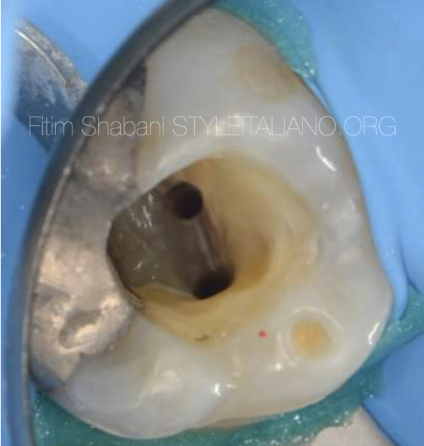
Prevalence of MB2 canal:
• Traditional methods (hand files, radiographs): ~50%
• Magnification (microscopes, loupes): 70–80%
• CBCT & micro-CT studies: 90–97% (Nosrat et al., 2015; Zhang et al., 2020)
The MB2 canal is commonly located mesiopalatal to MB1 and often remains unprepared without the use of enhanced visualization (Karabucak et al., 2016).

Fig. 4
The MB2 canal is commonly located mesiopalatal to MB1 and often remains unprepared without the use of enhanced visualization (Karabucak et al., 2016).

Fig. 5
The MB2 canal is commonly located mesiopalatal to MB1 and often remains unprepared without the use of enhanced visualization (Karabucak et al., 2016).
The MB2 canal is commonly located mesiopalatal to MB1 and often remains unprepared without the use of enhanced visualization (Karabucak et al., 2016).

Fig. 6
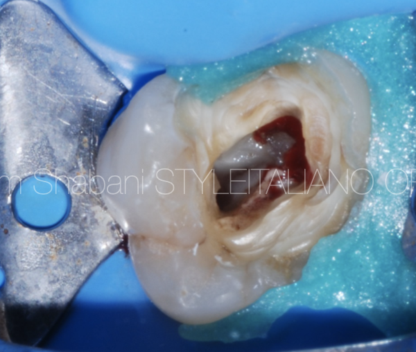
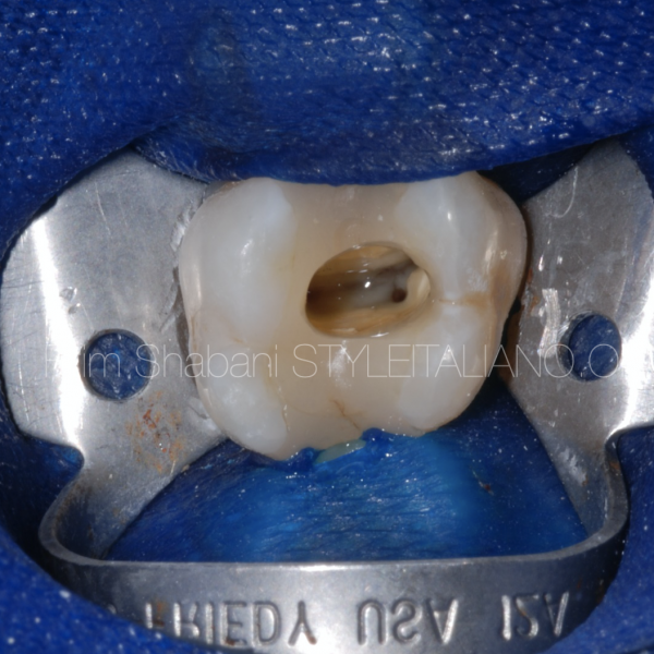
clinical case

Fig. 7
Distobuccal and Palatal Root Canal Morphology
•The distobuccal root contains one canal in >95% of cases (Weine, 1998), but bifurcation can occur in 2–5% (Cleghorn et al., 2006).
• The palatal root is relatively straightforward, with a single canal in 99% of cases (Pécora et al., 1992). However, reports exist of two separate palatal canals (Christie et al., 1991).
Advanced Diagnostic Imaging
Traditional radiographs are often inadequate in detecting the full complexity of the root canal system.
CBCT and micro-CT have greatly enhanced our ability to visualize hidden canal structures.
CBCT Advantages:
• Provides three-dimensional visualization of root canal morphology.
• Facilitates identification of MB2 canals and root fusions (Matherne et al., 2008).
•Reduces the risk of iatrogenic errors in canal preparation.
• Micro-CT in Morphological Studies:
•Gold standard for in vitro studies of root canal anatomy.
• Confirms the presence of additional lateral canals, apical deltas, and complex isthmuses (Zhang et al., 2020).

Fig. 8
MB2 Canal: A Key Factor in Treatment Success
Failure to detect and instrument MB2 is a leading cause of persistent apical pathology (Wolcott et al., 2002).
Strategies to enhance MB2 detection include:
• Pre-treatment CBCT scans for complex cases.
• Use of magnification and illumination to aid in locating hidden or calcified canals.
• Selective dentin removal and troughing techniques to expose MB2 without compromising tooth integrity.
Conclusions
maxillary first molar is a highly variable tooth with a complex root canal system. The mesiobuccal root, particularly the MB2 canal, represents a major challenge in endodontic therapy. CBCT and micro-CT studies confirm that MB2 is present in nearly all cases, underscoring the importance of advanced imaging, magnification, and proper access design to achieve optimal treatment outcomes.
Neglecting anatomical complexities, especially unprepared MB2 canals, significantly contributes to endodontic failure. Future research should continue exploring 3D-guided endodontics, artificial intelligence-assisted canal detection, and improved irrigation strategies to enhance treatment predictability.
Bibliography
1. Cleghorn BM, Christie WH, Dong CC. (2006). J Endod, 32(9):813-21.
2. Christie WH, Peikoff MD, Fogel HM. (1991). J Endod, 17(2):80-4.
3. Estrela C, Bueno MR, Sousa-Neto MD, et al. (2015). J Endod, 41(9):1545-50.
4. Kulild JC, Peters DD. (1990). J Endod, 16(7):311-7.
5. Matherne RP, et al. (2008). J Endod, 34(1):87-9.
6. Nosrat A, et al. (2015). J Endod, 41(9):1524-8.
7. Pécora JD, Woelfel JB, Sousa Neto MD, Issa EP. (1992). Braz Dent J, 3(1):27-30.
8. Vertucci FJ. (1984). Oral Surg Oral Med Oral Pathol, 58(5):589-99.
9. Wolcott J, et al. (2002). J Endod, 28(6):477-9.
10. Zhang R, et al. (2020). Int Endod J, 53(3):303-12.