Maxillary second molar with MB2 root canal system : an unusual variation
29/10/2026
Leena Rawal
Warning: Undefined variable $post in /home/styleendo/htdocs/styleitaliano-endodontics.org/wp-content/plugins/oxygen/component-framework/components/classes/code-block.class.php(133) : eval()'d code on line 2
Warning: Attempt to read property "ID" on null in /home/styleendo/htdocs/styleitaliano-endodontics.org/wp-content/plugins/oxygen/component-framework/components/classes/code-block.class.php(133) : eval()'d code on line 2
Maxillary second molars are often challenging to treat due to their limited accessibility and frequent variations in root canal anatomy. Early identification of additional canals is crucial for complete disinfection and predictable long-term success.
In this case, the patient reported with sensitivity to hot and cold, along with pain on chewing. Root canal treatment was completed in 2 sittings. MB2 was found using DTE ED3D endodontic tip by troughing dentin mesial and palatal to MB1 under DOM. Cleaning and Shaping was done by MG3 Blue Rotary files from Perfect Endo. The key highlight was the unusual root canal anatomy and the use of magnification and ultrasonic troughing.
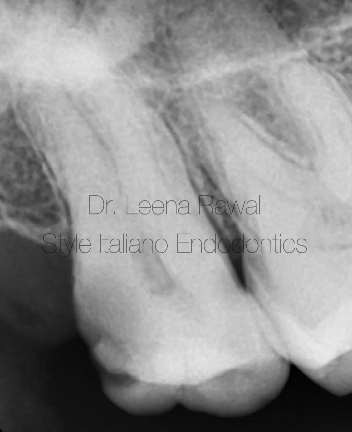
Fig. 1
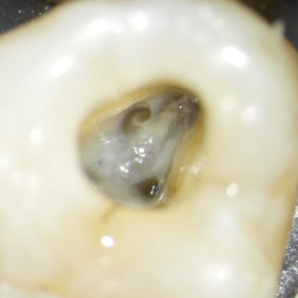
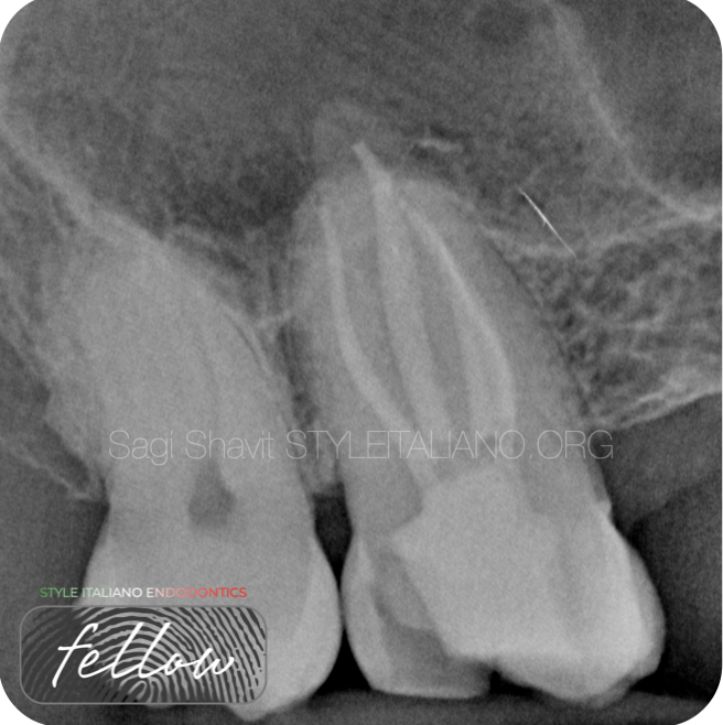
Pre-op IOPA shows caries approaching the pulp with partial calcification of the pulp chamber in 17. Calcification increases the likelihood of hidden or difficult-to-locate canals. MB2 detection may require ultrasonics and magnification.
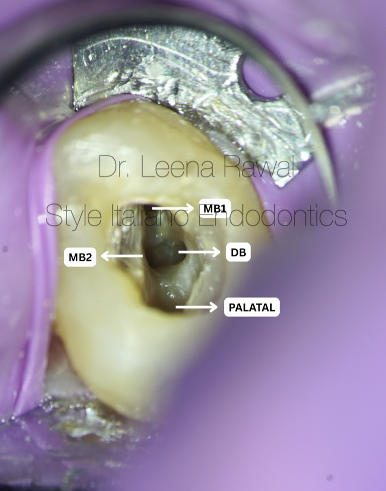
Fig. 2
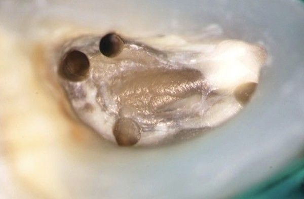
In this image, you can see all four prepared canals — MB1, MB2, DB, and Palatal — clearly shaped and ready for obturation.
MB2 was identified using the DTE ED3D ultrasonic tip, with conservative troughing to preserve dentin and maintain a controlled access.
Negotiation was completed under magnification, establishing a smooth and reproducible glide path before final shaping.
Video of the pulpal chamber
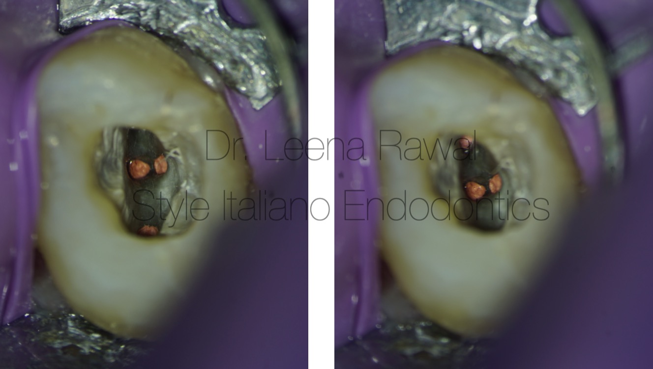
Fig. 3
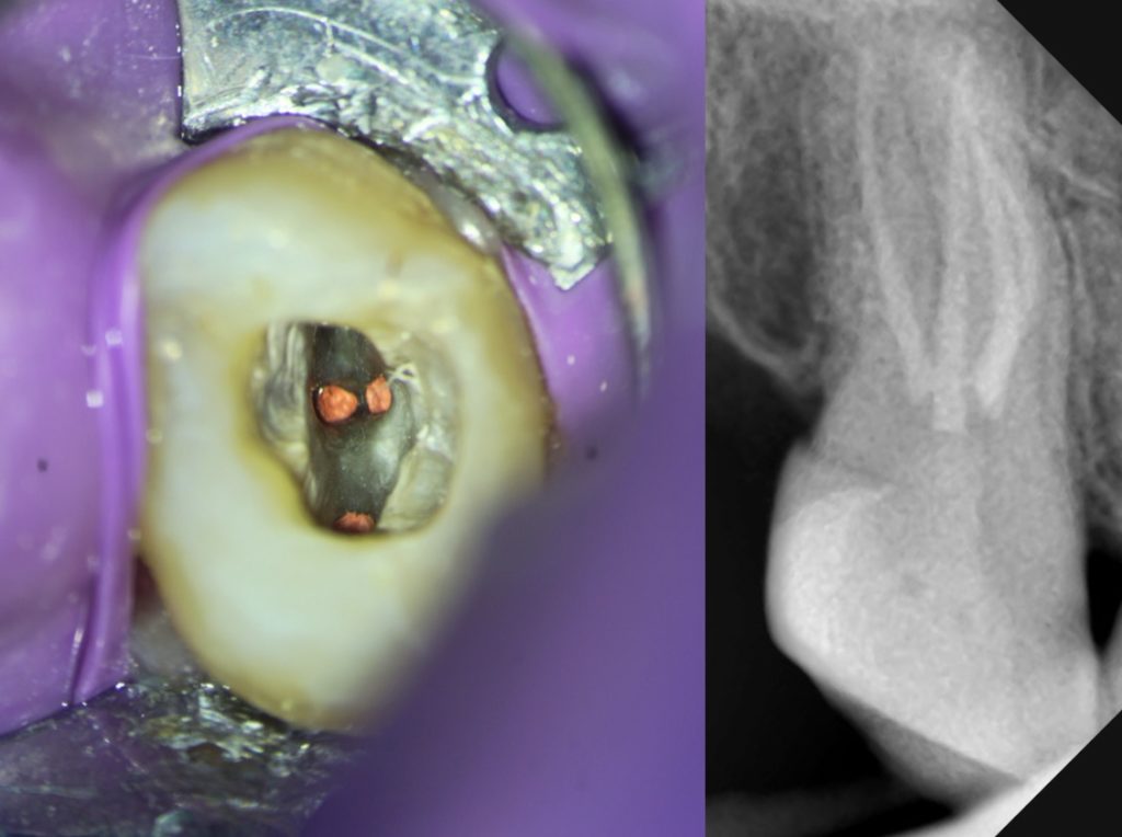
Single cone obturation done using Bio-Ceramic Root canal Sealer from Atyanta Dental.
Video after obturation
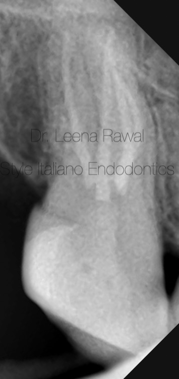
Fig. 4
Final xray
Conclusions
This case demonstrates that even in challenging maxillary second molars, the MB2 canal can be reliably identified with enhanced visualisation using magnification and precise ultrasonic refinement. Conservative dentin removal, thorough negotiation, and systematic preparation of all four canals contributed to a successful outcome.