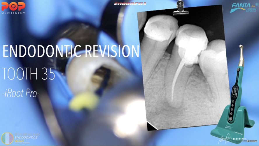
Management of the complex anatomy of lower premolars
17/03/2024
Fellow
Warning: Undefined variable $post in /home/styleendo/htdocs/styleitaliano-endodontics.org/wp-content/plugins/oxygen/component-framework/components/classes/code-block.class.php(133) : eval()'d code on line 2
Warning: Attempt to read property "ID" on null in /home/styleendo/htdocs/styleitaliano-endodontics.org/wp-content/plugins/oxygen/component-framework/components/classes/code-block.class.php(133) : eval()'d code on line 2
Variations in the root canal configurations are a great challenge for the endodontist during endodontic procedures. This necessitates the understanding of canal morphology before initiating the treatment.
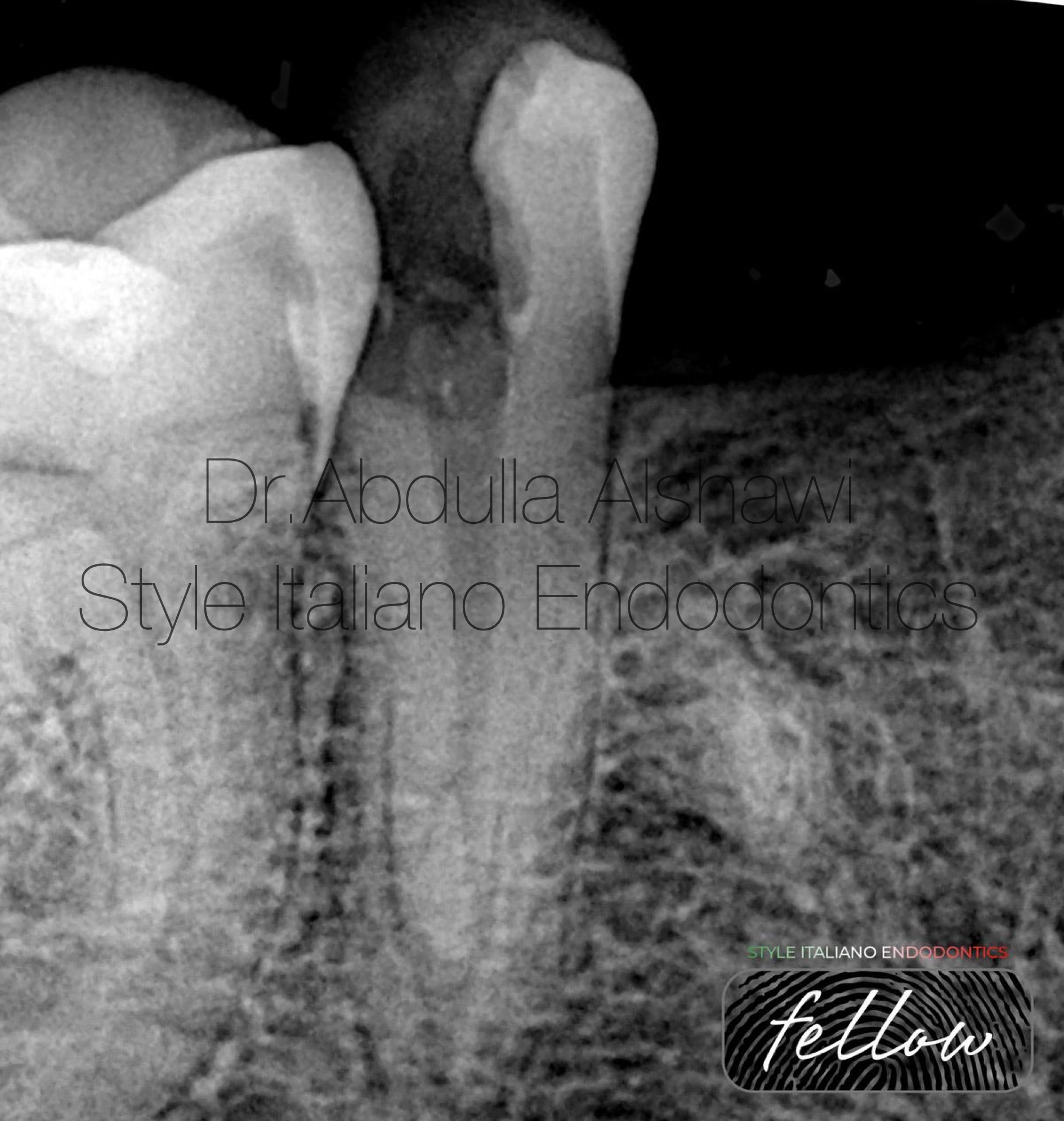
Fig. 1
First case: lower second premolar, patient came to the clinic suffering from pain on biting, pre op x-ray showing caires extend to the pulp and abnormal anatomy from the middle part of root.
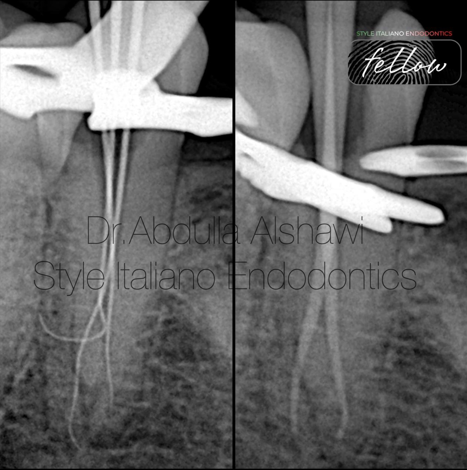
Fig. 2
After determining the W.L time for negotiation for lateral canals and take the M.C
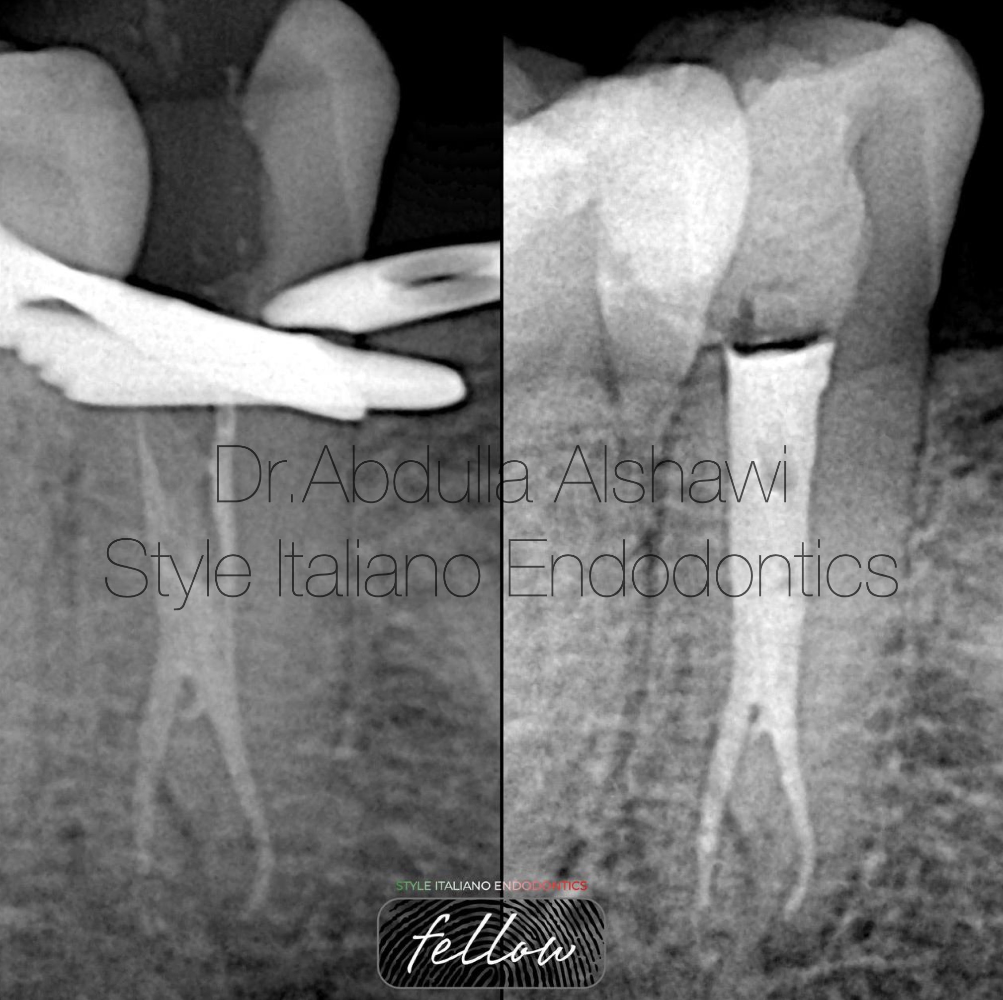
Fig. 3
Final x-ray modified WVC for obturation and to get seal for lateral canal.
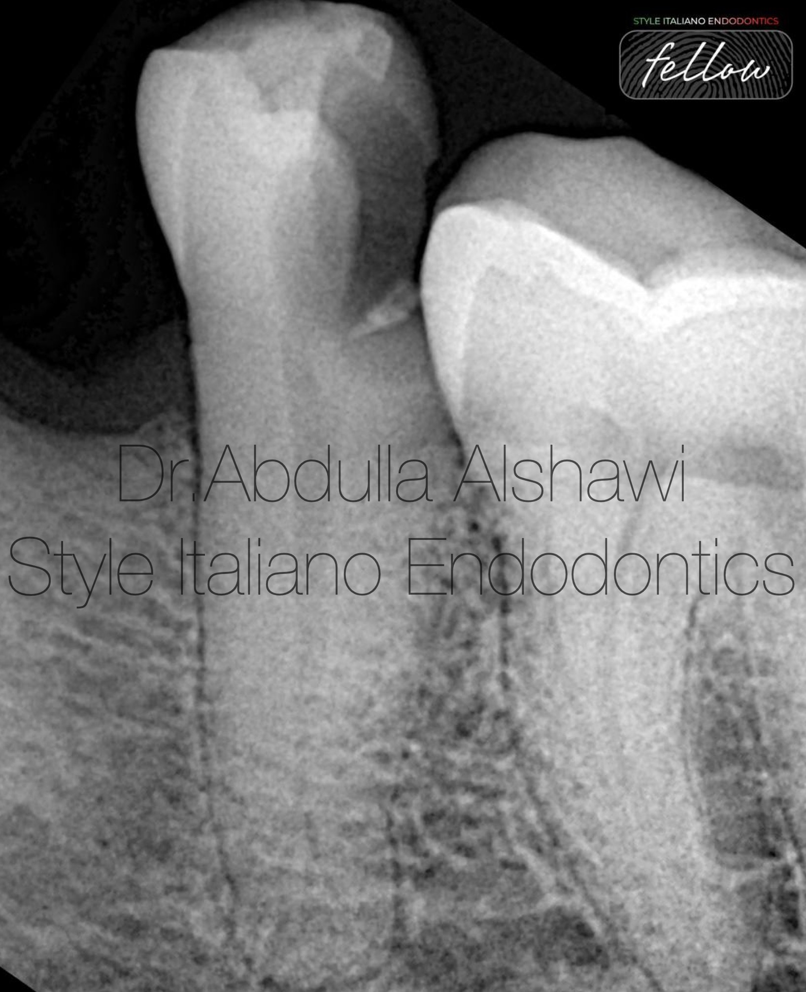
Fig. 4
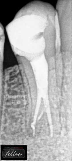
Second case: pre op x-ray showed abnormal shape of the apex for lower second premolar and the canal go lateral which means the apical delta.
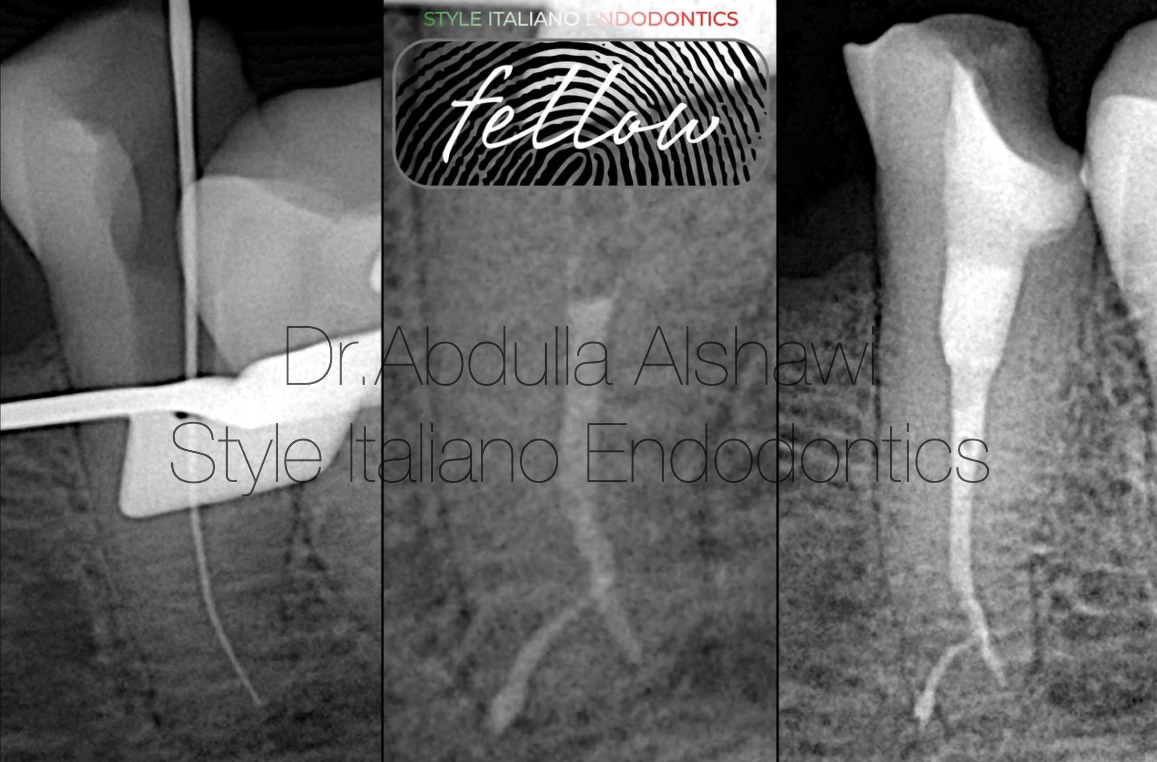
Fig. 5
After checking the W.L one more time file go lateral, and I failed to reach the other portal , so I depend on the irrigation protocol obturation done by modified WVC.
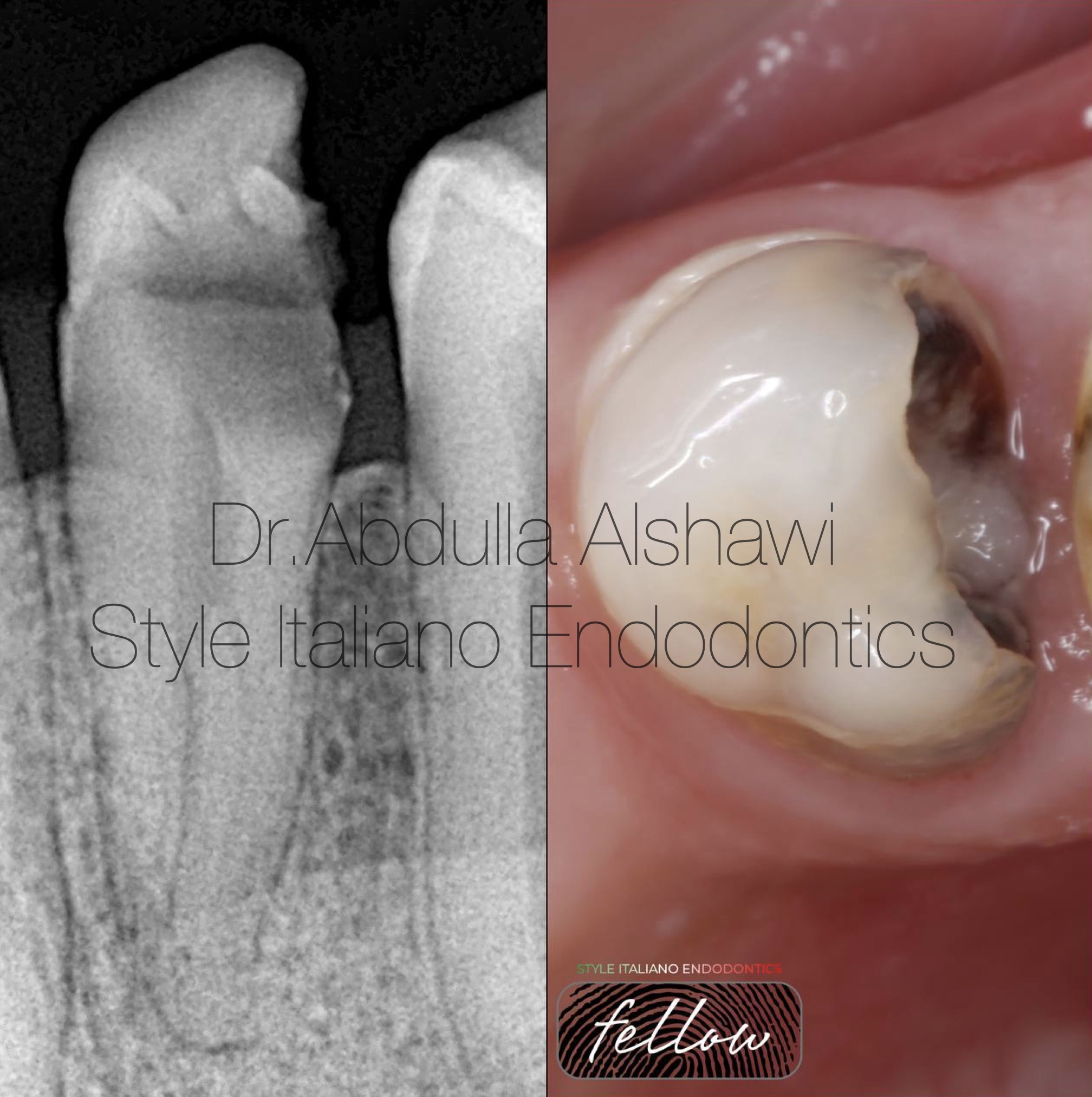
Fig. 6
3rd case: PreOp x-ray lower first premolar deep splitting from the middle part.
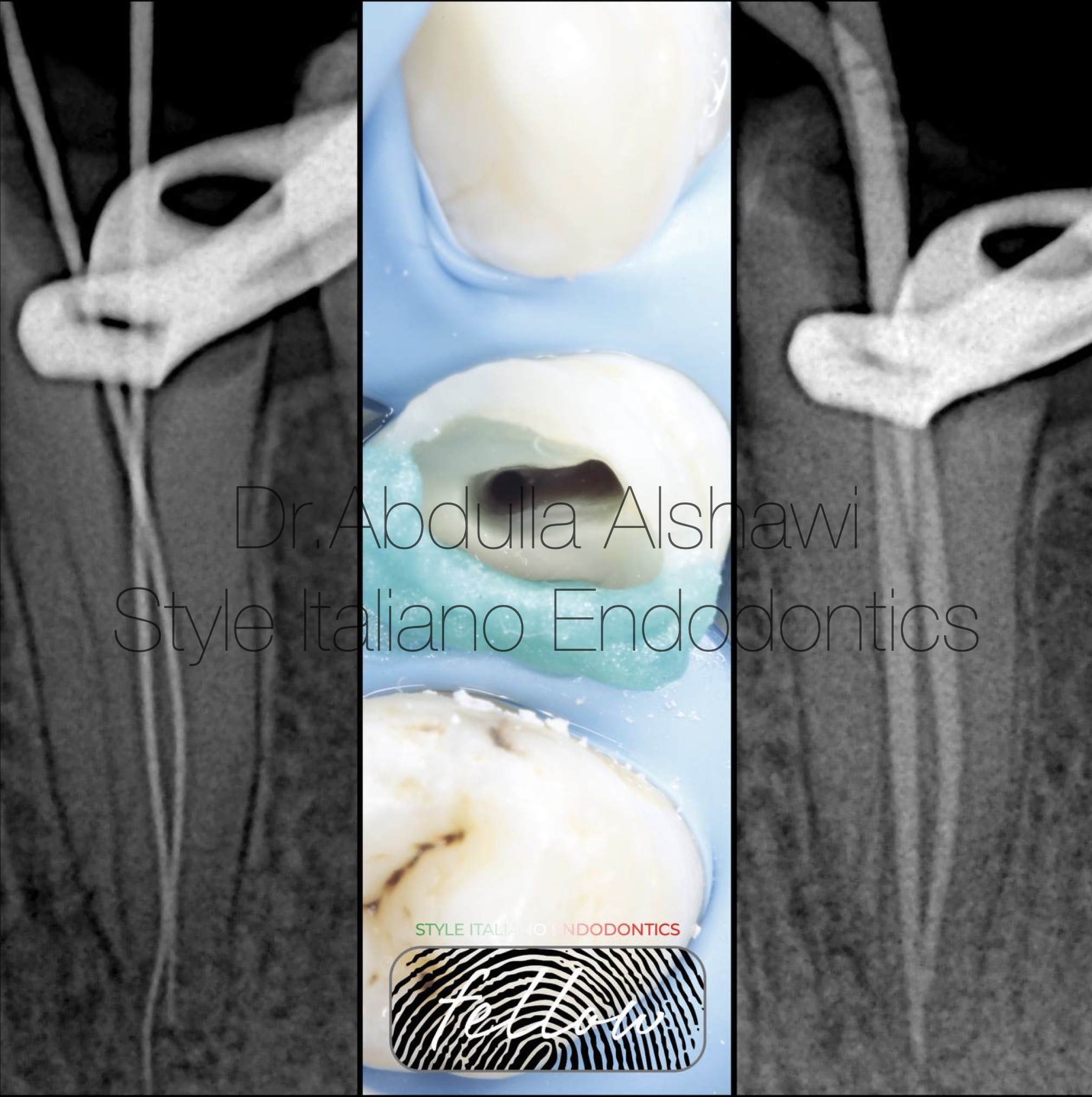
Fig. 7
Cleaning the asmus and determining W.L and check M.C
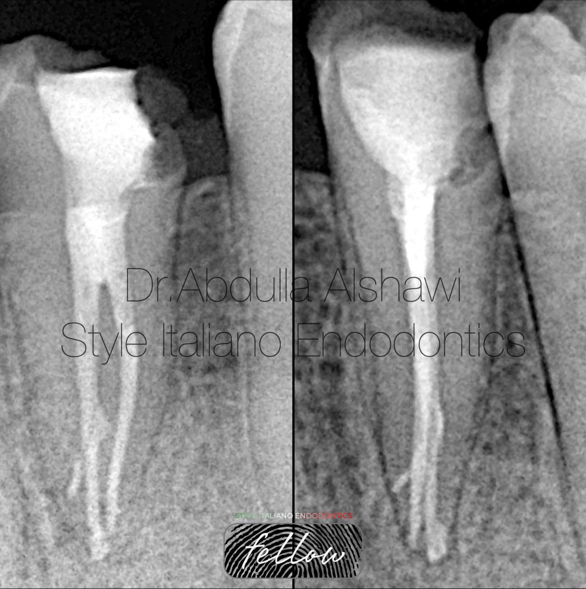
Fig. 8
Final x-ray obturation with BC sealer
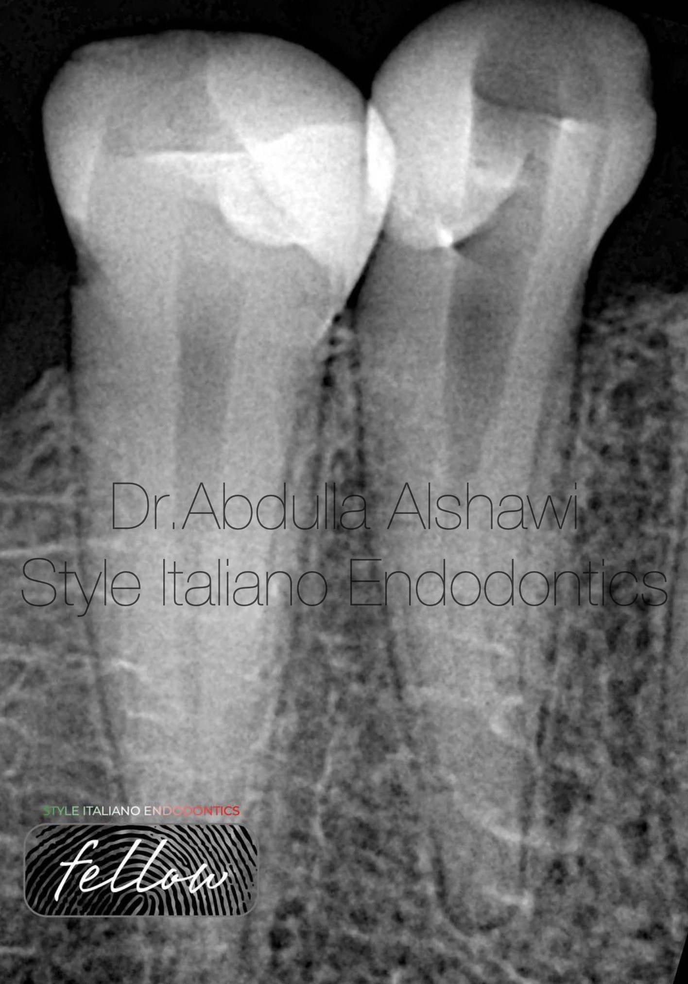
Fig. 9
Case 4th: lower first premolar, patient was refereed to my clinic after the previous dentist couldn’t reach the other canal .
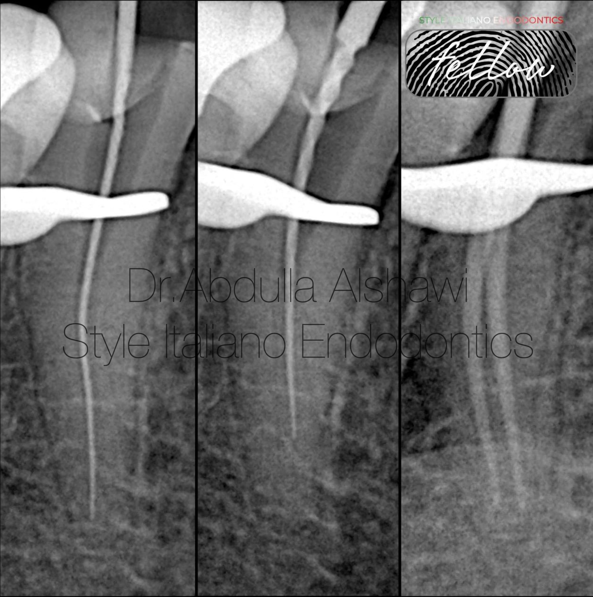
Fig. 10
Locate the second canal and check the M.C
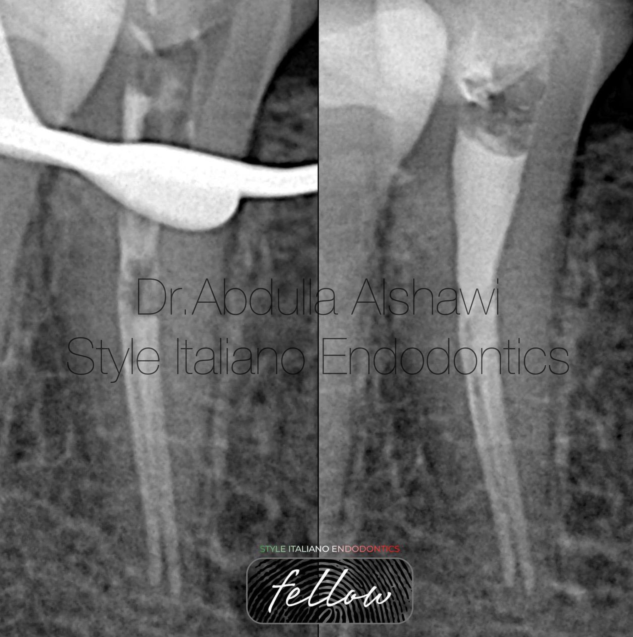
Fig. 11
Final x-ray for obturation with modified WVC and BC sealer.
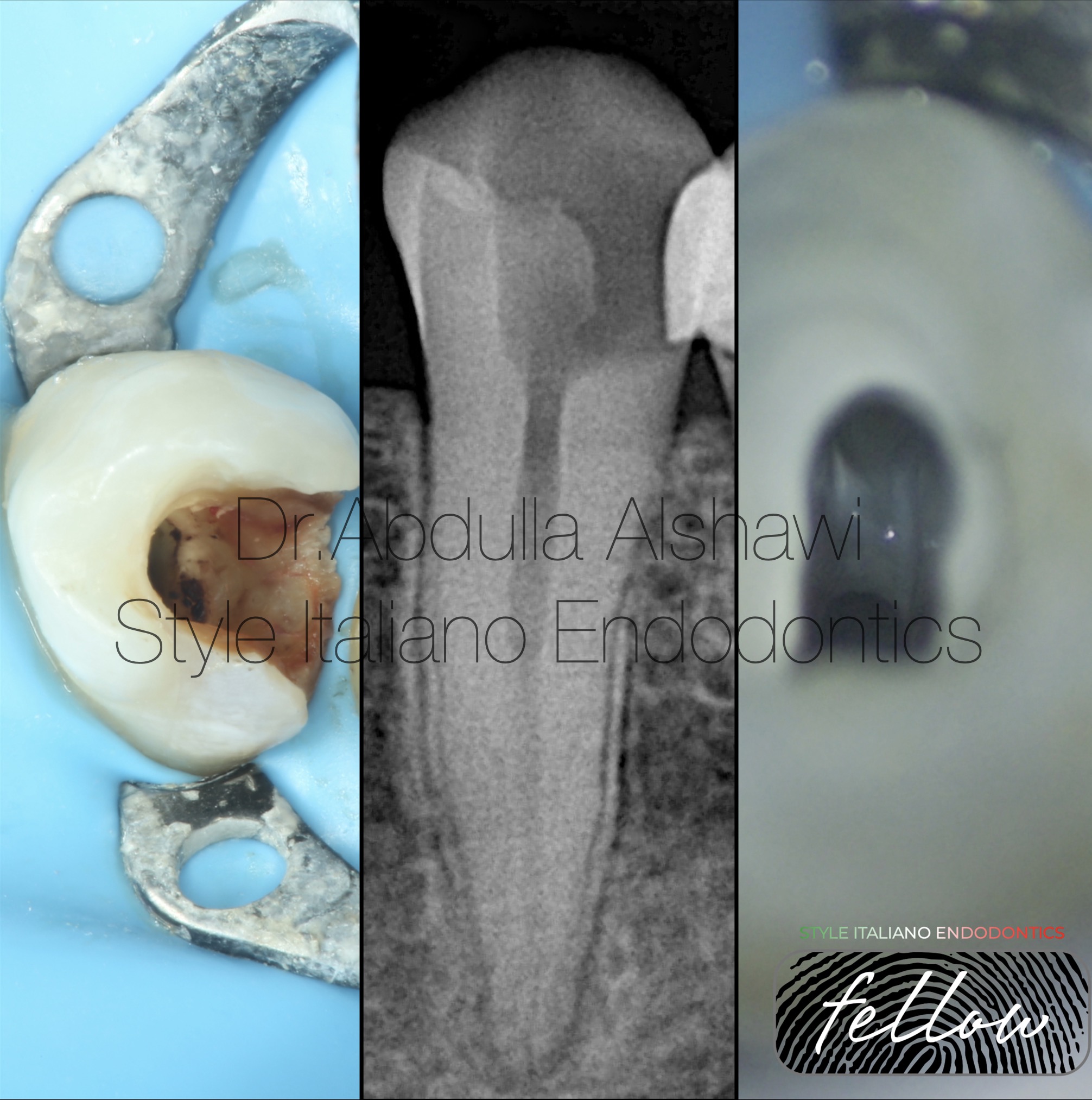
Fig. 12
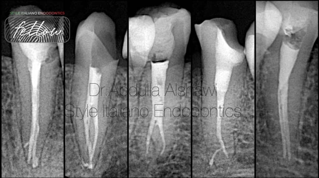
Case 5th. Lower first premolar, type 3 canal configuration.
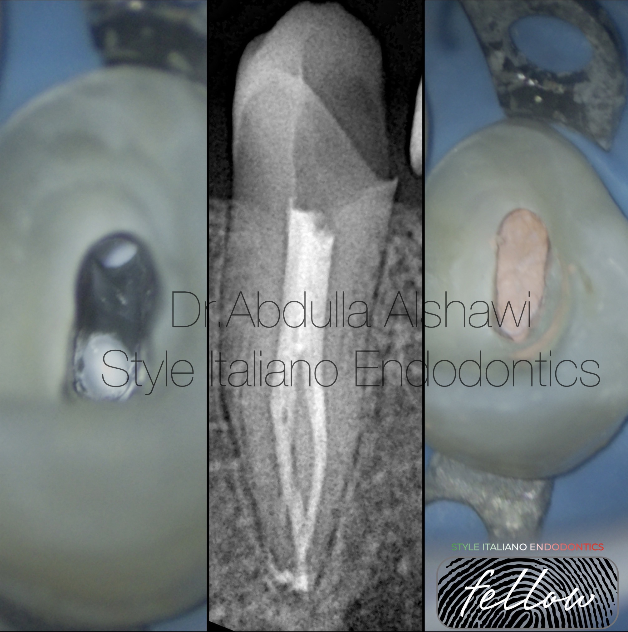
Fig. 13
Finalizzation

Fig. 14
Dr Abdulla Alshawi
B.D.S
Graduated from Uruk dental collage-Iraq 2018-2019
Fellow member in style Italiano endodontics.
Key opinion leader of MDclus
Conclusions
The clinician must be able to recognize the existence of unusual and unexpected variations in the root anatomy and canal configuration. To obtain a good clinical outcome and a complete understanding of the root canal anatomy.
Bibliography
11. Root anatomy and canal morphology of mandibular first premolars in a Chinese population. Dou L, Li D, Xu T, Tang Y, Yang D. Sci Rep. 2017;7:750.
2. Evaluation of the root canal anatomy of maxillary and mandibular premolars in a selected German population using cone-beam computed tomographic data. Bürklein S, Heck R, Schäfer E. J Endod. 2017;43:1448–1452.
3. Prevalence of complex root canal morphology in the mandibular first and second premolars in Thai population: CBCT analysis. Thanaruengrong P, Kulvitit S, Navachinda M, Charoenlarp P. BMC Oral Health. 2021;21:449