Management of large floor perforation
23/08/2023
Fellow
Warning: Undefined variable $post in /home/styleendo/htdocs/styleitaliano-endodontics.org/wp-content/plugins/oxygen/component-framework/components/classes/code-block.class.php(133) : eval()'d code on line 2
Warning: Attempt to read property "ID" on null in /home/styleendo/htdocs/styleitaliano-endodontics.org/wp-content/plugins/oxygen/component-framework/components/classes/code-block.class.php(133) : eval()'d code on line 2
Perforation (often of the floor) is one of the problem to solve that can happen when we start to do a retreatment.
There is a lot of confusion and question when we spoke about the management and the phases to solve this type of problem.
In this article I am describing all the phases step by step to simplify this type of procedure.
Into the world of endodontics, retreatments take up a large part of our clinical practice.
We know that we can meet up in front of a lot of difficulties to solve like the removal of old restoration and filling material, removal of metal and fiber post, hidden anatomy, broken instrument, perforation and so on.
Perforation is defined as “the mechanical or pathological communication between root canal system and the external tooth surface ( periodontal tissue). To try to avoid this type of problem the operator must always keep in mind the correct area to design the access cavity and see better thanks magnification during the endodontics procedures.
Today thanks a very useful instruments like microscope, Mta or bioceramics, ultrasounds and other instruments, that we must have in our clinics, it is possibile prevent and solve this type of problem in a better and simply way.
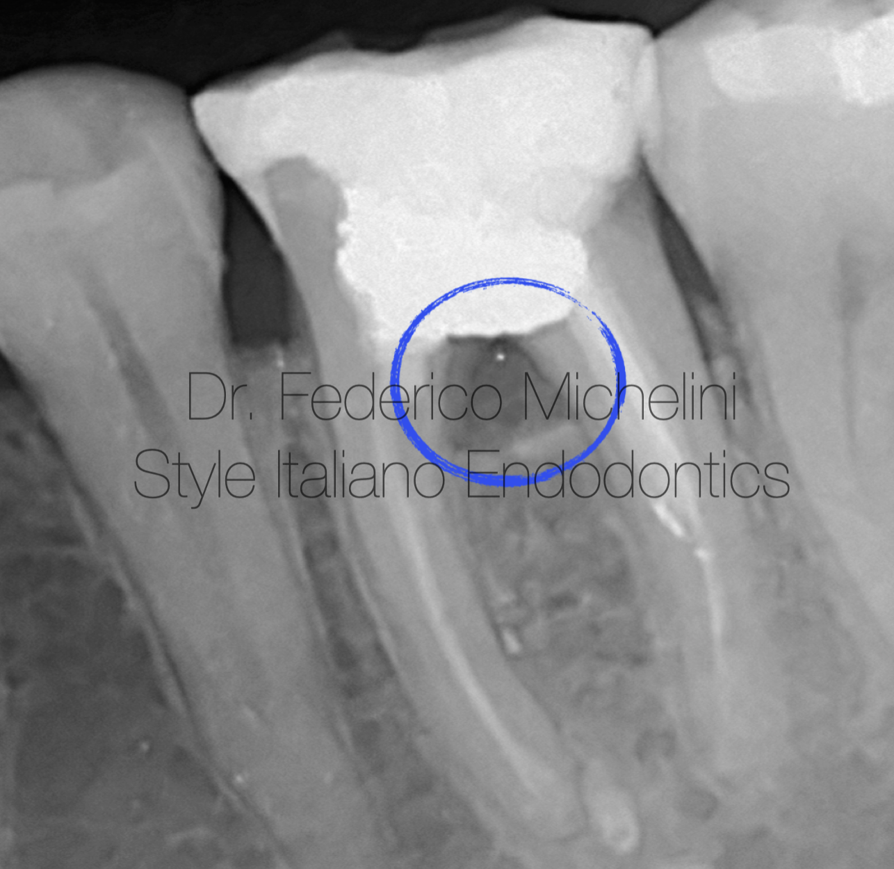
Fig. 1
Pre-operative X-ray
A 30 years old came to my clinic complaining with pain in a third quadrant especially to pressure and chewing.
After radiographic examination we see a poor root canal treatment and suspected floor and furcal perforation due to radiolucency in the furcal area.
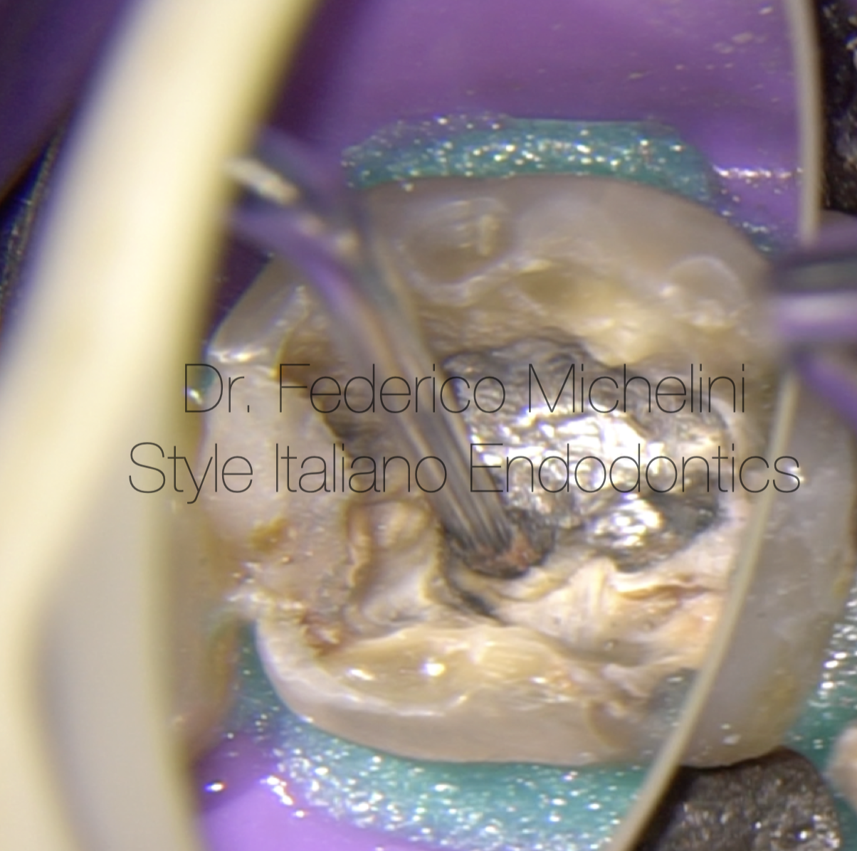
Fig. 2
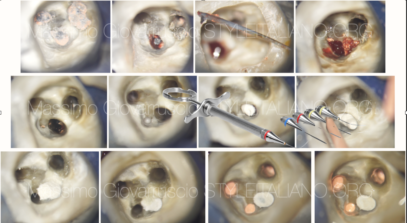
Re-access removing filling materials and metal post
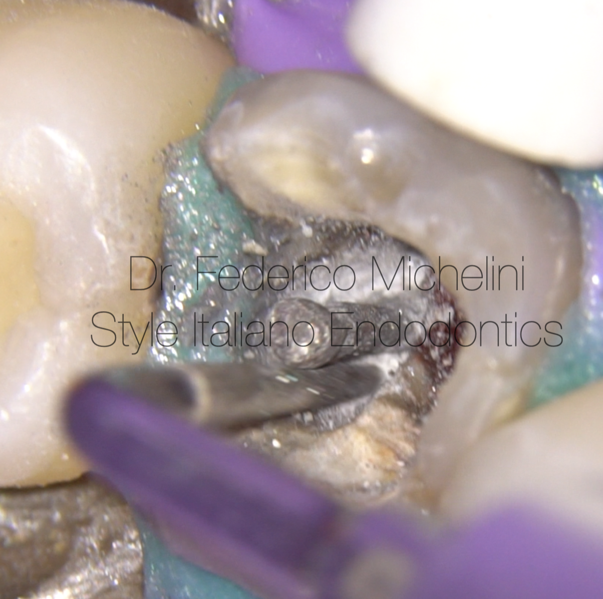
Fig. 3
Re-access removing filling materials and metal post
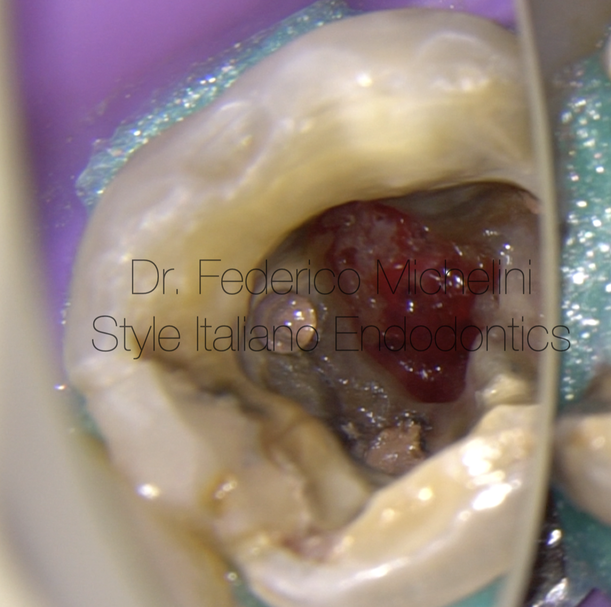
Fig. 4
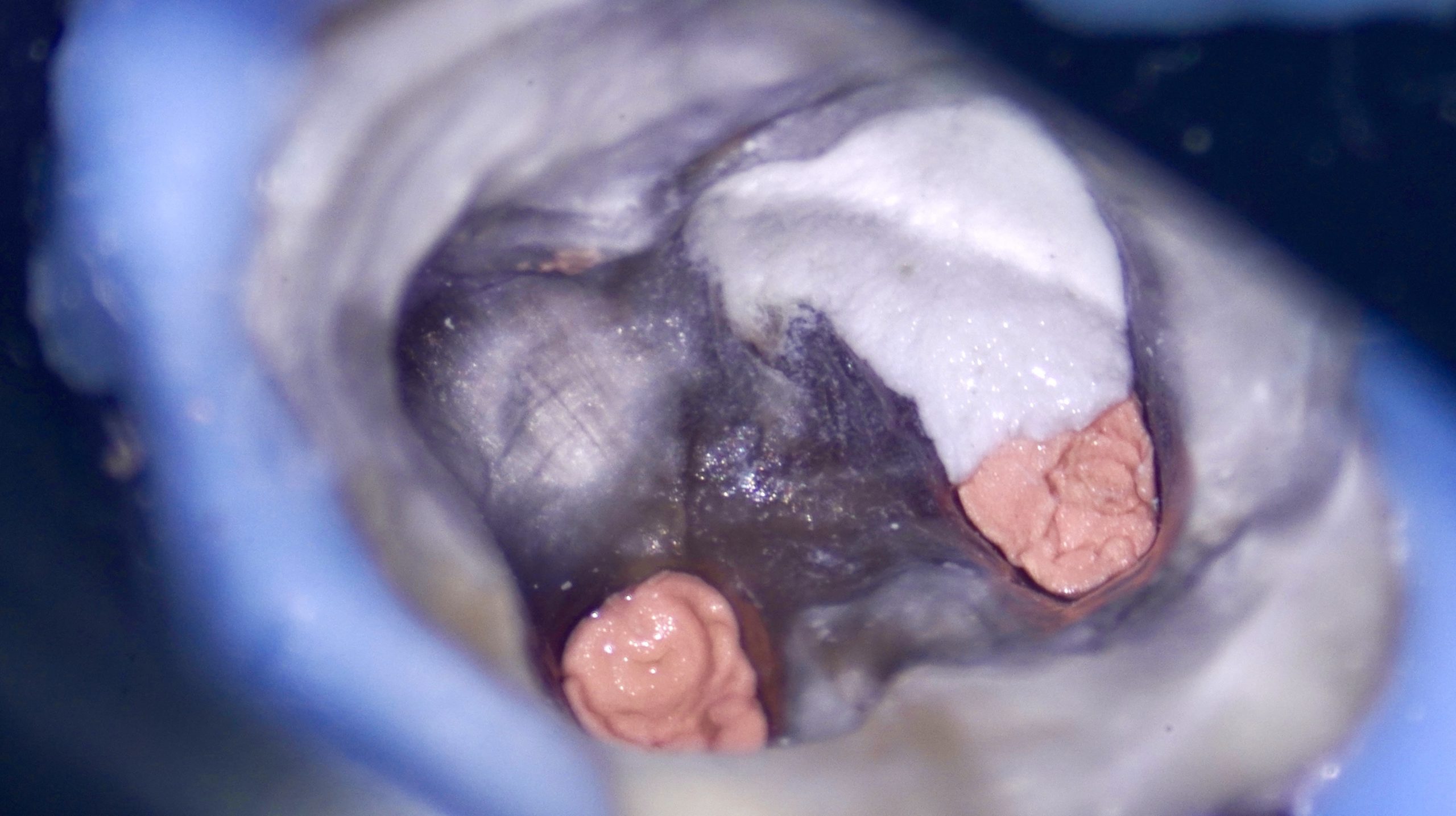
After re-design of access cavity and bleeding control thanks sodium hypochlorite it’s possibile see better the big perforation site.
The video shows all the steps: in big perforation its better to use sponge matrix (collagen of fibrin) to get some support for the material and to control the extrusion into periodontal tissue.
I put first of all bioceramic putty with a dedicated system in a vestibular part of perforation and than with a plugger and sterile wet cotton we close all the perforation.
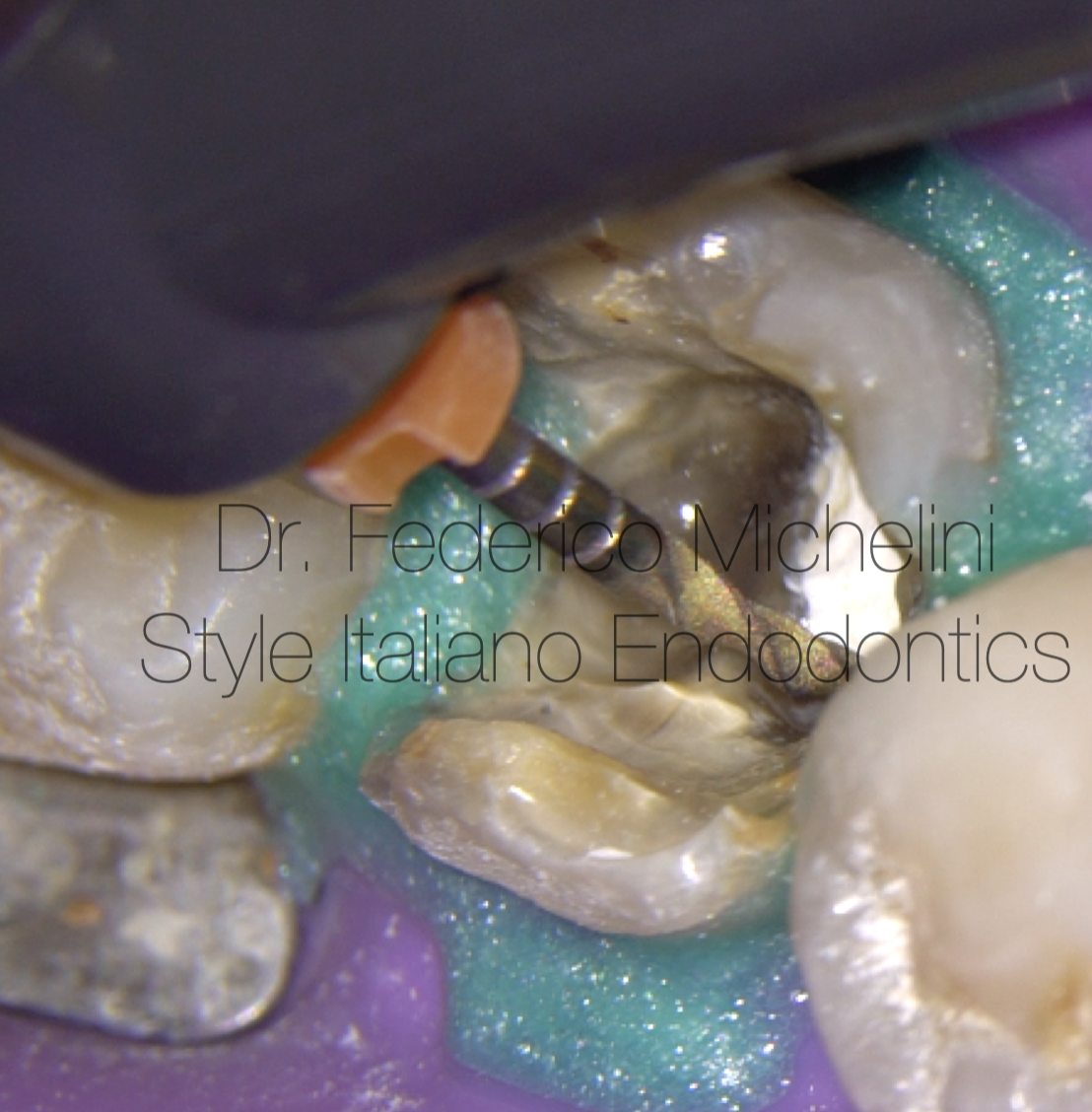
Fig. 5
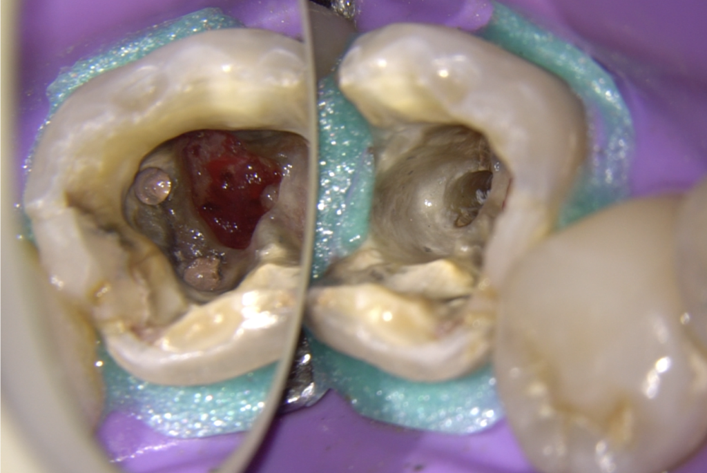
After the perforation I retreat and remove all the old filling material, re-shape the canals and do a second canal in a distal root that had been forgotten.
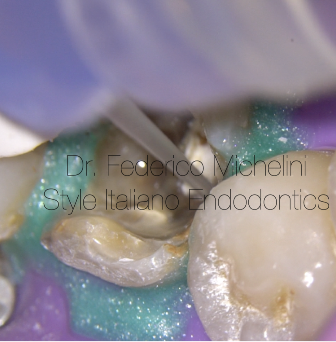
Fig. 6
Final irrigation protocol with a polymeric needle and sonic activation.
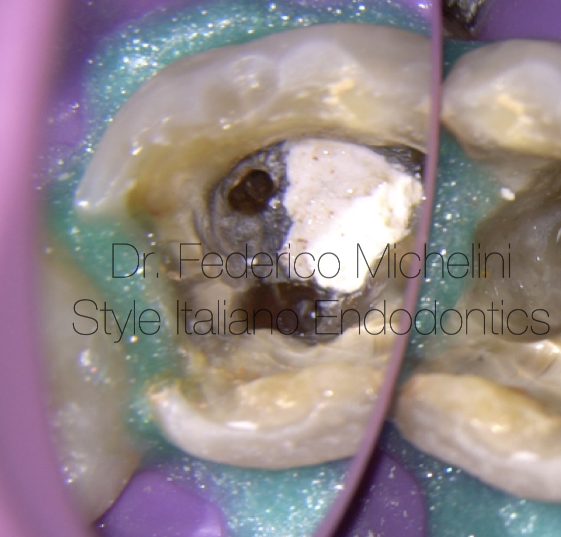
Fig. 7
Mesial canals.
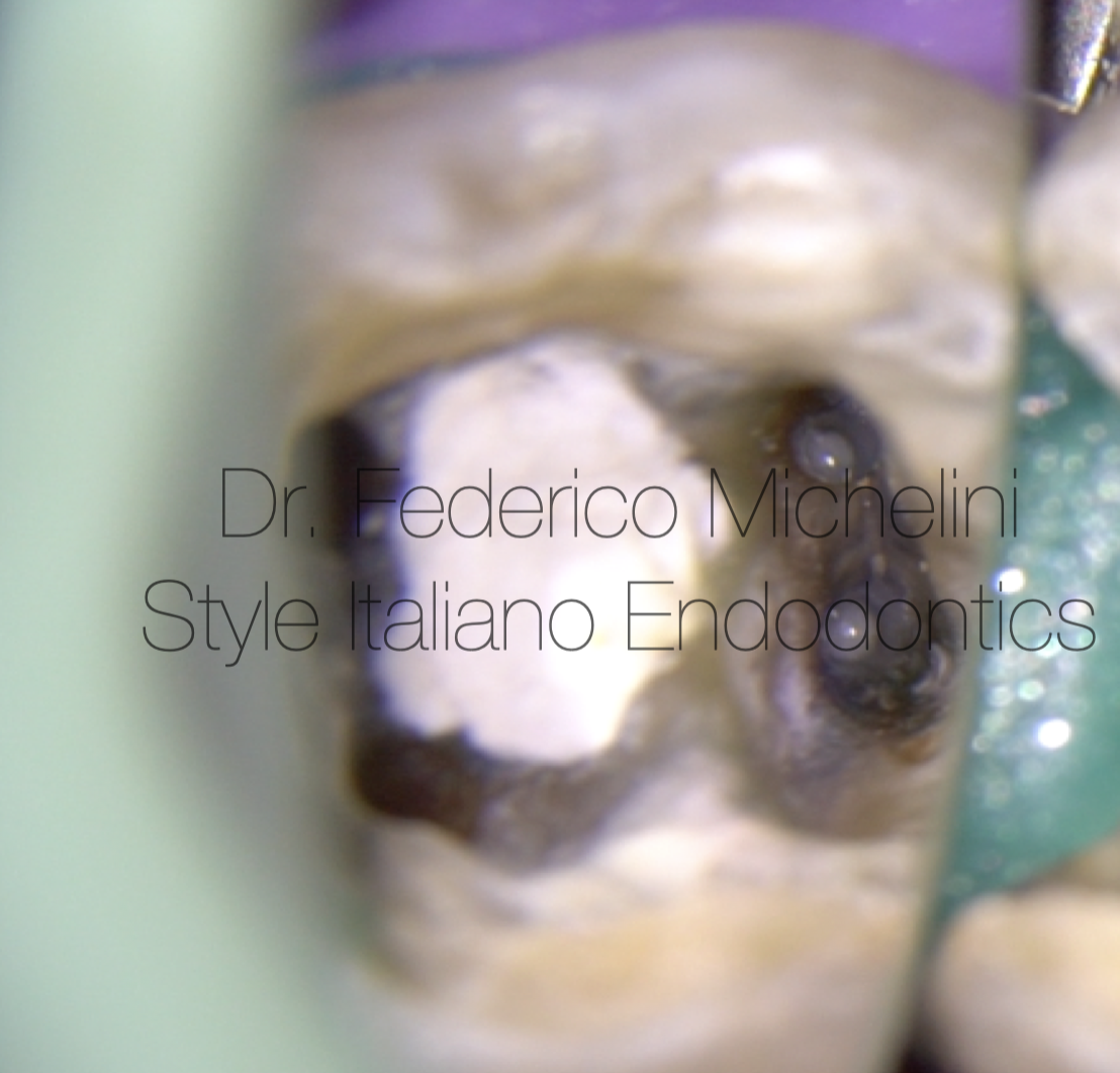
Fig. 8
Distal canals. Thanks to magnification became easier manage the two distal canals which divide deeply and have independent apex.
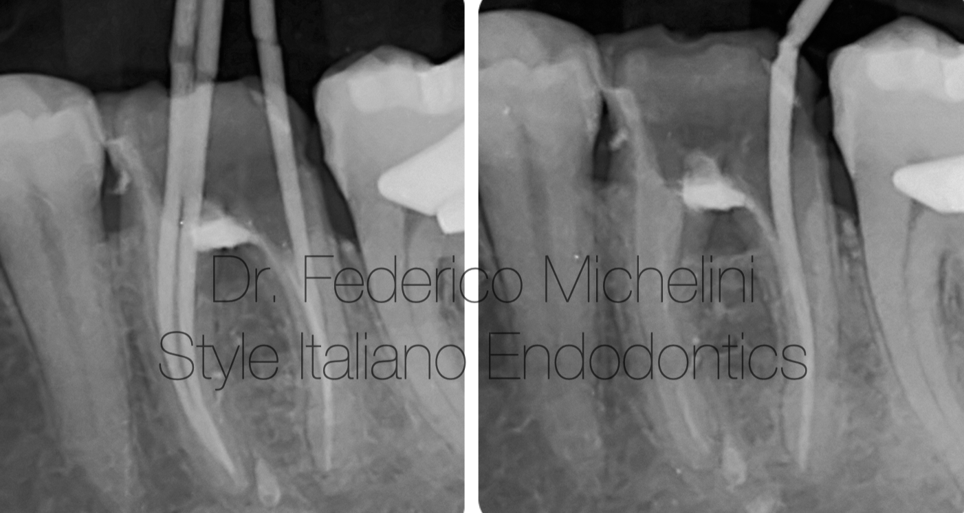
Fig. 9
Rx for checking the working lengths.
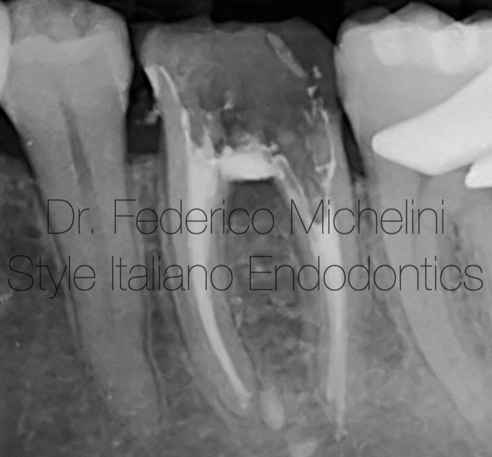
Fig. 10
Rx for checking the obturation.
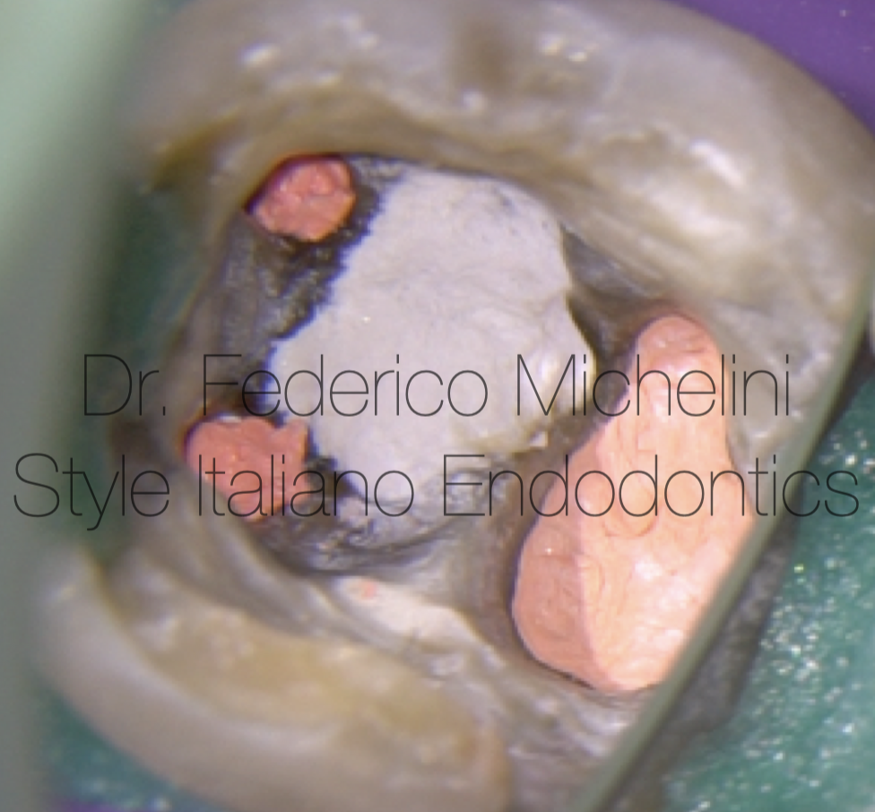
Fig. 11
Obturation of the 4 canals.
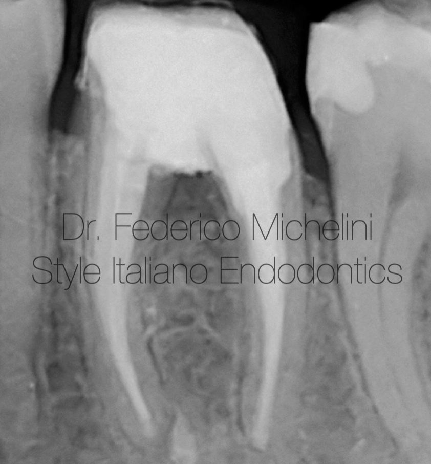
Fig. 12
Rx for checking the retreatment and the pre-prosthetic reconstruction with temporary crown.

Fig. 13
Dr. Federico Michelini
2015: Graduated in dentistry from the University of Valencia.
2017: Master in Endodontics from the University of Bologna.
Federico Michelini has been a speaker in national and international congresses regarding endodontics.
Tutor of the Master of Endodontics and of the Degree Course of Dentistry and Dental Prosthesis for the University of Bologna.
He won the Closed Meeting of Italian Society of Endodontics in 2019.
He attended the advanced course of retreatments and endodontics surgery of Italian Academy of Endodontics.
He attended the course of microscopy by Dott. Fabio Gorni.
Member of the Italian Society of Endodontics
Member Fellow of Style italiano Endodontics.
Opinion Leader and Speaker for Morita, Coltene and Labomed.
He works in Parma and Piacenza dedicating to microscopic endodontics.
Conclusions
Today thanks microscope and different types of materials, as in this case MTA or Bioceramic Putty, it is possibile manage difficult cases, avoid extraction and give “new life” to dental elements.
The purpose of this article is to give at all the clinicians a right and simple protocol to use in case of perforations following the philosophy of the group: feasible, teachable, repeatable.
Bibliography
1- Koc C, Aslan B, Ulusoy Z, Oruçoğlu H. Sealing ability of three different materials to repair furcation perforations using computerized fluid filtration method. J DentRes Dent Clin Dent Prospects. 2021 Summer;15(3):183-187.
2- Gorni FG, Andreano A, Ambrogi F, et al. Patient and clinical characteristics associated with primary healing of iatrogenic perforation after root canal treatment: results of a long-term Italian study. J Endod. 2016;42 (2): 211-215.
3- Estrela C, Decurcio DA, Rossi-Fedele G et al. Root perforations; a review of diagnosis, prognosis and materials. Braz Oral Res. 2018 Oct 18;32 (suppl 1):e73.