Management of deep root perforations
09/08/2023
Fellow
Warning: Undefined variable $post in /home/styleendo/htdocs/styleitaliano-endodontics.org/wp-content/plugins/oxygen/component-framework/components/classes/code-block.class.php(133) : eval()'d code on line 2
Warning: Attempt to read property "ID" on null in /home/styleendo/htdocs/styleitaliano-endodontics.org/wp-content/plugins/oxygen/component-framework/components/classes/code-block.class.php(133) : eval()'d code on line 2
Root perforation is a challenging situation when the root canal has a pathological communication with periradicular area. Perforations can occur as an iatrogenic accident during a root canal instrumentation and is the second cause of failed endodontic treatment. In some cases complications may lead even to an extraction. Thus, successful treatment of a root perforation depends on different crucial factors, like location, extent of the perforation, time from diagnosis to treatment and material used to seal the perforation area.
Root perforation is an artificial communication between the root canal system and the periradicular space.
Iatrogenic root perforations may occur during access cavity opening, root canal preparation or during post preparation. Procedural errors may cause the treatment fail.
Factors that may affect the prognosis of root perforations and its treatment are as follows, extent and location, timeline, material, technique and operator experience. Placement of the MTA apical plug in long curved root canals is quite sensitive, thus making it challenging in clinical practice.
Therefore, present case shows the simple technique for MTA placement in the deep root perforations during non-surgical root canal re-treatment of the upper right first premolar with chronic apical abscess
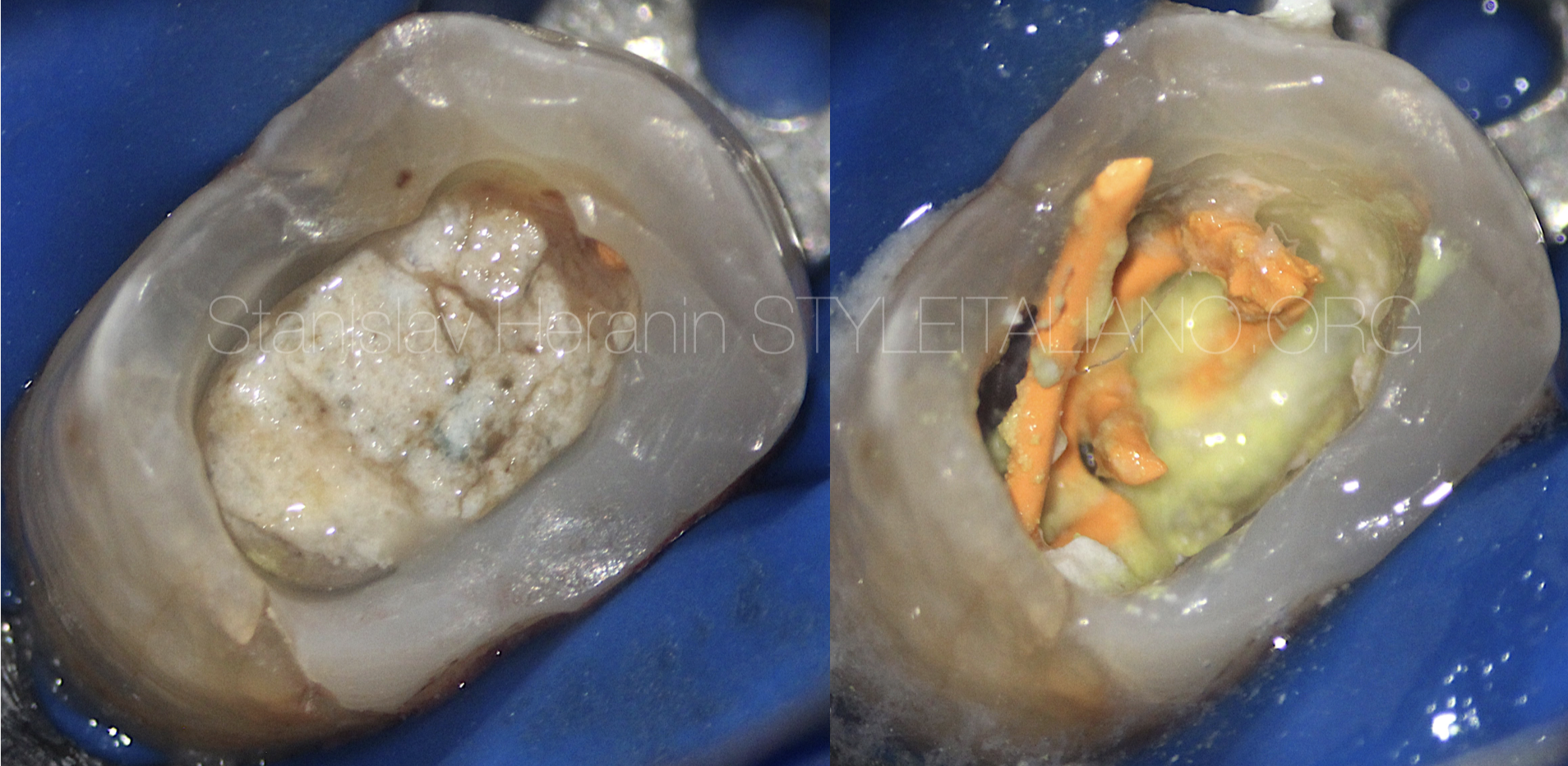
Fig. 1
Initial Appearance
Referred Patient, 32 yo, 1.4 Chronic Apical Abscess
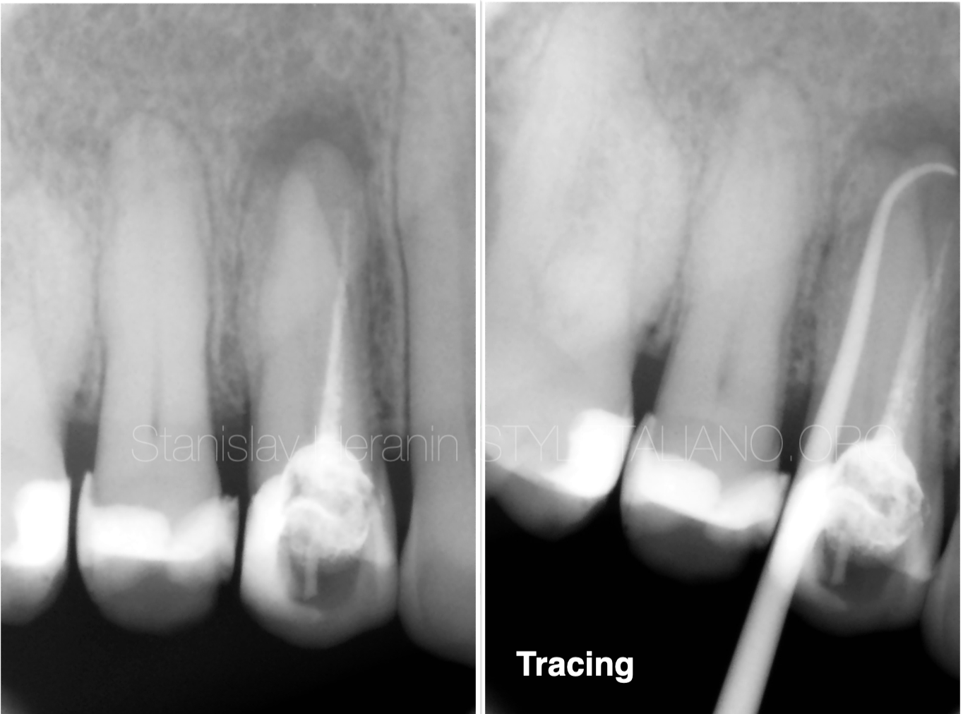
Fig. 2
Pre-operative X-Ray
Presence of the sinus tract next to the maxillary tooth on the right. Previously treated a week ago with unsuccessful attempts to finish the root canal treatment.
Diagnostic radiograph revealed the periapical radiolucent area. Tracing pointed the source of the sinus tract.
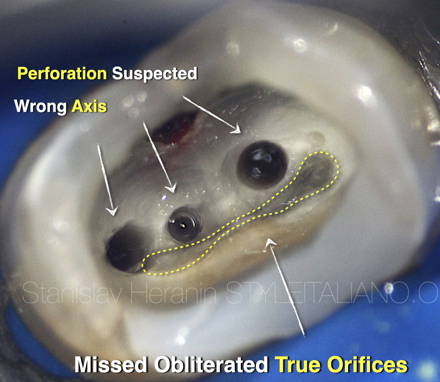
Fig. 3
Pulpal floor examination
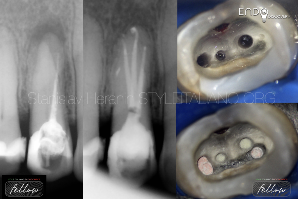
3 suspected pulpal floor perforations
Obliterated true orifices
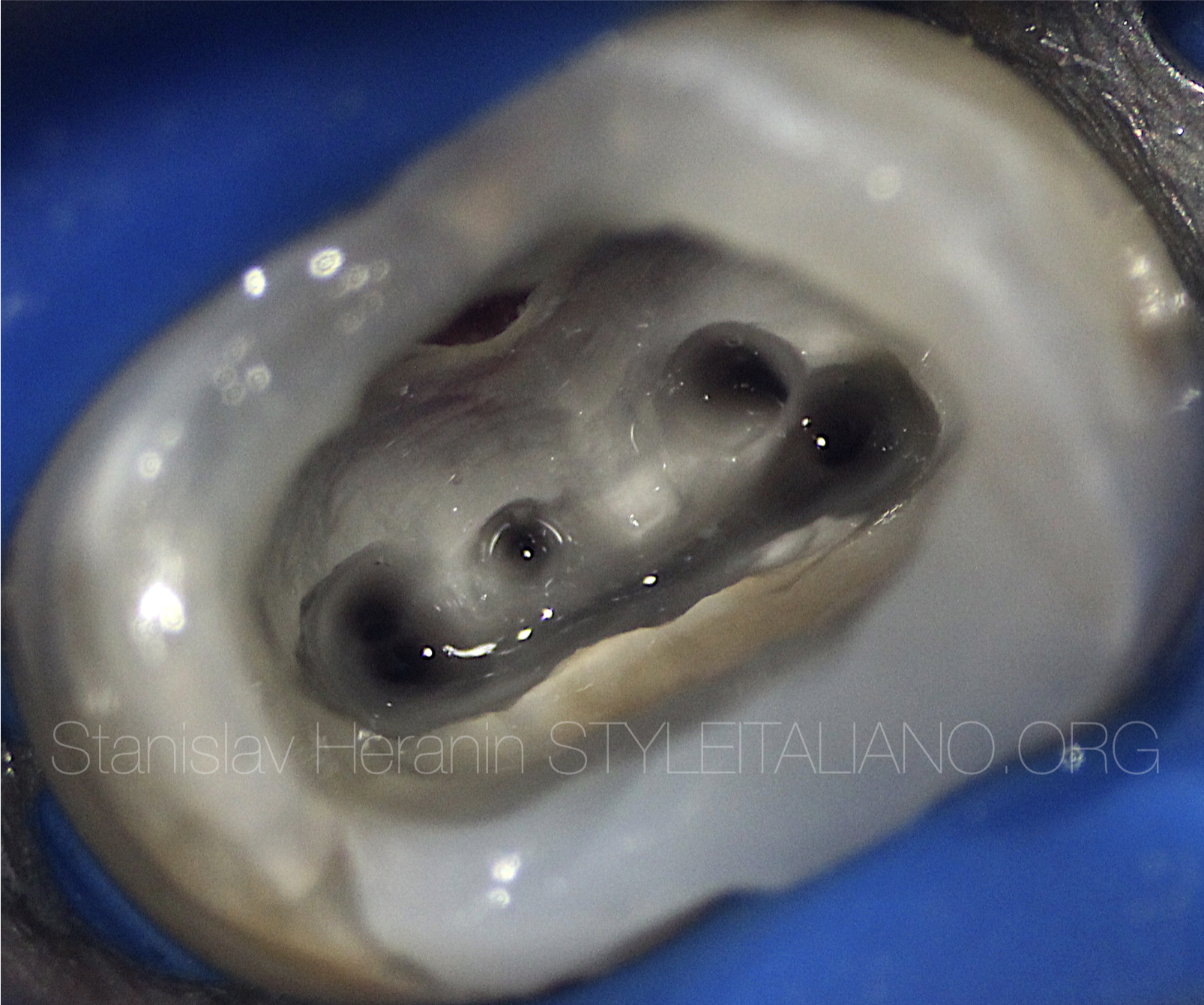
Fig. 4
True Orifice opening with carbide long-neck bur
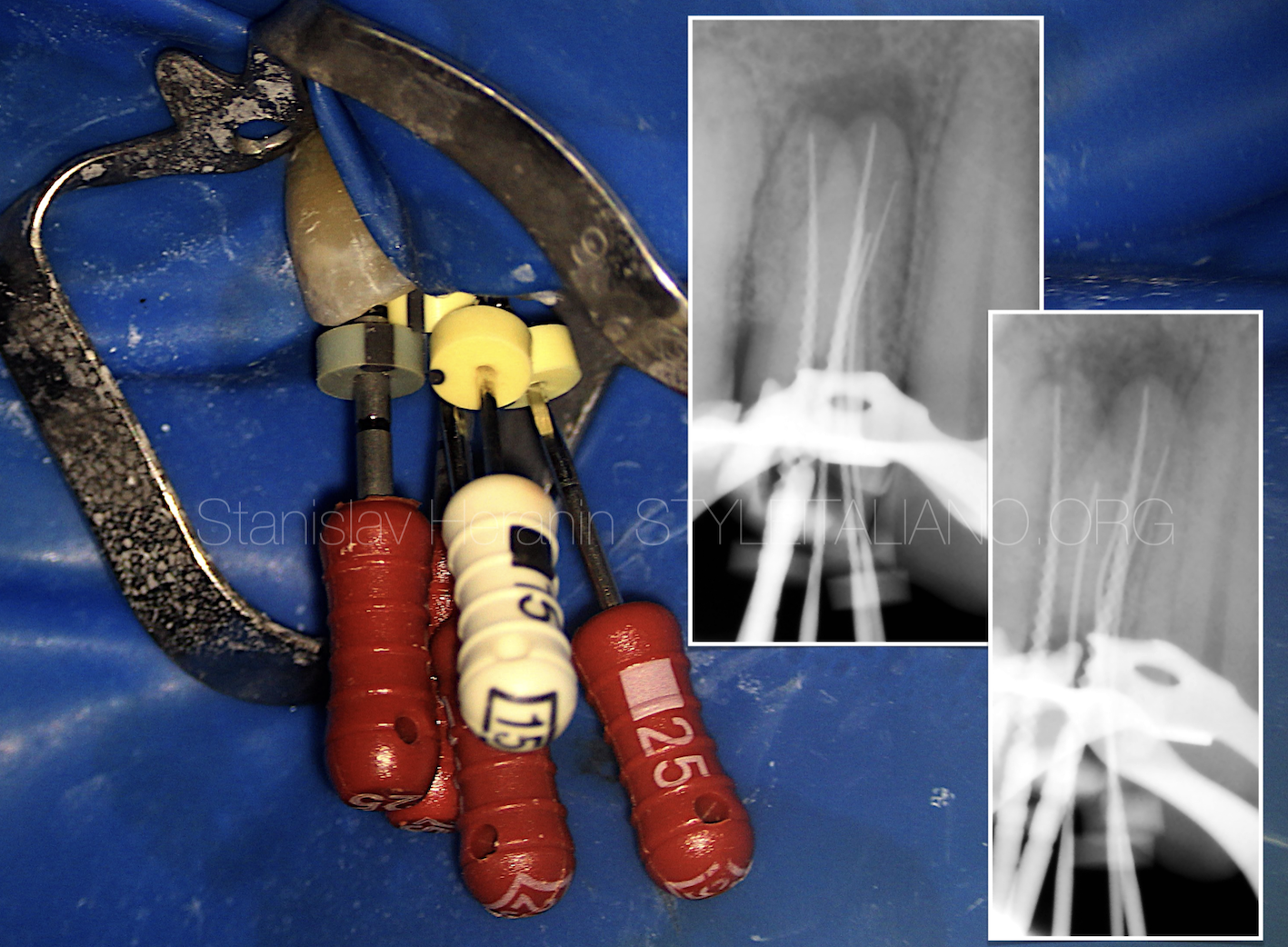
Fig. 5
Cleaning and Shaping of the root canal system
Revealing of the Perforations
Cleaning and Shaping of the root canal system
Final irrigation with 5.25% Sodium Hypochlorite and 17% EDTA
Final Appearance of the pulpal floor
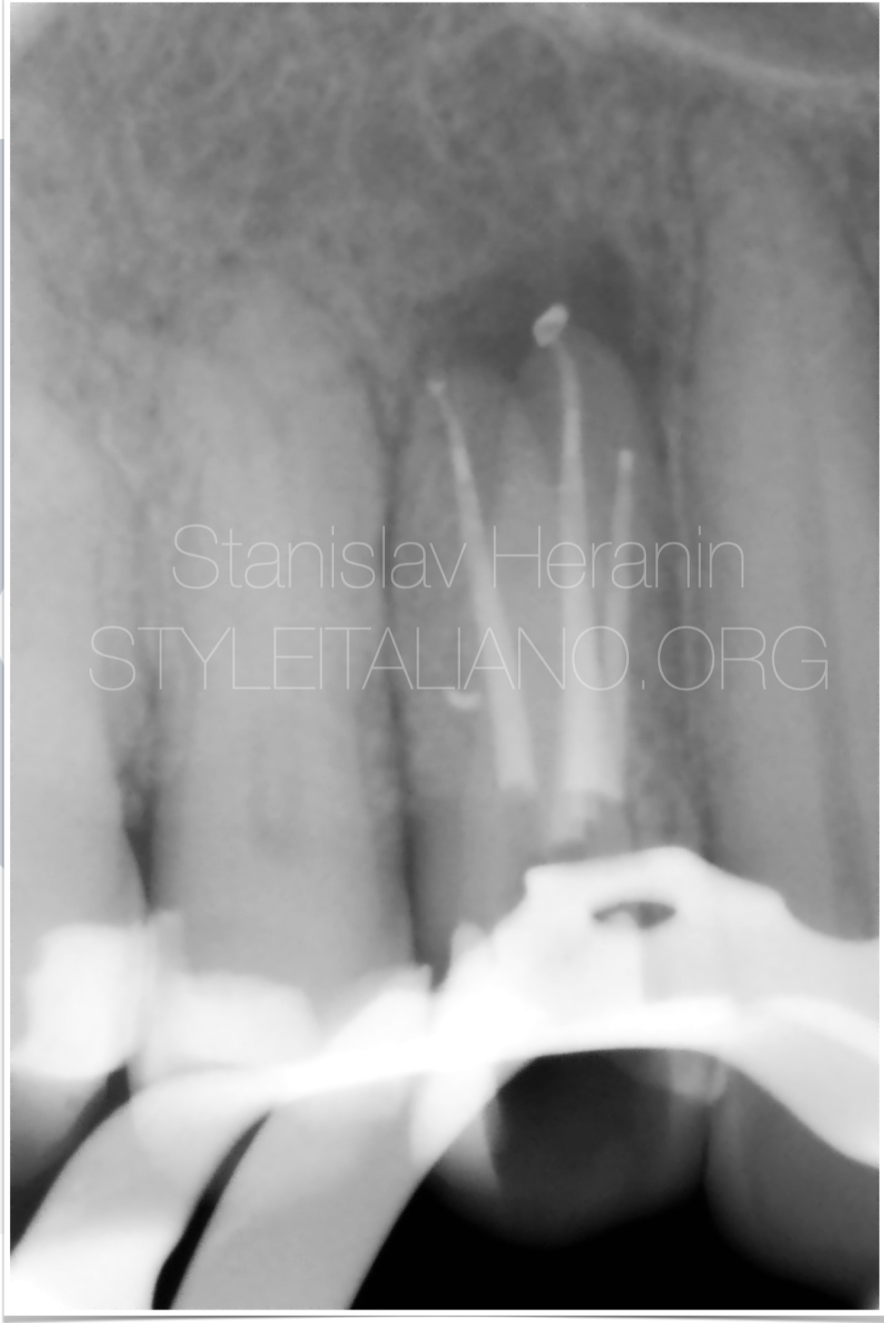
Fig. 6
Root canal filling (X-Ray control)
True Buccal & Lingual Canals - Continuous Wave Compaction
«Dead End» false Canal - Injection Molded Technique
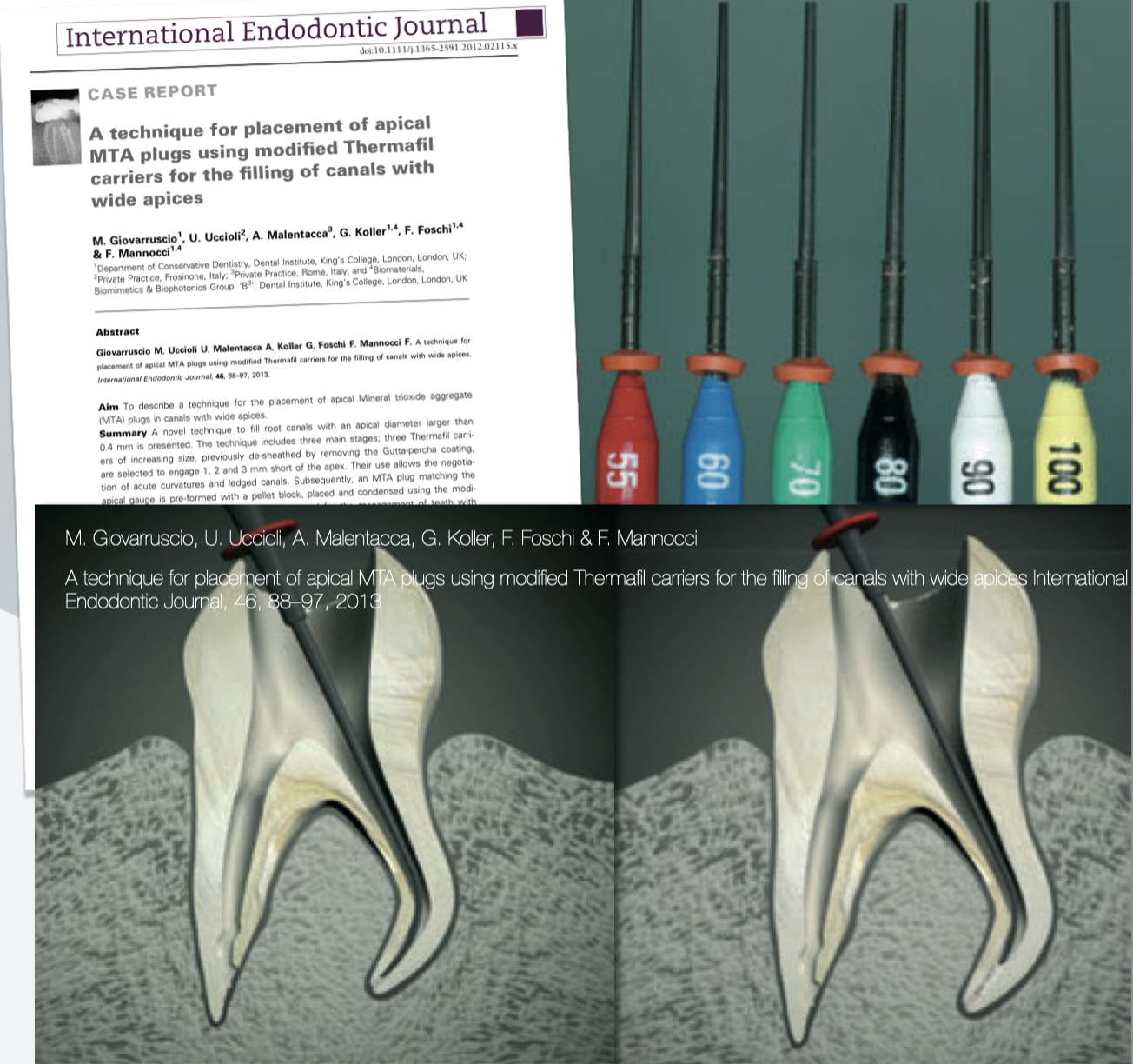
Fig. 7
MTA plugs & Thermafil carriers
Predictable placement of the MTA apical plug in long curved canals with plastic carriers
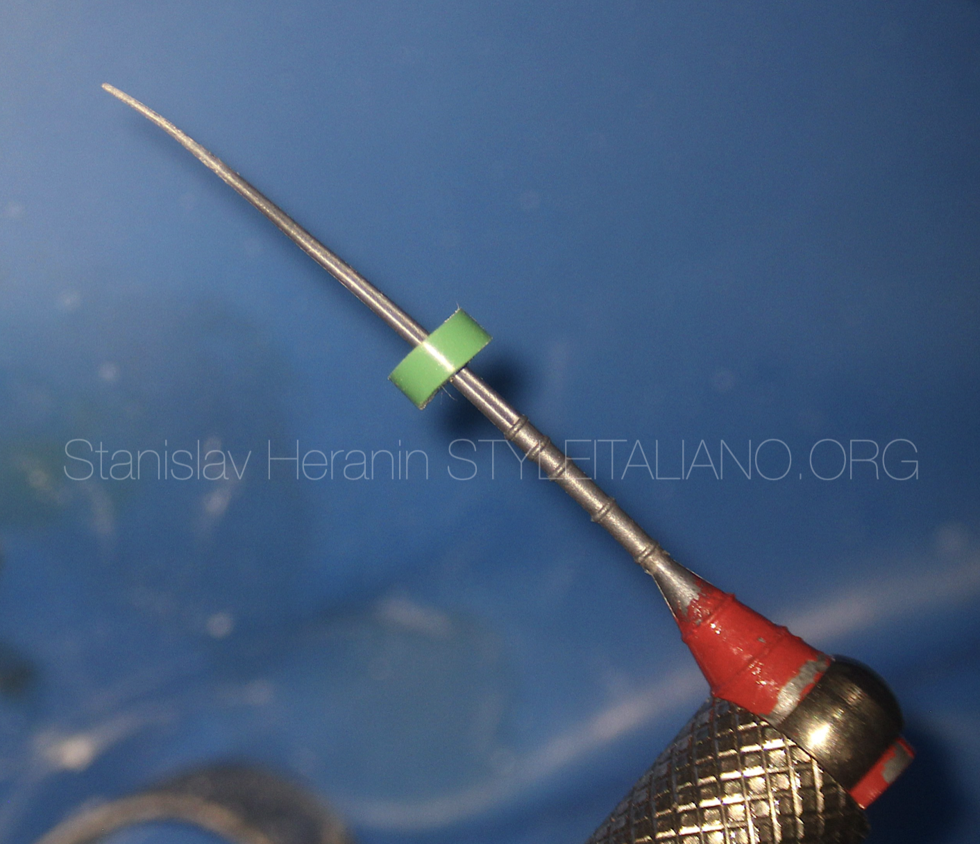
Fig. 8
MTA plugs & Thermafil carriers (Advanced Technique)
It is convenient to use special Endodontic file holder to facilitate the manipulations under the microscope
MTA plugs & Thermafil carriers (Advanced Technique)
Easy controlled apical placement of the MTA plug
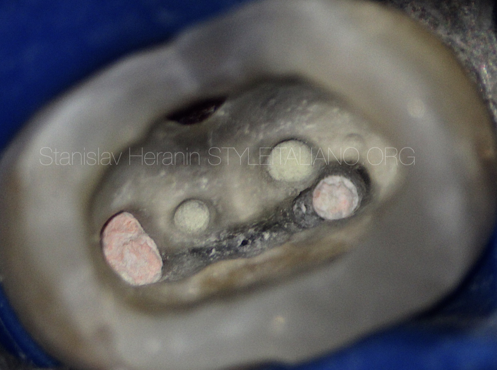
Fig. 9
Final appearance of the pulpal floor
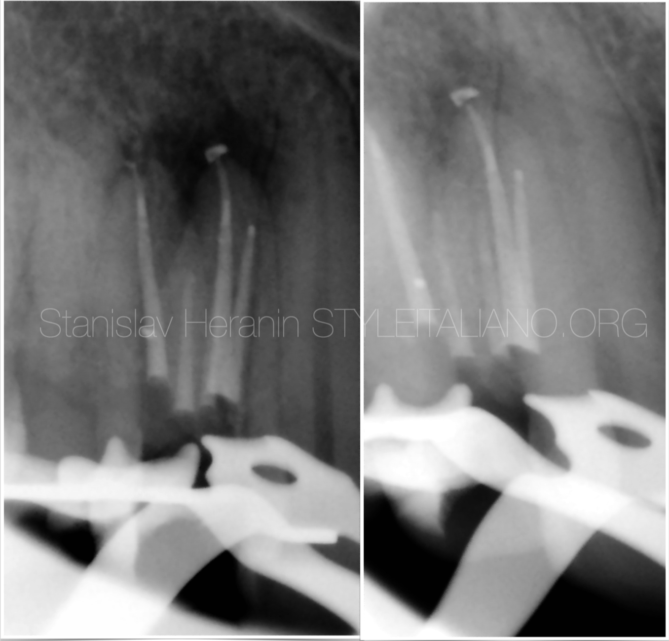
Fig. 10
X-Ray control
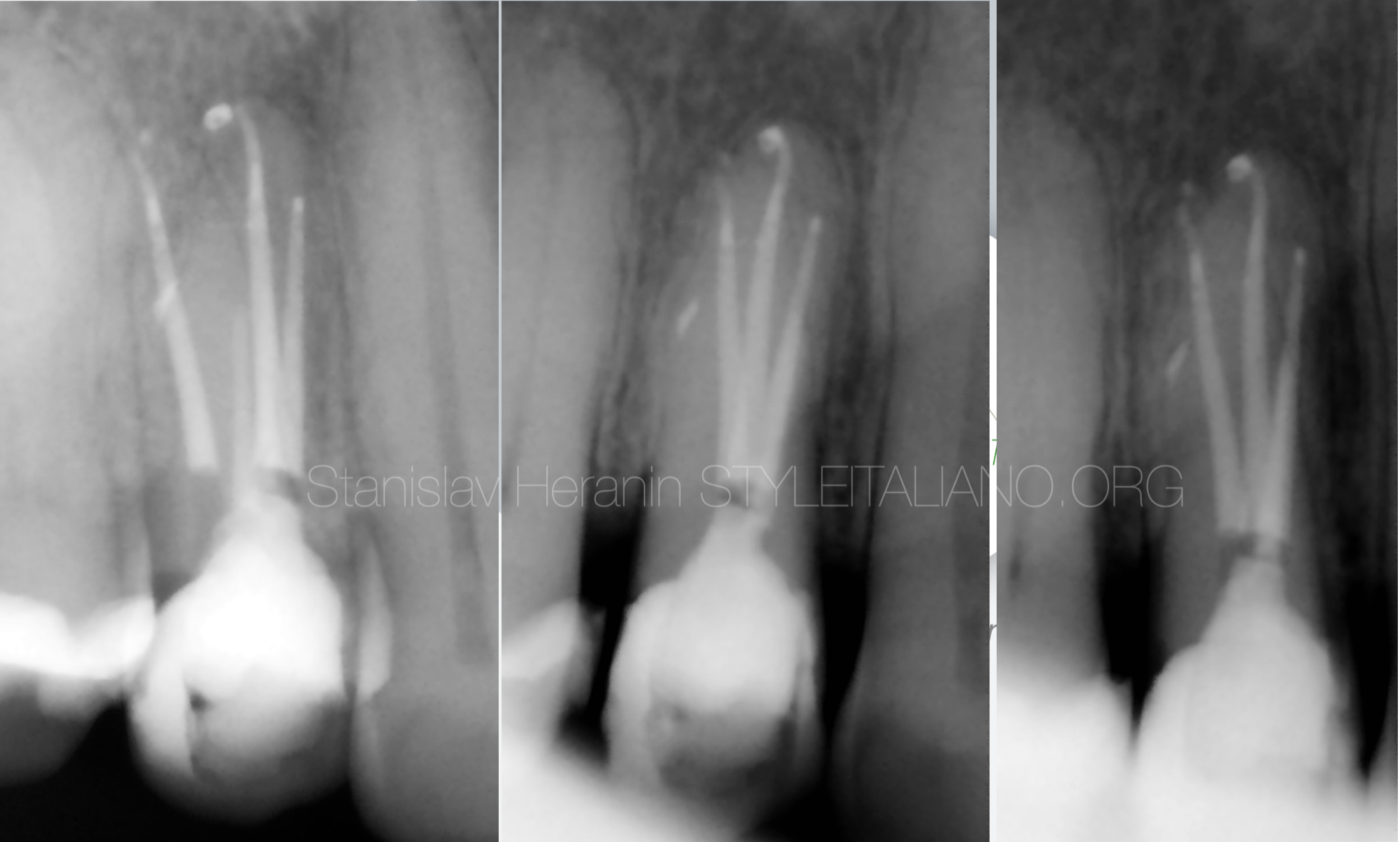
Fig. 11
10 m Follow-up

Fig. 12
1999: Graduated in Dentistry from the Ukrainian Medical Stomatological Academy - Faculty of Dentistry
1999-2001 - Postgraduate Education (UMSA)
2001-2012 - Professor Assistant - Department of Therapeutic Dentistry (UMSA)
2012-2019 - Professor Assistant - Department of Postgraduate Education of Dental Practitioners (UMSA)
2011: PhD in Dentistry (Ukrainian Medical Stomatological Academy)
2023 until now Associate Professor at The Department of Dentistry – School of Medicine - V.N.Karazin
Kharkiv National University
Private Dental Practice - Dental Centre “Machaon” (Poltava, Ukraine)
Founder of the Educational Centre EndoDiscovery
Past-President of the Ukrainian Academy of Esthetic Dentistry
Board Member of the Ukrainian Endodontic Association
Member of the Ukrainian Endodontic Society
Member of International Jury of Dental Restorative Contest ”Prisma-Championship”
Board member of the International Journal “Ukrainian Dental Journal”
Conclusions
Placement of the traditional MTA plug in a long curved root canals makes it quite challenging for the operator.
Success or failure depend on the ability to access the perforation area and promote an appropriate sealing.
Visual control of every single manipulation under the operational microscope gives the is the crucial factor in daily endodontics
Bibliography
- Giovarruscio M, Uccioli U, Malentacca A, Koller G, Foschi F, Mannocci F. A technique for placement of apical MTA plugs using modified Thermafil carriers for the filling of canals with wide apices. Int Endod J. 2013 Jan;46(1):88-97. doi: 10.1111/j.1365-2591.2012.02115.x. Epub 2012 Nov 9. PMID: 23137342.
- Fabio G. Gorni, DDS Andrei C. Ionescu, DDS, PhD, Federico Ambrogi, MSc, PhD Eugenio Brambilla, DDS Massimo M. Gagliani, MD, DDS Prognostic Factors and Primary Healing on Root Perforation Repaired with MTA: A 14-year Longitudinal Study DOI:https://doi.org/10.1016/j.joen.2022.06.005
- Clauder T. Present status and future directions - Managing perforations. Int Endod J. 2022 Oct;55 Suppl 4:872-891. doi: 10.1111/iej.13748. Epub 2022 Apr 28. PMID: 35403711.
- Torabinejad M, Parirokh M, Dummer PMH. Mineral trioxide aggregate and other bioactive endodontic cements: an updated overview - part II: other clinical applications and complications. Int Endod J. 2018 Mar;51(3):284-317. doi: 10.1111/iej.12843. Epub 2017 Oct 11. PMID: 28846134.
- Castellucci A. The use of mineral trioxide aggregate in clinical and surgical endodontics. Dent Today. 2003 Mar;22(3):74-81. PMID: 12705015.
- Castellucci A. The use of mineral trioxide aggregate to repair iatrogenic perforations. Dent Today. 2008 Sep;27(9):74, 76, 78-80; quiz 81. PMID: 18807954.
- Roda RS. Root perforation repair: surgical and nonsurgical management. Pract Proced Aesthet Dent. 2001 Aug;13(6):467-72; quiz 474. PMID: 11544819.


