Magnification & Ultrasonics: the perfect marriage
13/10/2024
Fellow
Warning: Undefined variable $post in /home/styleendo/htdocs/styleitaliano-endodontics.org/wp-content/plugins/oxygen/component-framework/components/classes/code-block.class.php(133) : eval()'d code on line 2
Warning: Attempt to read property "ID" on null in /home/styleendo/htdocs/styleitaliano-endodontics.org/wp-content/plugins/oxygen/component-framework/components/classes/code-block.class.php(133) : eval()'d code on line 2
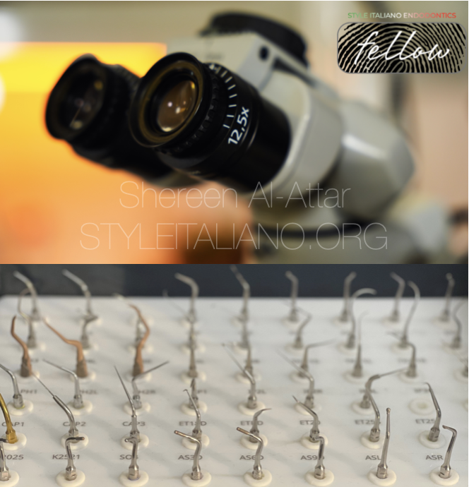
During the past few decades endodontic treatment has benefited from the development of new techniques and equipment. The synergistic use of these and other technologies has improved outcome and predictability. Important attributes such as the operating microscope and Ultrasonics (US) have found indispensable applications in primary root canal treatments and retreatment procedures. (Plotino et al, 2007)
Ultrasonics in endodontics has enhanced the quality of treatment and represents an important adjunct in the treatment of difficult cases. Since its introduction, ultrasonics has become increasingly more useful in applications such as gaining access to canal openings, cleaning and shaping, obturation of root canals, removal of intracanal materials and obstructions and endodontic surgery. (Park.E, 2013)
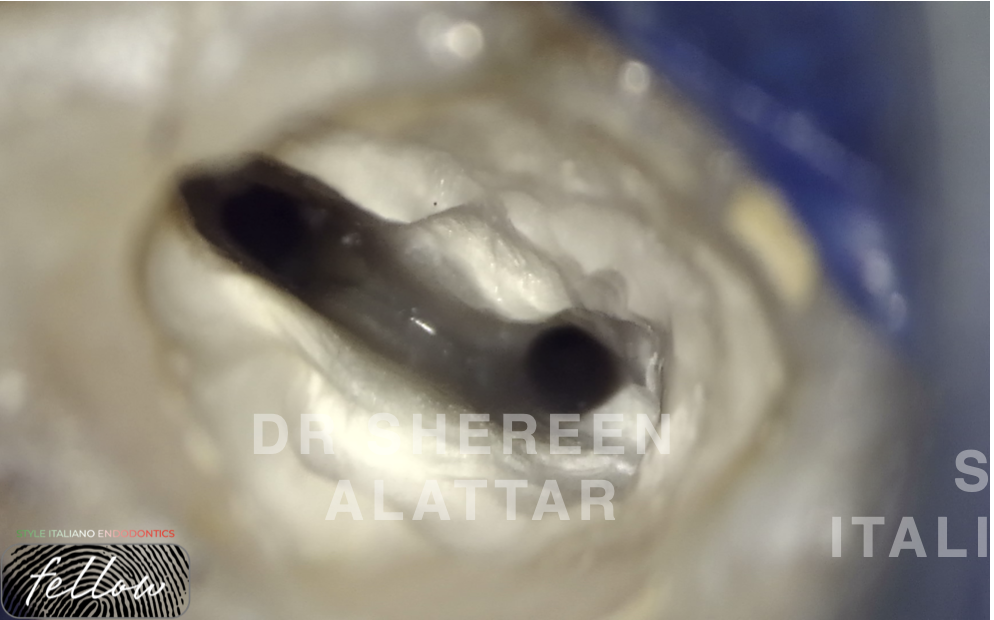
Fig. 1
It has been said that you can’t treat what you can’t see.
Seeing canals in appropriately prepared access cavities requires magnification and illumination.
In any event, intense light, good magnification through loupes or a microscope, and reliable ultrasonic technology are all a must if you are doing molar endodontics. (Gupta et al, 2021)
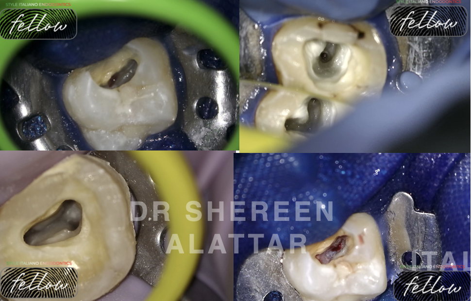
Fig. 2
1. Completing Access Cavity
Scaling tips are used to clean pulp chamber, and some special tips are used to extend access cavity and to remove dentin.
Ultrasonic systems importantly eliminate the bulky head of the conventional handpiece that notoriously obstructs vision. The working ends of specific ultrasonic instruments are 10 times smaller than the smallest manufactured round burs and their abrasive coatings allow them to precisely sand away dentin when exploring for missed canals. (Gupta et al, 2021)
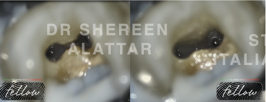
Fig. 3
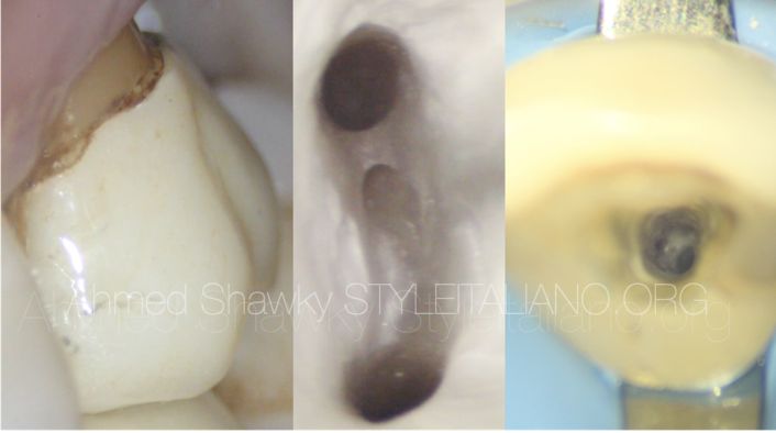
Root canal isthmus is a narrow ribbon-shaped communication between two root canals. It is an important anatomical feature because it may contain vital or necrotic pulp remnants if left untreated.
Instrumentation of an isthmus with burs can result in removal of precious dentin. The use of ultrasonics and magnification facilitates precise and selective cleaning of such areas.
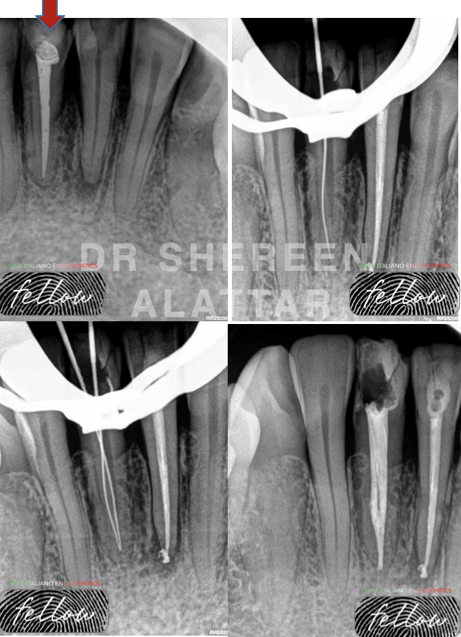
Fig. 4
2. Finding Canal Orifice
For both general practitioners and endodontists, the ultrasonic is a valuable tool to treat calcified and difficult to find canals, as long as it is complemented by the proper tip, and sufficient magnification and light.
It is less aggressive than a high-speed handpiece, and with high magnification the operator can always see where the tip is and where it is heading. (Paz et al, 2005).
Moreover, ultrasonic tips can be used to eliminate secondary dentin that slopes off the mesial wall and occlude the canal orifice
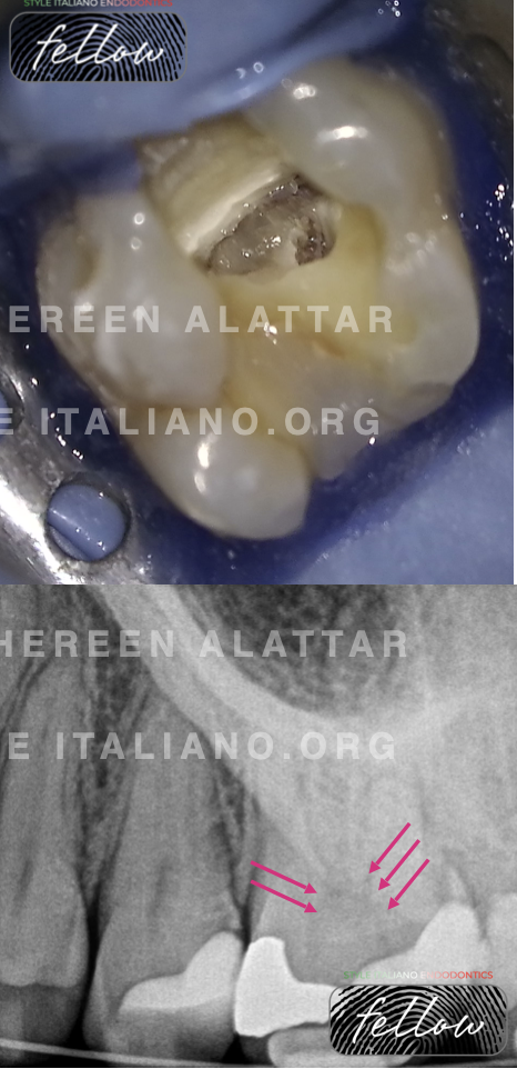
Fig. 5
Calcification of the root canal system occurs naturally as thetooth ages. However, if a tooth is subjected to traumatic events, increased deposition of tooth structure can occur occluding/blocking the root canal
The diamond coating of an ultrasonic tip makes it much more effective and abrasive.
When a pulp stone occludes the entrance to a canal orifice, the ultrasonic tip can be used to carefully smooth the pulp stone away.
It is very important to use, at this stage, tips that would not interfere with the floor of the pulp chamber not only to prevent iatrogenic damage (perforations), but also not to miss important references dictated by the floor itself on the position of the root canals.
This kind of tip, especially if used without irrigation, tend to get soiled with dentinal debris thus loosing cutting efficacy, therefore I prefer to use it with water irrigation.
The subsequent phase of finding canal orifices should be carried out with thinner and longer tips that facilitate working in deeper areas while maintaining clear vision and do not create iatrogenic injury such as coronal-middle perforations. Here I used ET 20 tip for this purpose
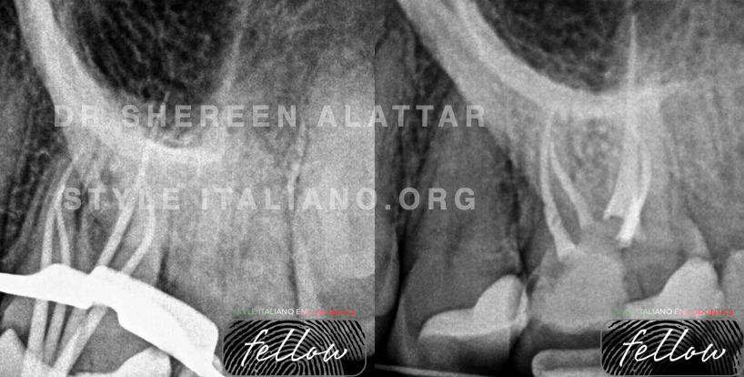
Fig. 6
ET 20 used to find orifices
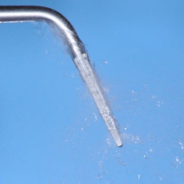
3. Increase Root Canal Disinfection
Ultrasonics use to activate irrigant in the canal has shown to be a clinically proven and efficient adjunct to cleaning and shaping instrument sequences and is used by many endodontic specialists.
Ultrasonic activation of irrigants produces at least 2 helpful effects: (1) cavitation, defined as the formation of thousands of tiny bubbles which rapidly implode, producing a "shock wave" removing biofilm, and (2) acoustic streaming which produces shear forces that will help extricate debris from instrumented canals. (Video 3)
An excellent ultrasonic tip instrument to create this type of passive ultrasonic irrigation is the Irrisafe file.
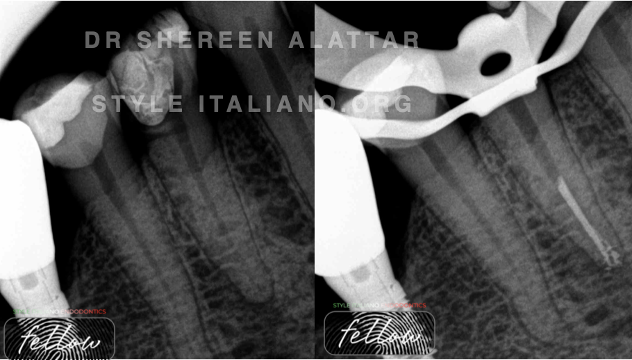
Fig. 7
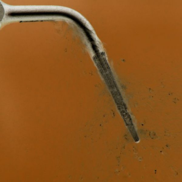
This clinically and scientifically accepted method of passive ultrasonic irrigation helps push the ultrasonic from luxury into necessity status!

Fig. 8
About the author:
Shereen Alattar
Dr Shereen Alattar (DDS, MSc) has graduated from Saint Joseph University in Beirut, Lebanon, where she received her Master’s Degrees in the specialisation of Endodontics. She works in a referral-based practice limited to endodontics in Amman, Jordan.
She is a fellow member in Style Italiano Endodontic Group and an active member in the Jordanian Scientific Committee of the Jordanian Dental Association and in the Jordanian endodontic society
Conclusions
This article aims at presenting the numerous uses of ultrasonics in clinical endodontics and emphasizes the broad applications in a modern-day endodontic practice.
Most dentists, who use ultrasonics for endodontic procedures under magnification, acknowledge that these technologies can help solve many clinical problems and challenges in treating complex root canal systems.
Habit converts luxury into necessity. A growing number of clinicians performing endodontics are adding the piezoelectric ultrasonic and tips to the list of endodontic "necessities."
Bibliography
- Plotino G, Pameijer CH, Grand NM, Somma F. Ultrasonics in Endodontics: a review of the literature. J Endod 2007;33:81-95.
- Park E. Ultrasonics in endodontics, Endod Topics 2013;29:125-159
- Gupta, A., Pareek, A., & Kapila, H. (2021). Ultrasonics in endodontics: A review. International Journal of Health Sciences, 5(S1), 264–277.
- Paz E, Satovsky J, Moldauer I. Comparison of the cutting efficiency of two ultrasonic units utilizing two different tips at two different power settings. J Endod. 2005;31:824-826.