Internal Bleaching of a Traumatized Non-Vital Maxillary Central Incisor: A Case Report
20/04/2026
Warning: Undefined variable $post in /home/styleendo/htdocs/styleitaliano-endodontics.org/wp-content/plugins/oxygen/component-framework/components/classes/code-block.class.php(133) : eval()'d code on line 2
Warning: Attempt to read property "ID" on null in /home/styleendo/htdocs/styleitaliano-endodontics.org/wp-content/plugins/oxygen/component-framework/components/classes/code-block.class.php(133) : eval()'d code on line 2
Internal bleaching, a minimally invasive dental procedure introduced in the 19th century, is an esthetic treatment indicated for discolored non-vital teeth. Its objective is to achieve intracoronal whitening by applying oxidizing agents within the pulp chamber.
The etiology of discoloration in non-vital teeth can be multifactorial, encompassing dental trauma resulting in intrapulpal hemorrhage, pulpal necrosis, pulp chamber calcification, the interaction between sodium hypochlorite and chlorhexidine, intracanal medicaments, inadequately removed root canal filling materials, coronal restorative materials such as amalgam, and endodontic bioceramic materials like mineral trioxide aggregate (MTA) containing bismuth oxide as a radiopacifying agent.
Internal bleaching offers a conservative, safe, and minimally invasive approach to restoring the esthetics of devitalized teeth that have lost their natural brightness.
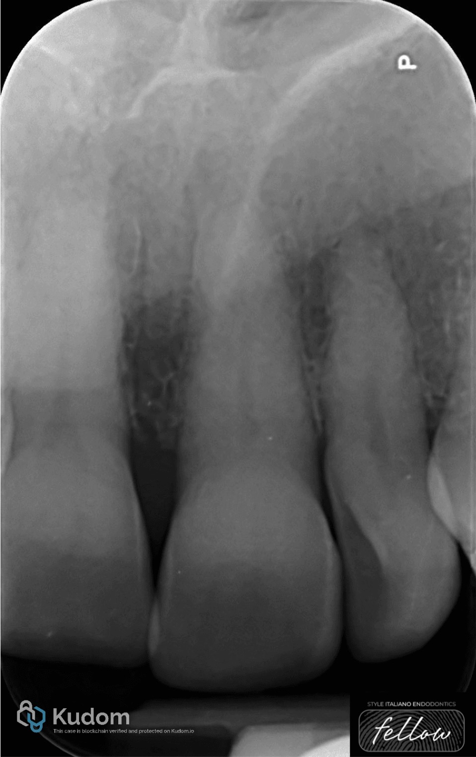
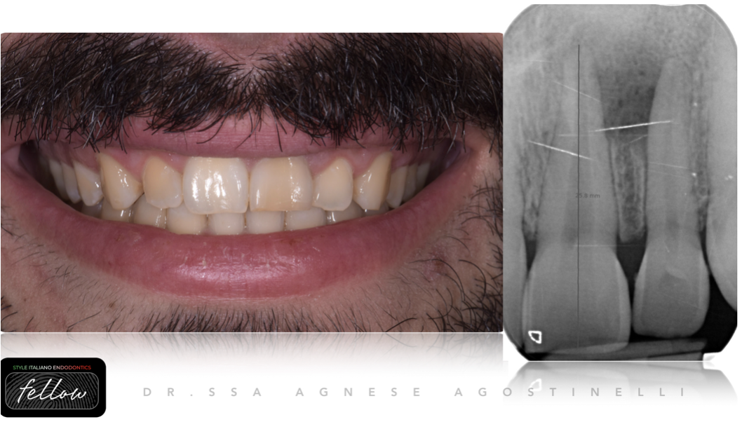
Fig. 1
A female patient presented with discoloration of tooth 21 and a history of dental trauma during childhood. The tooth was asymptomatic, with no clinical or radiographic signs of endodontic pathology. Pulp vitality testing performed with ethyl chloride confirmed the absence of vitality.
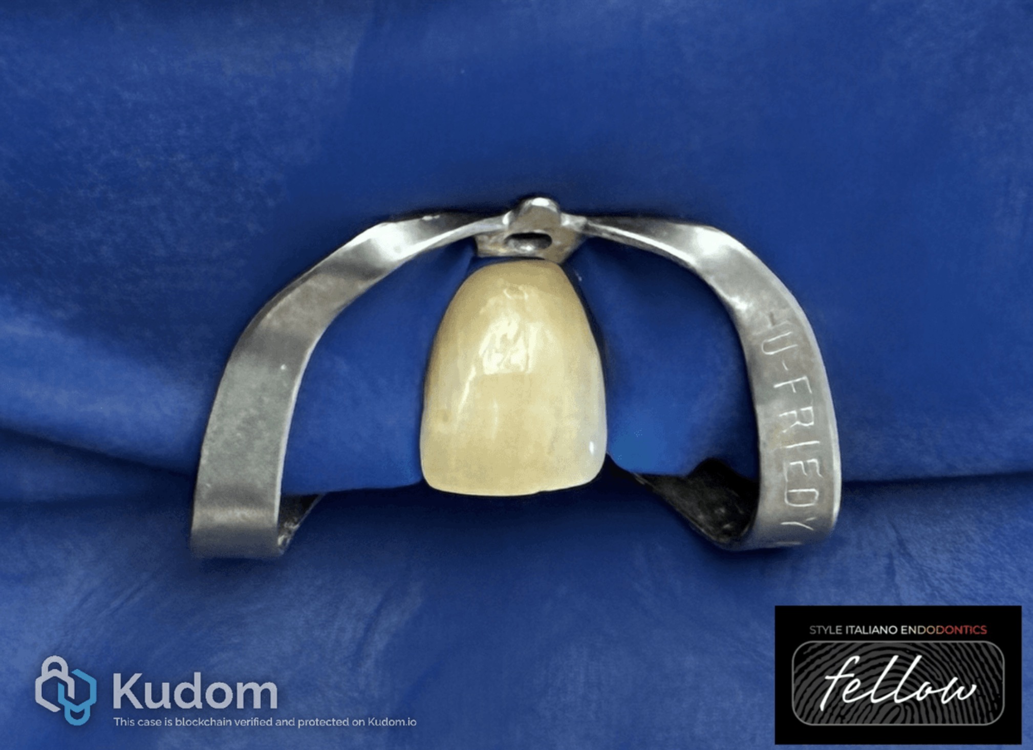
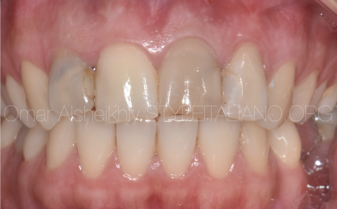
Fig. 2
The operative field was isolated under a rubber dam. An access cavity was prepared through the palatal surface, and necrotic pulp tissue was removed. Root canal treatment was performed with copious irrigation using 5.25% sodium hypochlorite (NaOCl), ultrasonically activated, followed by mechanical instrumentation with ProTaper Gold files.
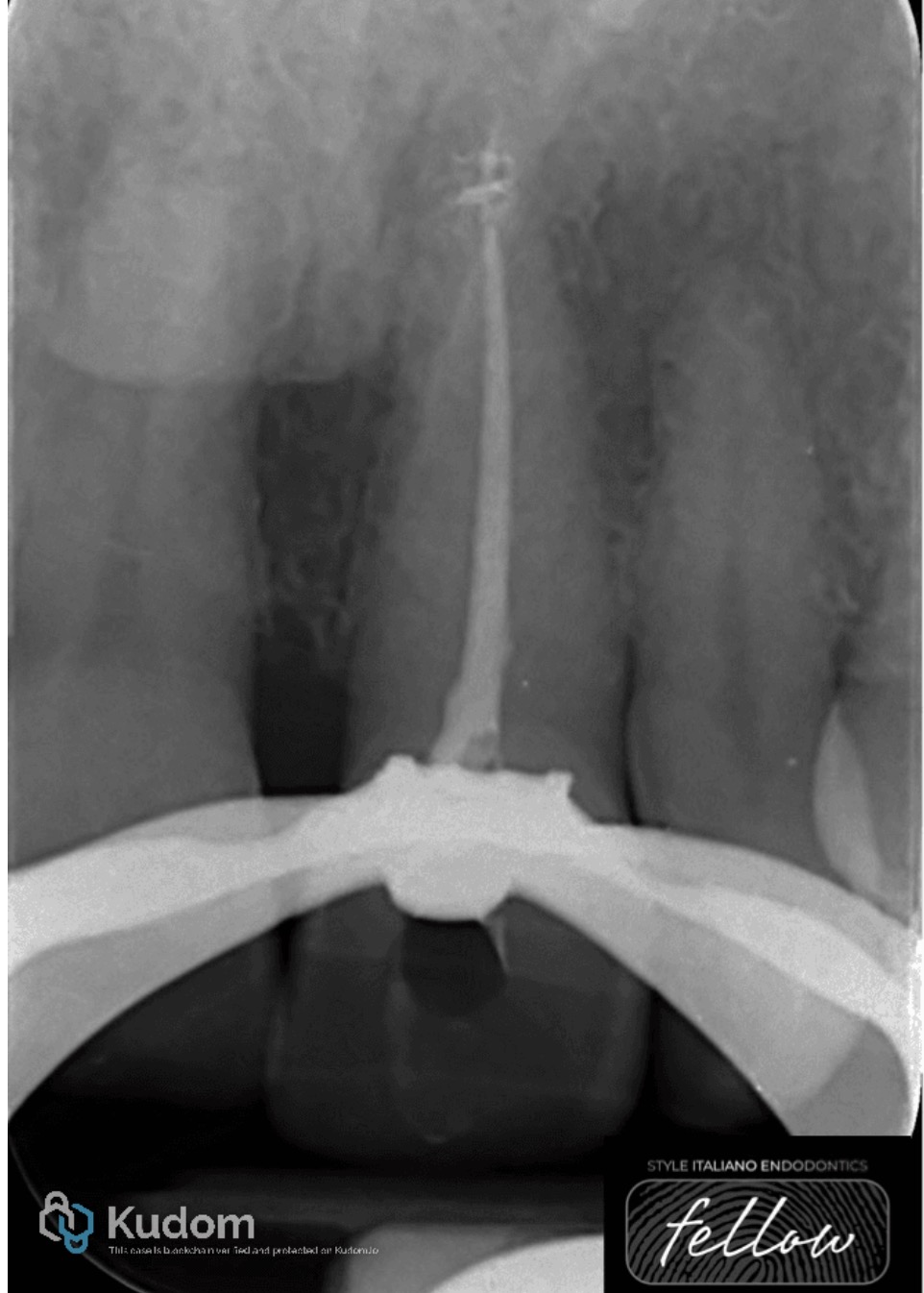
Fig. 3
Canal obturation was completed using a single-cone technique in combination with a bioceramic sealer.
A 2-mm vetroionomer barrier was positioned over the gutta-percha to establish a cervical seal. Subsequently, a 35% hydrogen peroxide bleaching gel was applied within the pulp chamber and left in place for a duration of seven days.
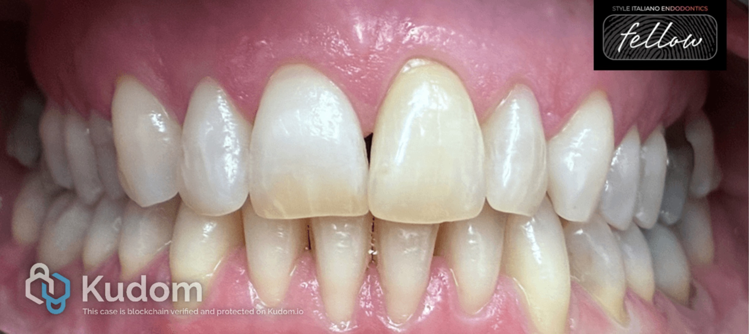
Fig. 4
After one week, the bleaching agent was removed by irrigation with saline solution. Subsequently, the access cavity was resealed with glass ionomer cement for an additional two weeks to mitigate the potential interference of residual oxygen with the bonding performance of the definitive composite restoration.
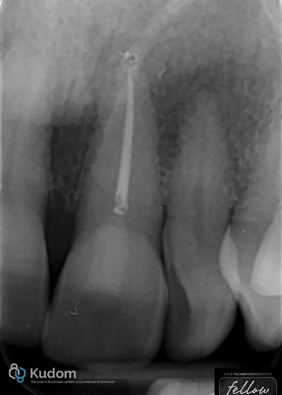
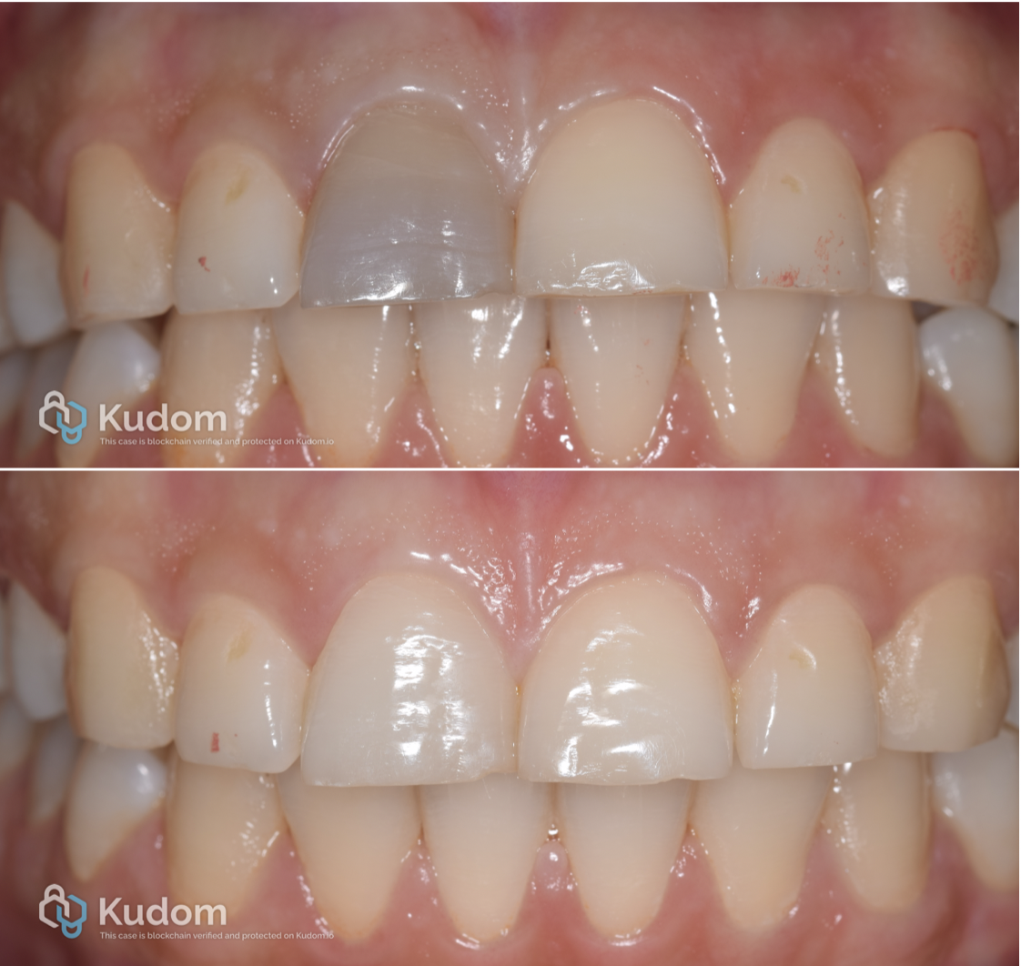
Fig. 5
A final postoperative radiograph was acquired.

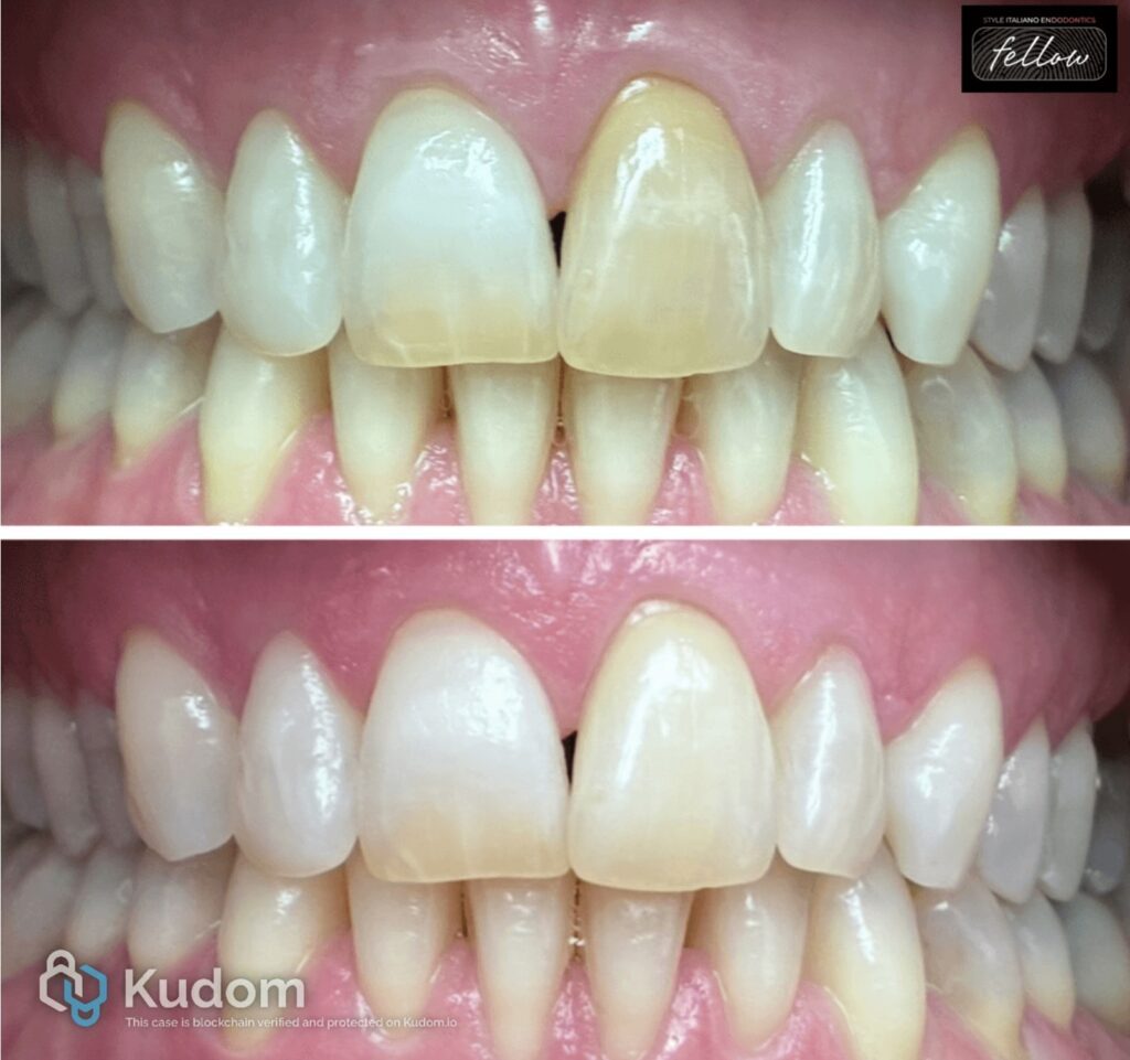
Fig. 6
Born in Agrigento on 22/04/1985
Graduated with honours in dentistry and dental prosthesis from the "Magna Graecia" University of Catanzaro. She mainly dedicates herself to conservative dentistry and endodontics. She works as a consultant in Tolmezzo (UD). Fellow of Style Italiano Endodontics.
Conclusions
Internal bleaching is a conservative, simple, effective, and low-cost procedure, with good esthetic results, in the treatment of non-vital tooth discolorations
Bibliography
- Joiner, A.; Luo, W. Tooth colour and whiteness: A review. J. Dent. 2017, 67S, S3–S10.
- Montero, J.; Gomez-Polo, C.; Santos, J.A.; Portillo, M.; Lorenzo, M.C.; Albaladejo, A. Contributions of dental colour to the physical attractiveness stereotype. J. Oral Rehabil. 2014, 41, 768–782.
- Al-Zarea, B.K. Satisfaction with appearance and the desired treatment to improve aesthetics. Int. J. Dent. 2013, 2013, 912368.
- Zimmerli, B.; Jeger, F.; Lussi, A. Bleaching of nonvital teeth. A clinically relevant literature review. Schweiz. Mon. Zahnmed. 2010, 120, 306–320.
- Plotino, G.; Buono, L.; Grande, N.M.; Pameijer, C.H.; Somma, F. Nonvital tooth bleaching: A review of the literature and clinical procedures. J. Endod. 2008, 34, 394–407.