Endodontic–restorative management of a periapical lesion involving tooth 25: diagnosis, treatment, and clinical outcome
29/12/2025
Fellow
Warning: Undefined variable $post in /home/styleendo/htdocs/styleitaliano-endodontics.org/wp-content/plugins/oxygen/component-framework/components/classes/code-block.class.php(133) : eval()'d code on line 2
Warning: Attempt to read property "ID" on null in /home/styleendo/htdocs/styleitaliano-endodontics.org/wp-content/plugins/oxygen/component-framework/components/classes/code-block.class.php(133) : eval()'d code on line 2
A patient reported pain in the upper jaw, left side. Pulp vitality and electric tests were negative. Radiographs revealed a periapical lesion on tooth 25. Following local anesthesia, the pulp chamber was opened, revealing purulent exudate. The root canal was cleaned using irrigation and activation of sodium hypochlorite and EDTA, then filled with bioceramic material. The tooth was subsequently restored with an indirect overlay. Follow-up radiographs at 6 and 11 months showed progressive healing.
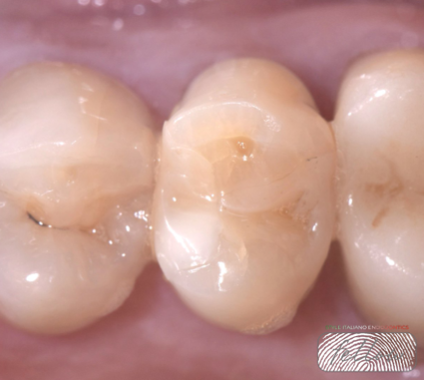
Fig. 1
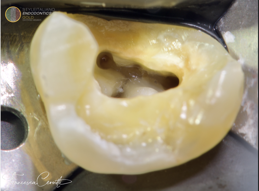
Clinical image of tooth 25 showing a previous and extensive direct composite restoration.
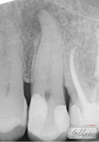
Fig. 2
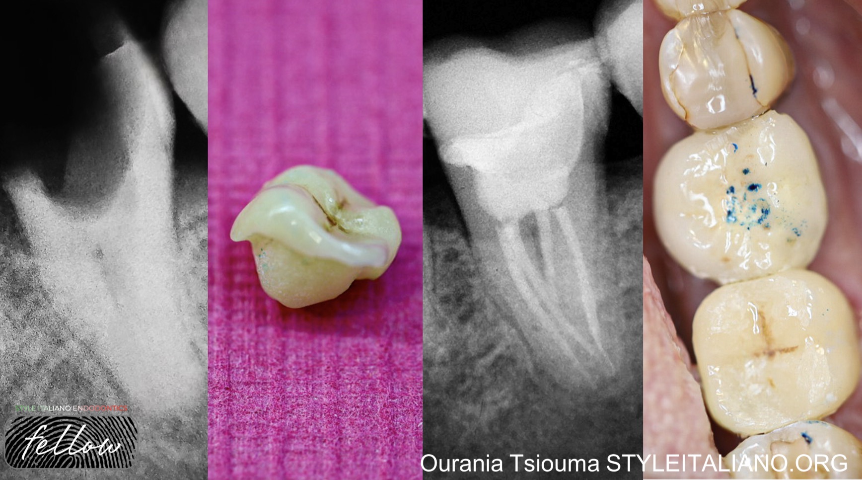
The X-ray shows the presence of a periapical lesion on tooth 25. Pulp vitality and electric tests were negative.
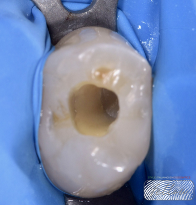
Fig. 3
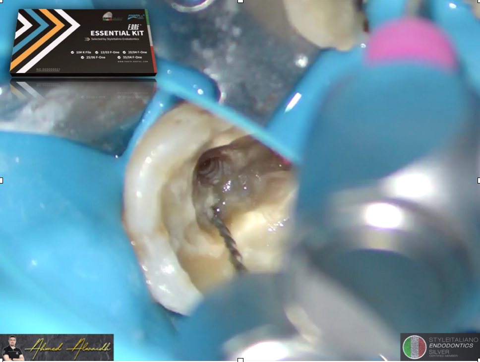
Following local anesthesia, the pulp chamber was opened, revealing purulent exudate. Proper root canal shaping is crucial, particularly in necrotic teeth, as it allows irrigants to penetrate deeply, exert antimicrobial effects, and dissolve organic debris. Proper shaping also facilitates the effectiveness of sodium hypochlorite, enhancing pulp tissue dissolution and thorough canal cleaning.
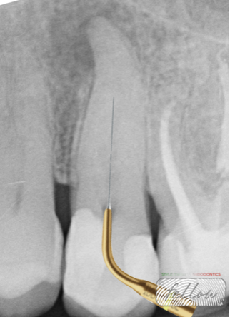
Fig. 4
Root canal shaping was performed using a combined rotary system to achieve optimal shaping and apical enlargement while preserving the original canal anatomy: Mtwo rotary files and ProTaper Gold finishing files.
- Mtwo Purple 10/.04 – initial shaping of coronal and middle thirds
- Mtwo White 10/.05 – shaping of coronal and middle thirds
- ProTaper Gold F1 – apical finishing
- ProTaper Gold F2 – final apical enlargement and continuous tapering
In this case, the importance of proper irrigation is clear. The protocol employed a carefully sequenced use of irrigating solutions to achieve thorough chemical cleaning of the root canal system, with each solution performing a specific function.
Sodium hypochlorite 5.25% (NaOCl) served as the primary irrigant, effectively dissolving organic tissue and eliminating microbes. Ethylenediaminetetraacetic acid (EDTA) was used as a final rinse to remove the smear layer.
To enhance the effectiveness of irrigation, each solution was ultrasonically activated for 20 seconds using IrriSafe™ passive ultrasonic tips, ensuring deeper penetration and thorough cleaning throughout the canal system.
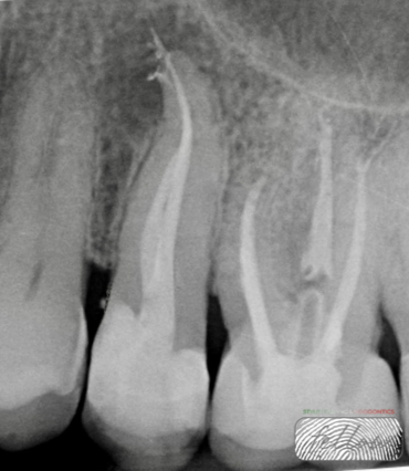
Fig. 5
Root canal obturation was performed for each canal using a bioceramic sealer with a single-cone technique.
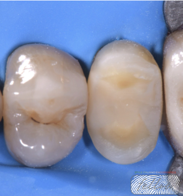
Fig. 6
The tooth was prepared for an indirect overlay restoration, preserving healthy tooth structure through a minimally invasive approach.
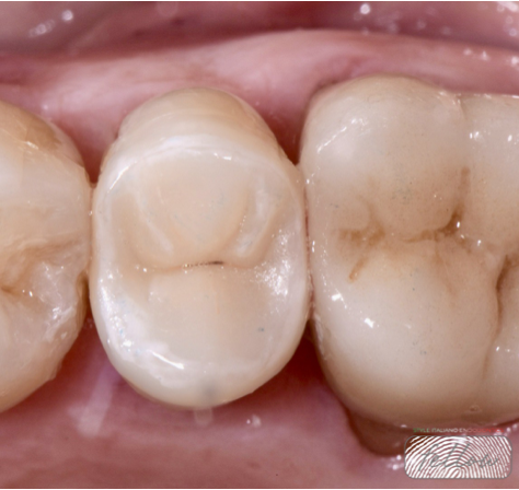
Fig. 7
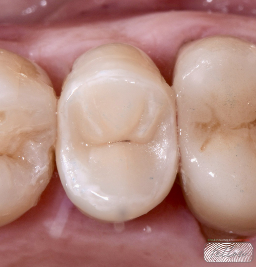
Final cementation of the indirect overlay restoration was carried out.Indirect overlays generally show better fracture resistance and higher survival rates compared to large direct fillings on endodontically treated teeth.
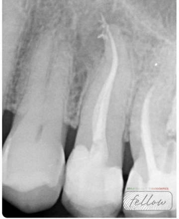
Fig. 8
Radiographs at 6 months showed the initial signs of healing.
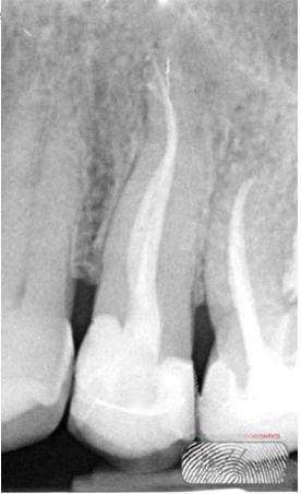
Fig. 9
At 11 months, radiographs confirmed progressive and complete periapical healing. The patient will continue to be monitored with yearly follow-ups.

Fig. 10
About the author:
Dr Agnese Agostinelli
Specialization in Oral Surgery at the Vita-Salute San Raffaele University, Milan. Post Graduate: Reconstructive surgery of periodontal and peri-implant tissues, course director Prof. Massimo de Sanctis, periodontology department of Vita-Salute San Raffaele University. Collaboration with Dr. Fabio Gorni and Partners at Milano Face Institute and with Professor Tiziano Testori at the Lake Como Institute.
Conclusions
Accurate diagnosis, effective canal shaping, and an irrigation protocol based on activated sodium hypochlorite were key factors in the successful management of the periapical lesion. The use of a bioceramic sealer with a single-cone technique supported periapical healing, while the final indirect overlay restoration ensured adequate coronal sealing and structural support. Radiographic follow-up showed initial healing at 6 months and periapical healing at 11 months. Annual follow-ups are planned.
Bibliography
1. Gorni, Fabio. "Shaping&Cleaning of the endodontic space: the Style Italiano Endodontic Philosophy and techniques.
2-Gomes BP, Aveiro E, Kishen A. Irrigants and irrigation activation systems in endodontics. Braz Dent J. 2023;34(4):1–33.
3- Cai C, Chen X, Li Y, Jiang Q. Advances in the role of sodium hypochlorite irrigant in chemical preparation of root canal treatment. Biomed Res Int. 2023;2023:8858283.
4- Nagendrababu V, Jayaraman J, Suresh A, Kalyanasundaram S, Neelakantan P. Effectiveness of ultrasonically activated irrigation on root canal disinfection: a systematic review of in vitro studies. Clin Oral Investig. 2018 Mar;22(2):655-670. doi: 10.1007/s00784-018-2345-x. Epub 2018 Jan 25. PMID: 29372445.
5- Zamparini F, Prati C, Taddei P, Spinelli A, Di Foggia M, Gandolfi MG. Chemical-Physical Properties and Bioactivity of New Premixed Calcium Silicate-Bioceramic Root Canal Sealers.
6. Estivalet, M.S.; de Araújo, L.P.; Immich, F.; da Silva, A.F.; Ferreira, N.d.S.; da Rosa, W.L.d.O.; Piva, E. Bioactivity Potential of Bioceramic-Based Root Canal Sealers: A Scoping Review. Life 2022, 12, 1853.
7-Shu X, Mai QQ, Blatz M, Price R, Wang XD, Zhao K. Direct and Indirect Restorations for Endodontically Treated Teeth: A Systematic Review and Meta-analysis, IAAD 2017 Consensus Conference Paper. J Adhes Dent. 2018;20(3):183-194. doi: 10.3290/j.jad.a40762. PMID: 29984369.
8.G.M.G.Hommez, C.R.M.Coppens, R.J.G.De Moor : Periapical health related to the quality of coronal restorations and root fillings, International endodontic journal, august 2002/volume 35, issue 8/ pp.680-689.