Controlled preparation of the Endodontic Access Cavity
20/11/2021
Fitim Shabani
Warning: Undefined variable $post in /home/styleendo/htdocs/styleitaliano-endodontics.org/wp-content/plugins/oxygen/component-framework/components/classes/code-block.class.php(133) : eval()'d code on line 2
Warning: Attempt to read property "ID" on null in /home/styleendo/htdocs/styleitaliano-endodontics.org/wp-content/plugins/oxygen/component-framework/components/classes/code-block.class.php(133) : eval()'d code on line 2
The access cavity must make the succeeding steps easier and safer. It must therefore meet the following requirements
- Permit the removal of all the chamber contents
- Permit complete, direct vision of the floor of the pulp chamber and the canal openings
- Facilitate the introduction of canal instruments into the root canal openings
- Provide access as direct as possible to the apical one third of the canal for both preparation instruments and canal filling instruments
- Always have four walls
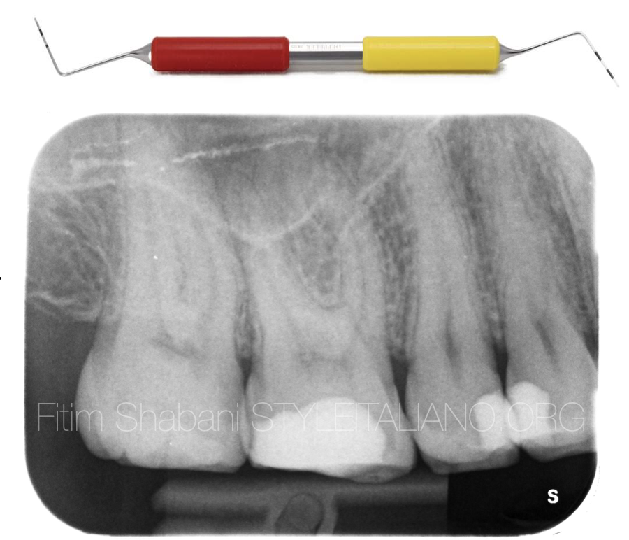
Fig. 1
MisurEndo: Double-ended probe – One side has been designed for conventional diagnostic probing. The other side is a guide to access cavity design correctly, with laser markings 4, 6, 9 and 10MM.
Clinical case
The first upper right molar was diagnosed with irreversible pulpitis
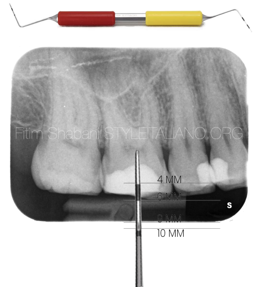
Fig. 2
The distance between the occlusal surface and the tooth floor is 6MM.
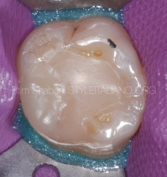
Fig. 3
Initial situation after the isolation of the operative field.
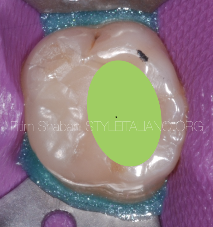
Fig. 4
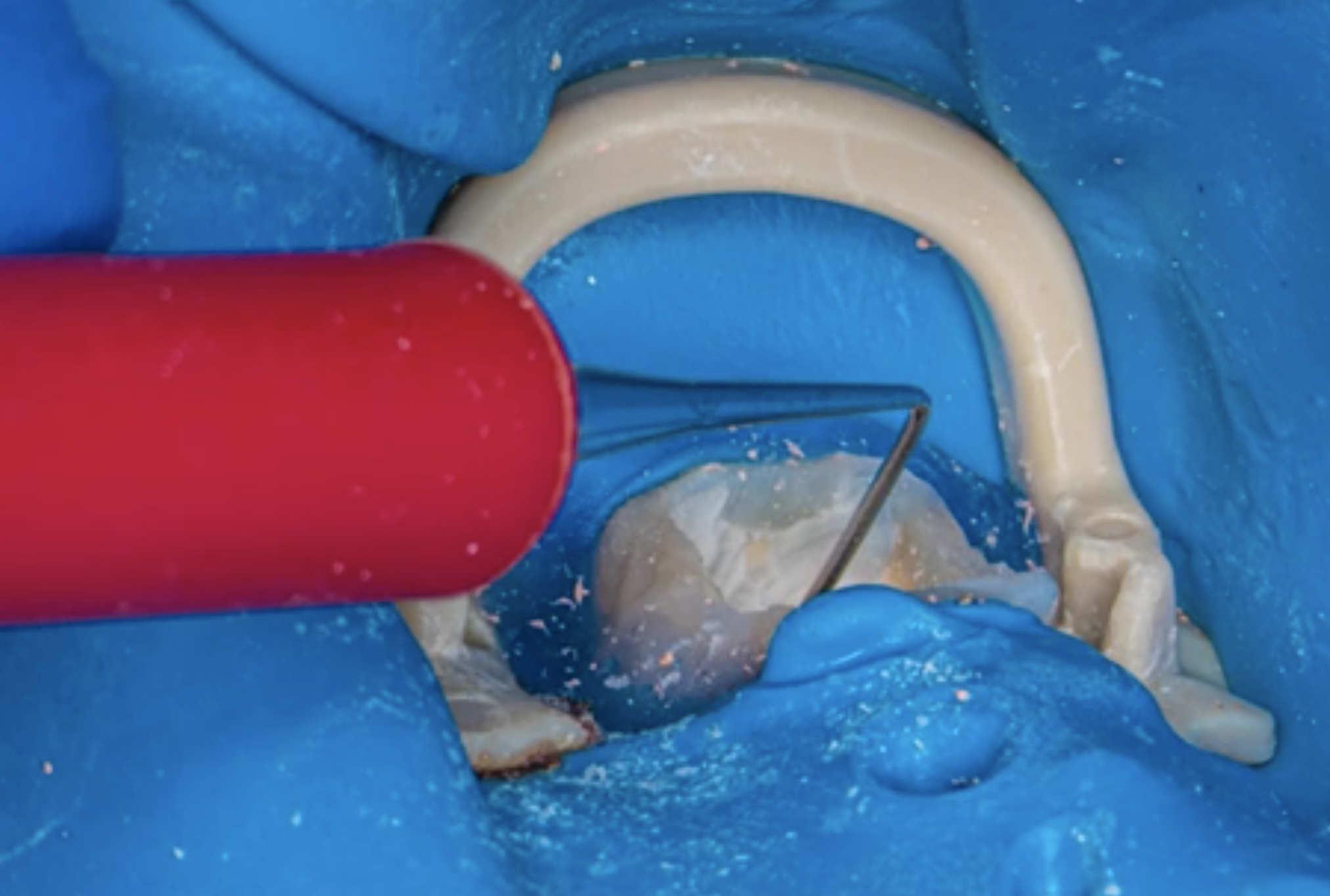
In this area of the tooth we need to prepare and search for the canals.
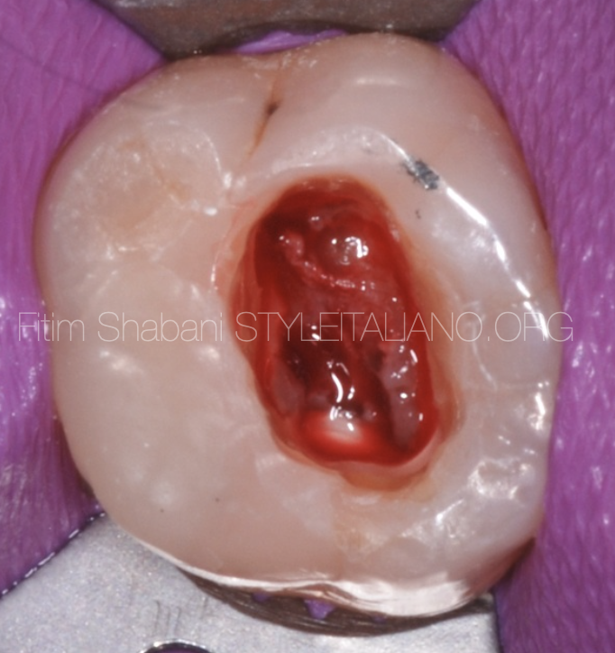
Fig. 5
Initial preparation of the endodontic access cavity.
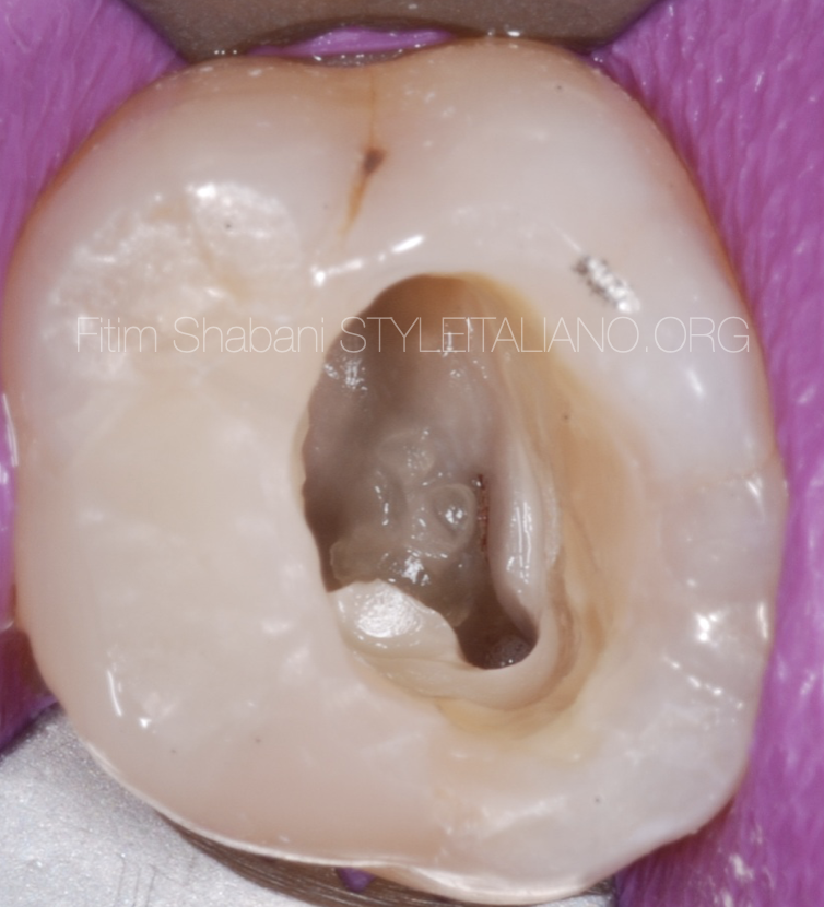
Fig. 6
Opening the correct endodontic access cavity allows us to more easly explore, prepare and fill the canals.
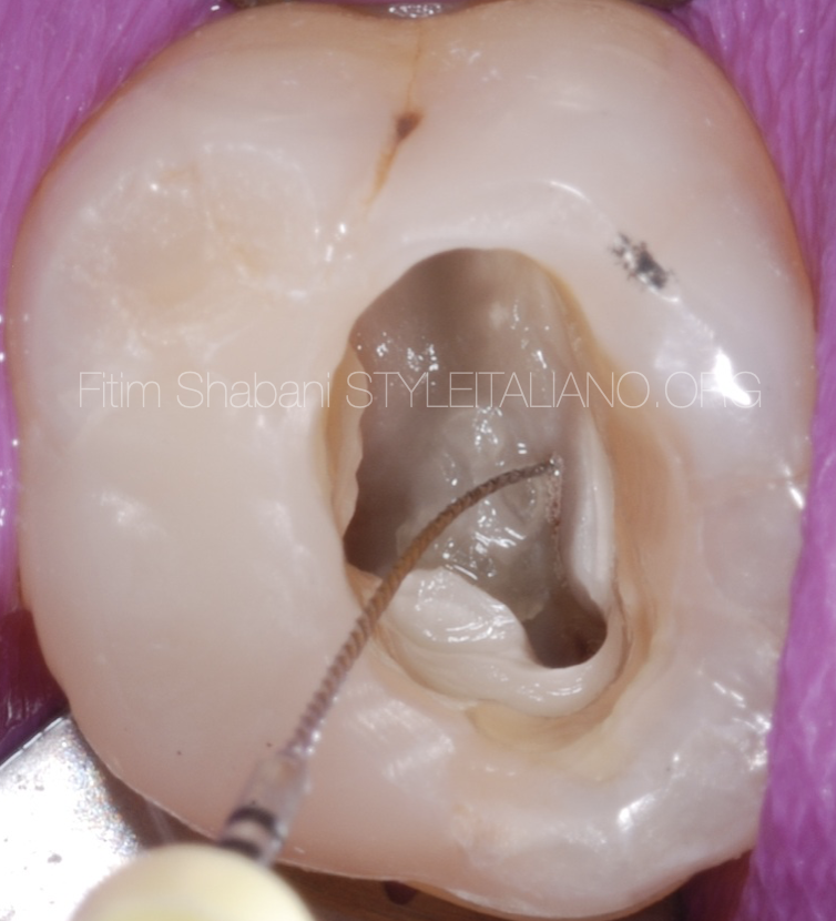
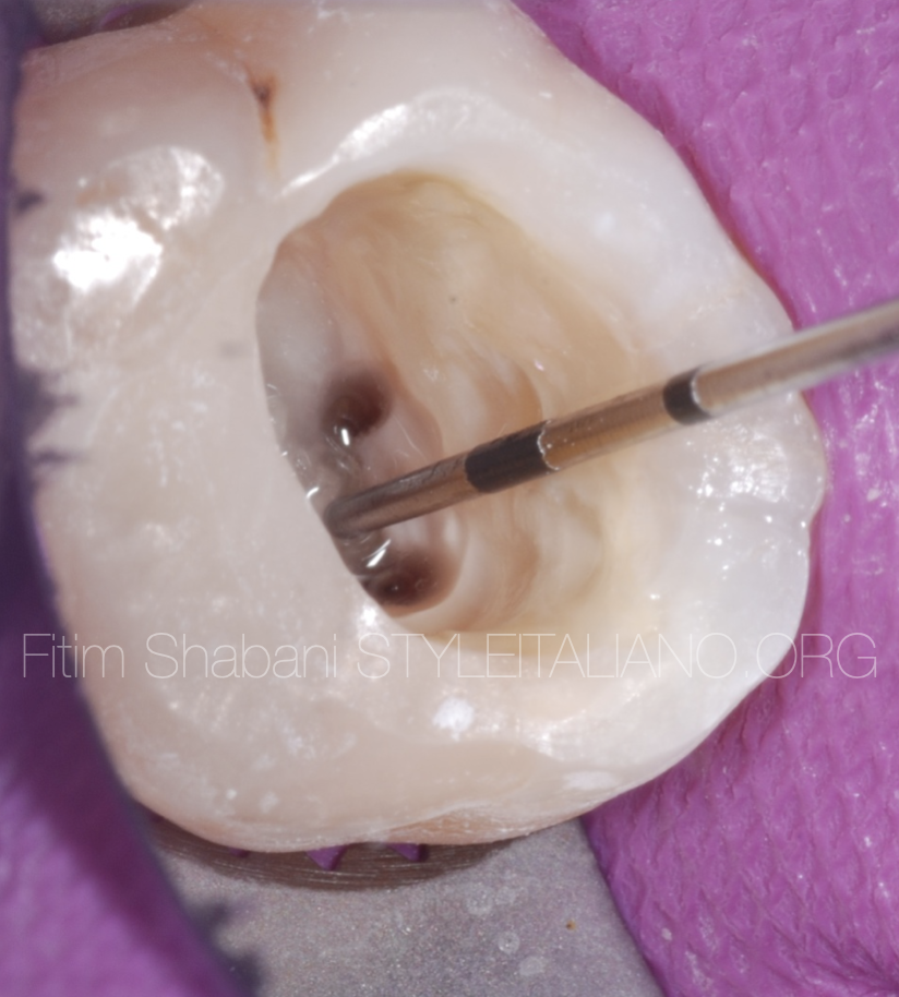
Fig. 7
Localisation of the MB2 canal, thanks to the good view, which is the result of correct endodontic access cavity.
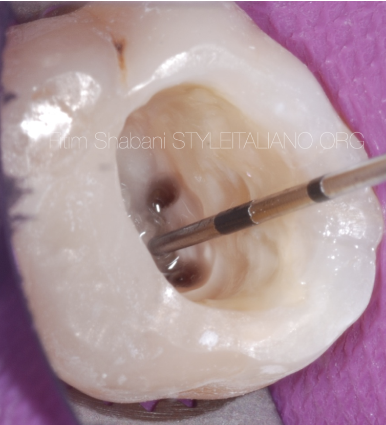
Fig. 8
The distance between the occlusal surface and the tooth floor is 6MM.
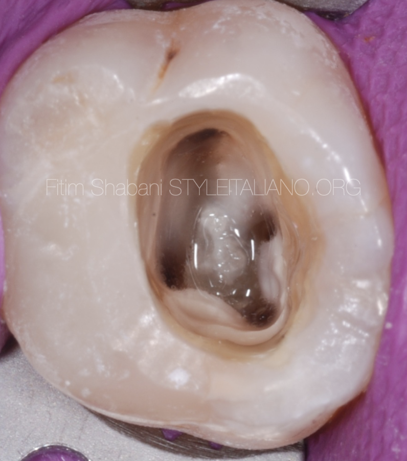
Fig. 9
The view after preparation and cleaning of the canals.
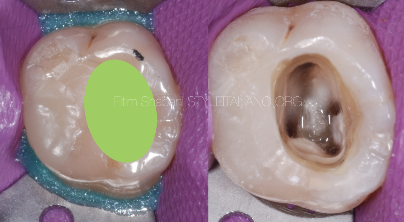
Fig. 10
Pre and post preparation
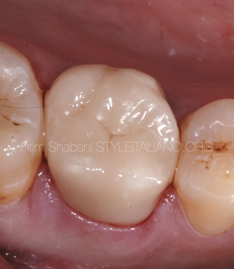
Fig. 11
The view after Crown cementation.
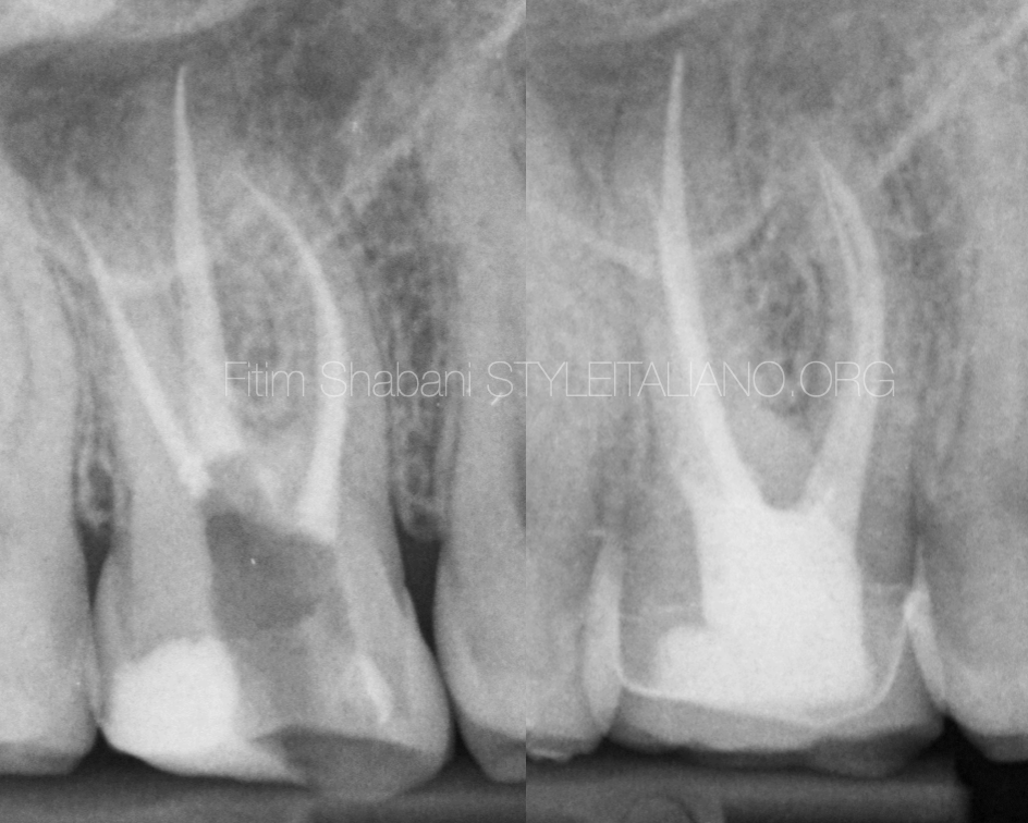
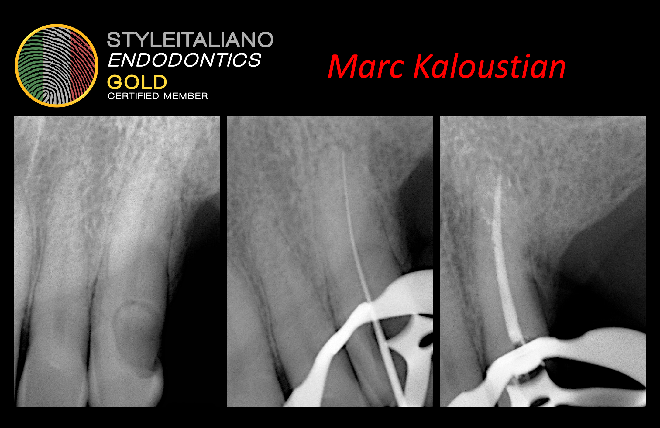
Fig. 12
Final X-ray
Conclusions
The initial and crucial step of endodontic treatments is undoubtedly the endodontic access cavity, from which we find, prepare, clean and fill the endodontic spaces. Therefore, respect the principles and use the right instruments which facilitate our work and ensure good progress of the treatment and its prognosis in general.
Bibliography
- TahaÖzyürekDDS, PhD Özlem ÜlkerDDS et all. The Effects of Endodontic Access Cavity Preparation Design on the Fracture Strength of Endodontically Treated Teeth: Traditional Versus Conservative Preparation, Journal of endodontics 2018.
- S. Patel & J. Rhodes, A practical guide to endodontic access cavity preparation in molar teeth, British dental journal 2007.
- Janik JM, Access cavity preparation, Dental Clinics of North America, 01 Oct 1984.
- Michael E.RampadoDDS, MSc, FRCD (Canada) Leo TjäderhaneDDS, PhD Shimon FridmanDMD Stanley J.HamstraPhD, The Benefit of the Operating Microscope for Access Cavity Preparation by Undergraduate Students, Journal of endodontics December 2004.