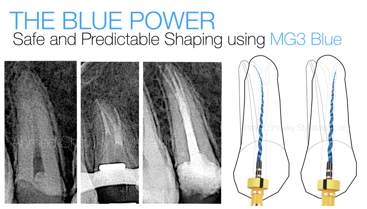
Conquering complex Anatomy with MG3
16/11/2023
Ahmed Shawky
Warning: Undefined variable $post in /home/styleendo/htdocs/styleitaliano-endodontics.org/wp-content/plugins/oxygen/component-framework/components/classes/code-block.class.php(133) : eval()'d code on line 2
Warning: Attempt to read property "ID" on null in /home/styleendo/htdocs/styleitaliano-endodontics.org/wp-content/plugins/oxygen/component-framework/components/classes/code-block.class.php(133) : eval()'d code on line 2
Encountering a complex anatomy is a associated with a lot of liabilities in case the clinician didn’t pay attention to the details of the case in the pretreatment phase.
Skipped details may lead, not only to failure of anatomy management, but also to a series of aggressive mishaps that can take the case difficulty to a whole new level.
Therefore, the clinician must be able to analyze all the case details in the pre-treatment phase to avoid mishaps during the treatment course.
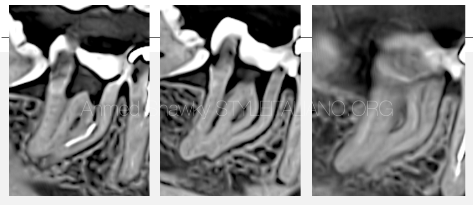
Fig. 1
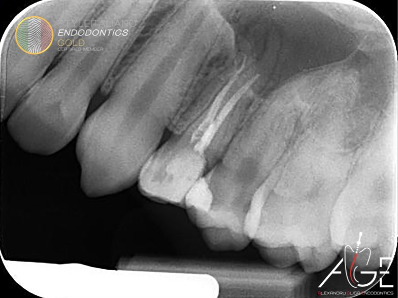
In this article, a case is presented that was referred for a separated instrument retrieval and completion of the incomplete primary treatment.
Pretreatment evaluation involved clinical and radiographic examination, which confirmed a symptomatic apical periodontitis in the mandibular right second molar.
2D and CBCT evaluation revealed a small hand file separated in the ML canal, CBCT was mandatory to plan the retrieval approaching relevance to the respective anatomy.
The configuration of the mesial root canal system was shown to have multi-planar curvatures, highlighting the reason for file separation
The instrument probably separated due to the improper shaping strategy followed for shaping of the double curved mesial root canals.
It has be emphasized that, For safe and predictable shaping of double curvatures, the clinician should adopt a “Pressureless Crown-down approach”, to address different parts of the multi-planar curvature. The most important step in that approach, is the coronal pre-flaring, which can be safely performed mechanically to eliminate severe coronal dentin resistance. Such dentinal resistance, can be a reason for file separation in the coronal and middle portions, as in this case.
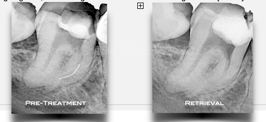
Fig. 2
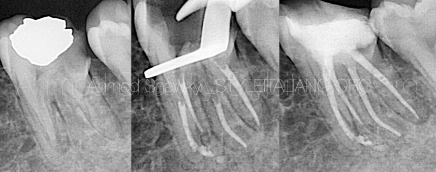
Treatment was performed over two visit, where the first visit was dedicated to retrieval of the separated instrument using loop technique.
Briefly, management of the separated instrument involved the use of thin ultrasonic tips operated on low power setting, to avoid any possible secondary fracture of the small size, yet long fragment, considering minimal loss of dentin during retrieval.
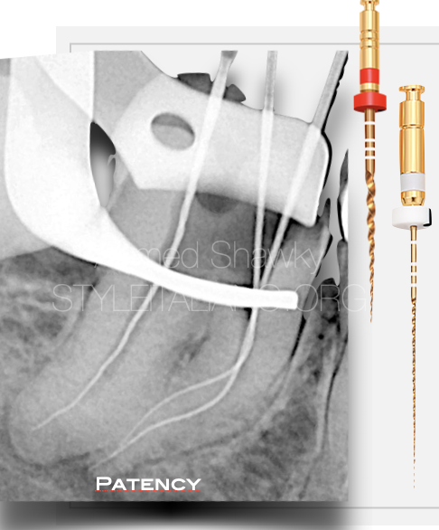
Fig. 3
The shaping strategy
Shaping of the mesial root canal system was performed, combining two different alloys and two different kinematics;
- The MG3 Gold Instruments, [25/.06, & 20/.06] were used in the coronal portion of the root canal, “on two levels” to benefit from the cutting efficiency during mechanical pre-flaring. These instruments were used in ATR mode “adaptive motion” in a BR Rap motor, to adapt the instrument to the condition of the canal in that portion.
Patency was achieved using K files after coronal pre-flaring Followed by Mechanical Glide path using MG3 Gold 15/.03
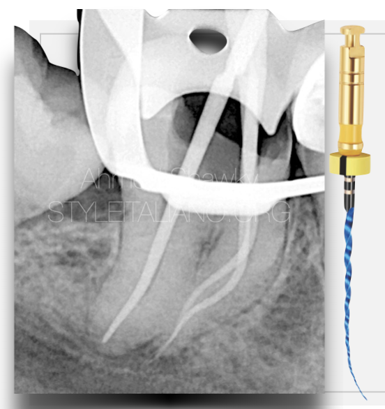
Fig. 4
- The MG3 Blue instruments, were used in the Body portion and for apical finishing, to benefit from the controlled memory behavior avoiding alteration of the anatomy “transportation”.
- Another benefit from using MG3 Blue is the enhanced cyclic fatigue resistance owing to the predominantly martensitic micro-structure
These instruments were used in interrupted rotation [REC mode] with a net clockwise motion (CW 170 / CCW 50)
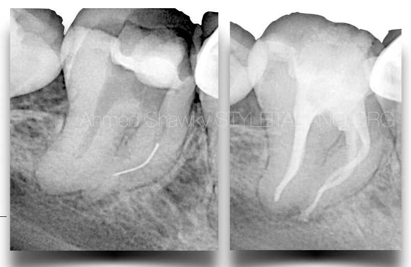
The Final Shaping size in the mesial root canals were MG3 Blue #20/.04
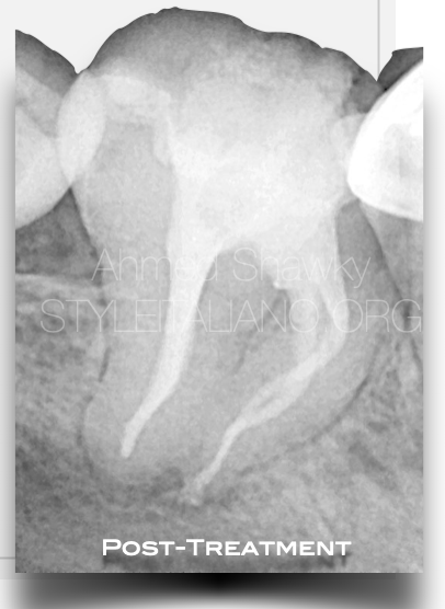
Fig. 5
The Distal root canal was shaped using a single file in interrupted rotation [MG3 Gold, 30/.04]
The irrigation protocol involved full strength NaOCl throughout instrumentation along with continuous chelation using 17% EDTA solution
The final rinse protocol involved activation of both solutions using Ultrasound
Obturation was performed utilizing Warm compaction of gutta percha and heat compatible calcium silicate based sealer
Conclusions
Endodontic Treatments are strategy based treatments rather than just procedural steps
The clinician must integrate knowledge about Instrument metallurgy and motions to be able to address the complex anatomy encountered in daily practice
MG3 Gold and Blue instruments provides versatility in clinical uses and can be adapted to manage complex root canal systems
Bibliography
1- Scarfe WC, Levin MD, Gane D, Farman AG. Use of cone beam computed tomography in endodontics. Int J Dent. 2009;2009:634567. doi: 10.1155/2009/634567. Epub 2010 Mar 31. PMID: 20379362; PMCID: PMC2850139.
2- Patel S, Durack C, Abella F, Shemesh H, Roig M, Lemberg K. Cone beam computed tomography in Endodontics - a review. Int Endod J. 2015;48(1):3-15. doi:10.1111/iej.12270
3- Bertani, P., Gagliani, M. & Gorni, F. 2020, Retreatments. Solutions for Periapical Diseases of Endodontic Origin, Edra.
4- Salehrabi R, Rotstein I. Endodontic treatment outcomes in a large patient population in the USA: an epidemiological study. J Endod 2004;30:846–50.
5- Outcome of Endodontic Treatment through Existing Full Coverage Restorations: An Endodontic Practice Case Series. https://doi.org/10.1016/j.joen.2021.11.008


