An endodontic retreatment case with multiple challenges
16/12/2021
João Meirinhos
Warning: Undefined variable $post in /home/styleendo/htdocs/styleitaliano-endodontics.org/wp-content/plugins/oxygen/component-framework/components/classes/code-block.class.php(133) : eval()'d code on line 2
Warning: Attempt to read property "ID" on null in /home/styleendo/htdocs/styleitaliano-endodontics.org/wp-content/plugins/oxygen/component-framework/components/classes/code-block.class.php(133) : eval()'d code on line 2
A complex case can be diagnosed pre-operatively with radiographs and cone-beam computed tomography systems (CBCT) that provide the exact root canal configuration allowing a deep understanding of the canal anatomy as well as other complications from previous treatments. Endodontic specialists usually treat complex cases which needed advanced materials and techniques. Also is expected that a skilled specialist with technical and anatomical knowledge, time and equipment solve these cases doing all efforts possible to make teeth again available for their functional oral status with a long term prognosis.
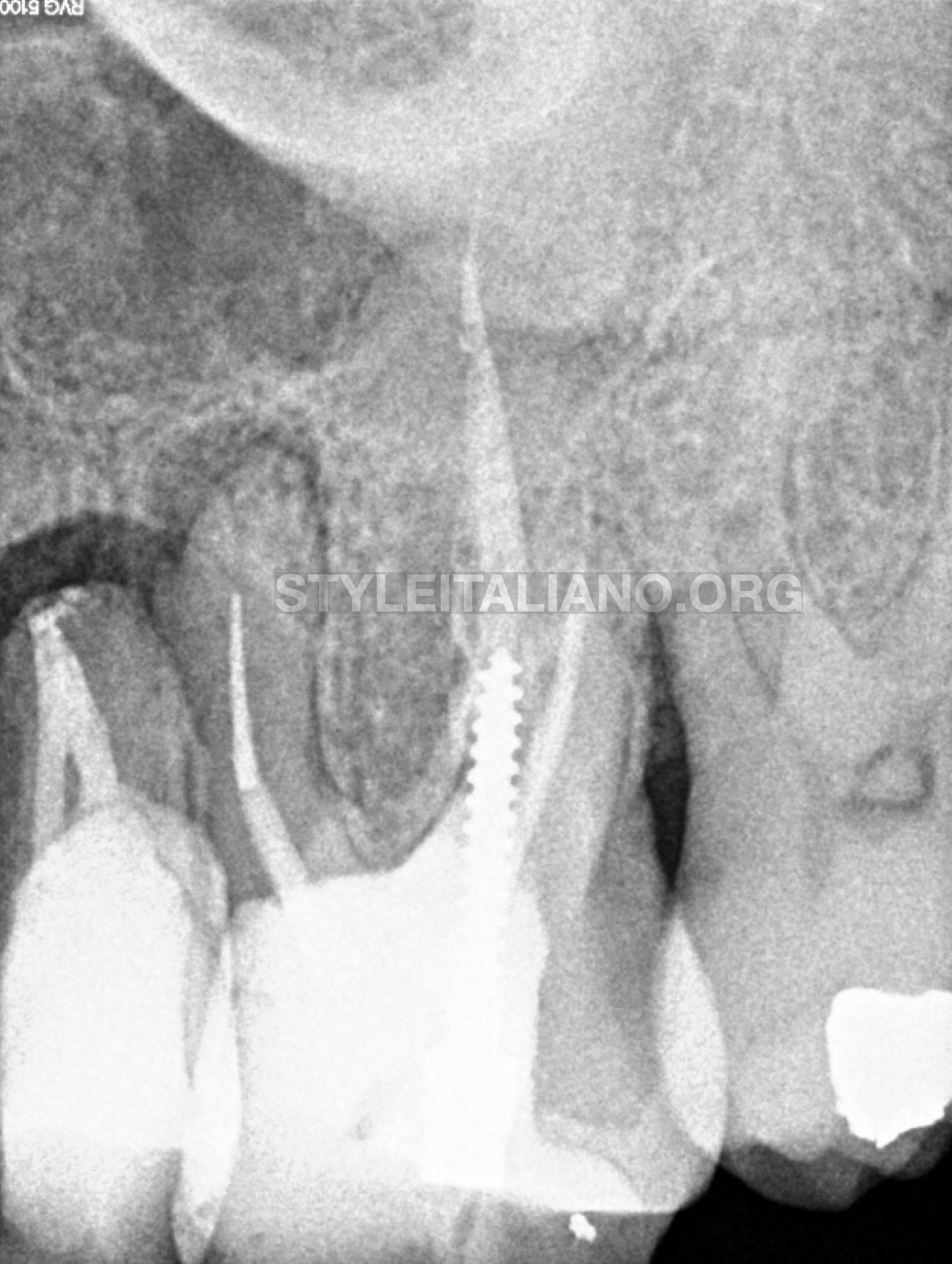
Fig. 1
Preoperative radiographic analysis
A 45-year-old female patient was referred by a colleague. In the radiographic examination, the first left upper molar presented a metal post, a separated instrument located in the middle third of the mesiobuccal canal and no MB2 canal was found on the previous treatment.
The tooth 26 was diagnosed with a symptomatic apical periodontitis and a non-surgical endodontic retreatment was suggested.
Metal post removal
Ultrasonic vibration breaks the bond between the post and the canal walls, facilitating its removal. There are many advantages of using ultrasonics for this procedure, including speed, conservation of tooth structure, and minimizing risk of tooth fracture and root perforation.
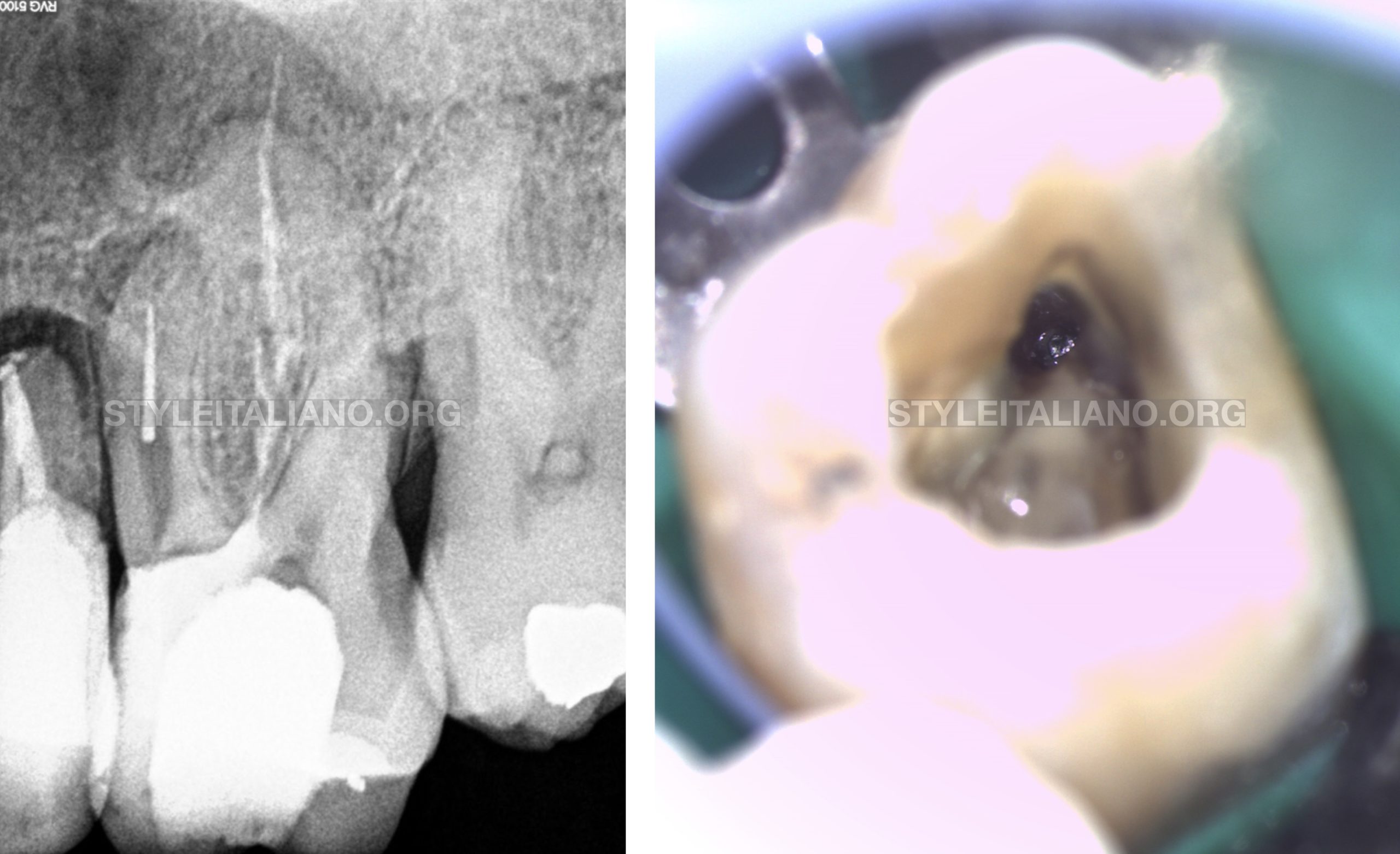
Fig. 2
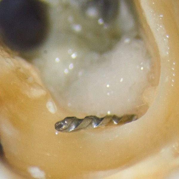
After old restoration and gutta percha removal, the separated instrument on the mesiobuccal canal was exposed.
Separated instrument removal
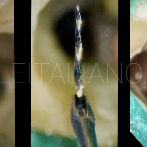
The bypass of the instrument was attempted with no success. Using a K files (Dentsply Maillefer, Ballaigues, Switzerland) coupled to Endo-chuck (SybronEndo, Orange, California), it was possible to remove the fragment.
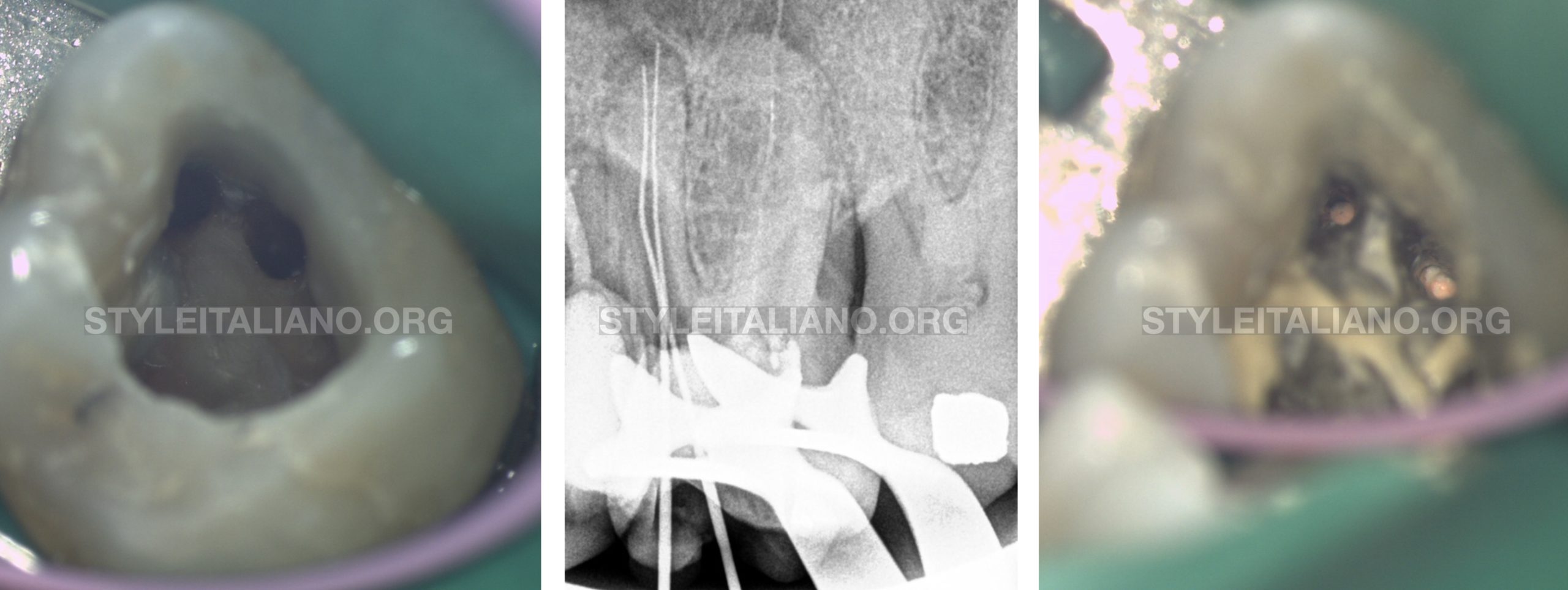
Fig. 3
Missed MB2 canal
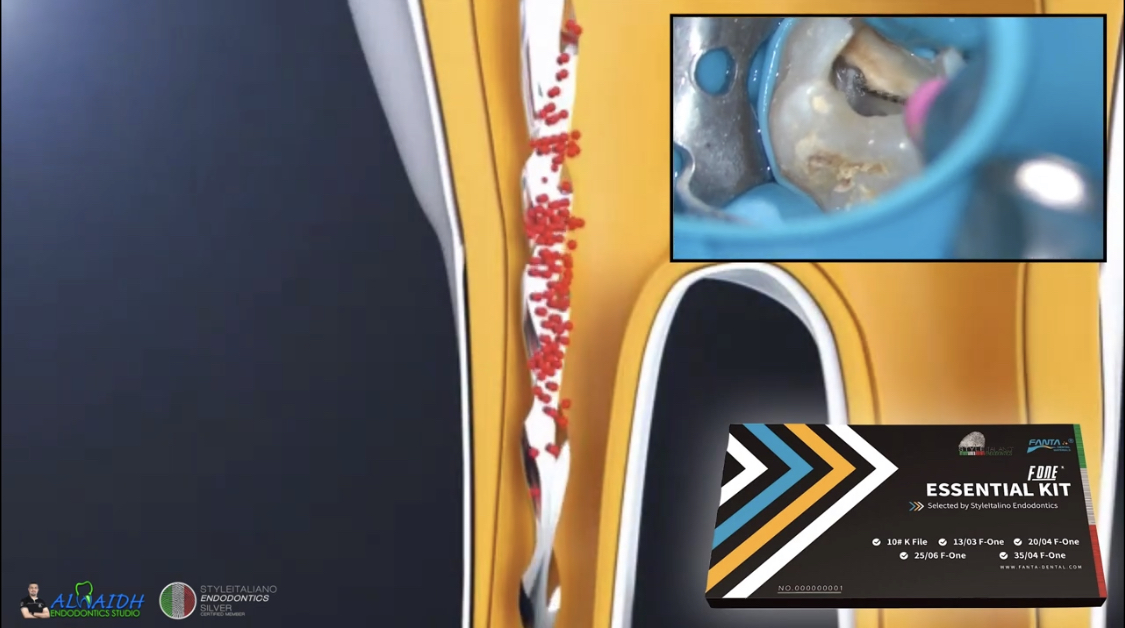
The use of microscope and ultrasonics is especially valuable in canal location. In this particular case, it was used to confirm that there was a second mesiobuccal canal in the mesial root. An untreated MB2 canal is the main reason for endodontic failures in maxillary molars.
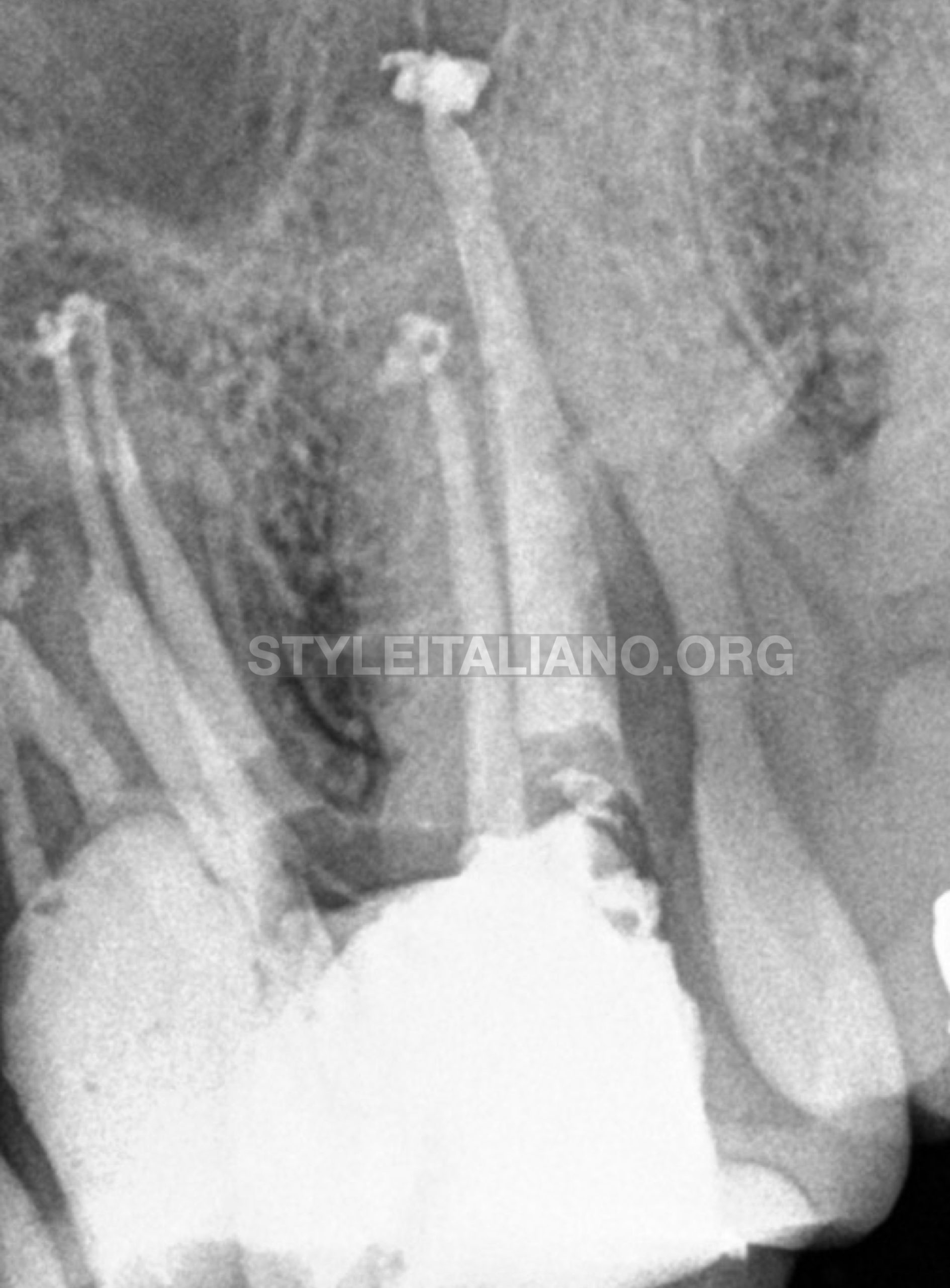
Fig. 4
The tooth was completely instrumented, disinfected, filled with gutta percha and subsequently temporally restored. It was recommend a cuspal coverage restoration.
Conclusions
The use of the 3D diagnostic tools, microscope, ultrasonics devices, repair materials and all the technical advancements available today will help the dentist in solving complex clinical cases with multiple pathologies. The level of case difficulty mainly depends on the presence of iatrogenic errors, such as ledges, perforations or separated instruments, or the existence of persistent infections. Regarding initial treatment complexity is often related to anatomy variations. The decision to proceed or not with a more complex case may depend on the operator training / expertise and obviously, a good planning.
Bibliography
- American Association of Endodontists. Glossary of Endodontic Terms, 8th ed. Chicago: American Association of Endodontists; 2012.
- Hauman, C. H. J., Chandler, N. P., & Purton, D. G. (2003). Factors influencing the removal of posts. International Endodontic Journal, 36(10), 687–690.
- Parashos P, Messer HH. Rotary NiTi Instrument Fracture and its Consequences. Journal of Endodontics. 2006;32:1031–1043.
- Panitvisai P, Parunnit P, Sathorn C, Messer HH. Impact of a Retained Instrument on Treatment Outcome: A Systematic Review and Meta-analysis. Journal of Endodontics.
- Baruwa, A. O., Martins, J. N. R., Meirinhos, J., Pereira, B., Gouveia, J., Quaresma, S. A., … Ginjeira, A. (2019). The Influence of Missed Canals on the Prevalence of Periapical Lesions in Endodontically Treated Teeth: A Cross-sectional Study. Journal of Endodontics.


