A simple trick for a sharp apical barrier placement
03/06/2016
Riccardo Tonini
Warning: Undefined variable $post in /home/styleendo/htdocs/styleitaliano-endodontics.org/wp-content/plugins/oxygen/component-framework/components/classes/code-block.class.php(133) : eval()'d code on line 2
Warning: Attempt to read property "ID" on null in /home/styleendo/htdocs/styleitaliano-endodontics.org/wp-content/plugins/oxygen/component-framework/components/classes/code-block.class.php(133) : eval()'d code on line 2
One step apexification with MTA is nowadays a widely diffused procedure, although the major problem in cases of a wide open apex is the need to limit and confine the apexification material at the apex, avoiding its extrusion into the periodontal tissue. The use of a biocompatible matrix is advisable because its placement in the area of bone destruction provides a base on which the sealing material, especially MTA, can be placed and packed.
The major challenge the operator can find is the accurate placement of the matrix, completely outside anatomic apex, but always in close contact with it. One of the most common mistakes is to leave part of the resorbable matrix inside the canal, but this causes a subsequent filling with MTA in a defect of adhesion between the material and the root because the matrix is interposed between them.
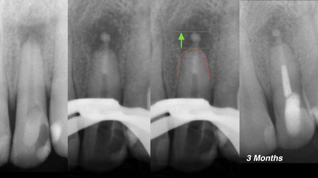
Fig. 1
Another common mistake is to place the matrix too far beyond the apex and this leads to a obturation with overfilling.
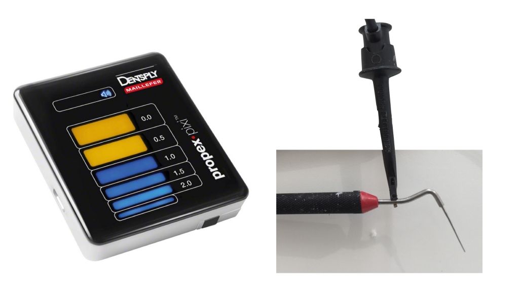
Fig. 2
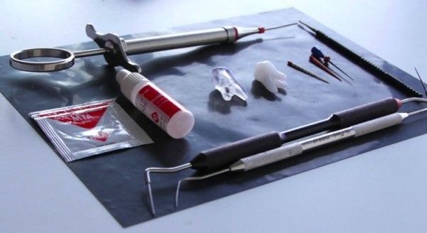
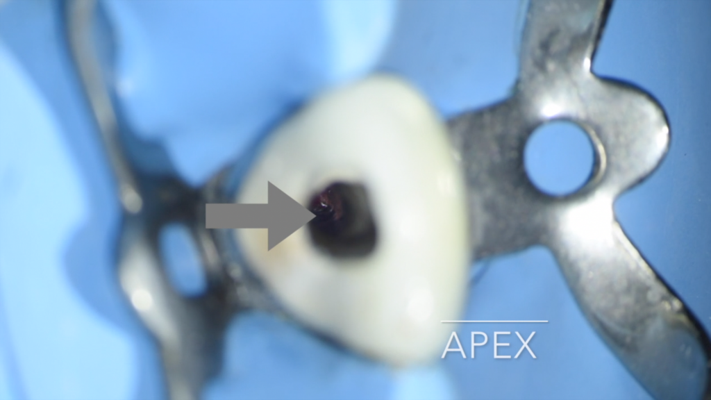
To test what is the best method for positioning the resorbable matrix has been created an experimental model which simulates a large lesion, namely the worst situation that the operator can meet. In addition creating this model it was born the idea of being able to use the apex locator connected to the plugger to position the matrix in the best way possible.
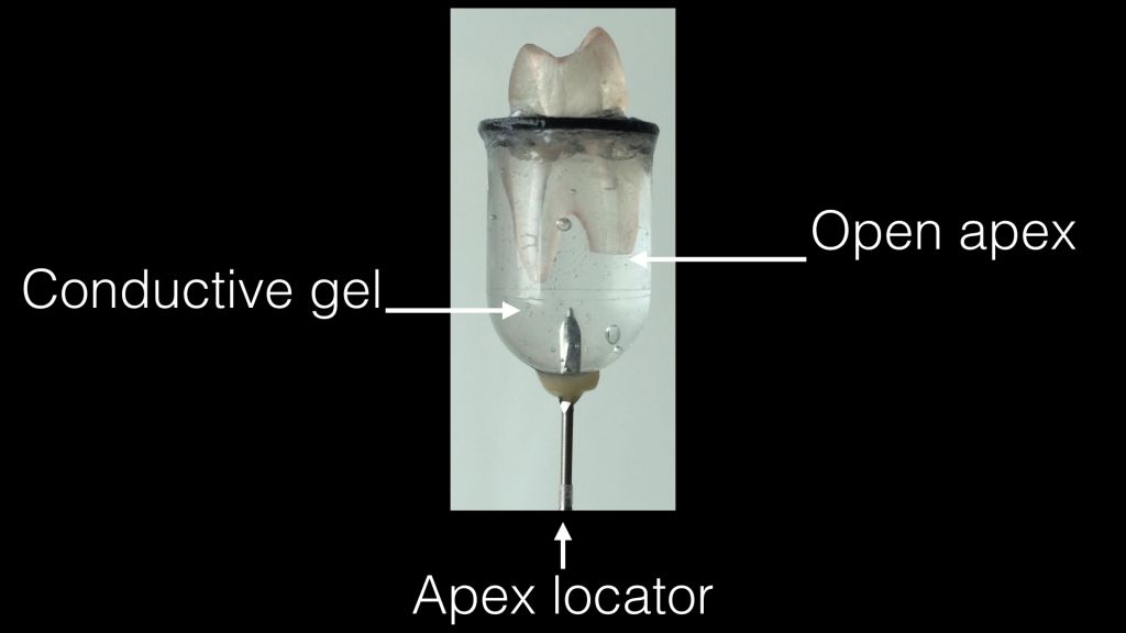
Fig. 3
A 3D printed was selected and one root was cut simulating an open and reabsorbed apex. The final prepared diameter of the apex was carried to 0.70. The tooth was subsequently inserted in a closed system in order to put in contact rootsct with a high-density elettroconductive gel. Through a metal rod positioned in touch with the gel, apex locator is now able to work as in vivo conditions.
Conclusions
The attached video shows how placement of the matrix can be simplified by the use of an apex locator and how the operator is facilitated during this challenging procedure which is not always so predictable. In the second part of video the first test is performed in vivo to demonstrate how this procedure is feasible and repeatable with very little effort.
Bibliography
- Sheehy EC, Roberts GJ(1997). Use of calcium hydroxide for apical barrier formation and healing in non-vital immature permanent teeth: a review. Br Dent J. 1997 Oct 11;183(7):241-6.
- Rafter M(2005)Apexification:a review. Dental Traumatology21.1-8.
- Stephen Cohen, Kenneth M. Hargreaves. Pathways of the pulp, St Louis, Missouri, Mosby 2006, 622.
- Giuliani V, Baccetti T, Pace R, Pagavino G. The use of MTA in teeth with necrotic pulps and open apices. Dent Traumatol. 2002 Aug;18(4):217-21.
- Hachmeister DR, Schindler WG, Walker WA 3rd, Thomas DD.The sealing ability and retention characteristics of mineral trioxide aggregate in a model of apexification. J Endod. 2002 May; 28(5):386-90.


