A Challenging Clinical Case
13/06/2025
Massimo Giovarruscio
Warning: Undefined variable $post in /home/styleendo/htdocs/styleitaliano-endodontics.org/wp-content/plugins/oxygen/component-framework/components/classes/code-block.class.php(133) : eval()'d code on line 2
Warning: Attempt to read property "ID" on null in /home/styleendo/htdocs/styleitaliano-endodontics.org/wp-content/plugins/oxygen/component-framework/components/classes/code-block.class.php(133) : eval()'d code on line 2
Three-rooted maxillary first premolars represent a significant anatomical variation and a considerable challenge in Endodontic practice. This article aims to analyze the clinical management strategies pertaining to this specific tooth morphology, using a new reciprocation file: R-One Mini Kit.
The reported prevalence of three-rooted maxillary first premolars ranges from 0% to 9.2%. In all documented instances of this variant, the presence of three distinct root canals has been consistently observed.
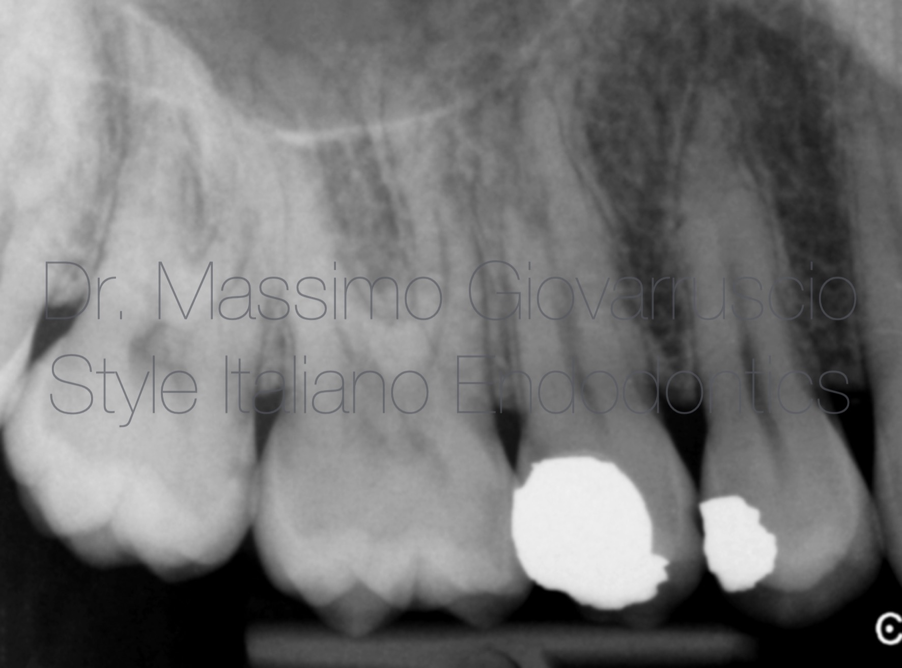
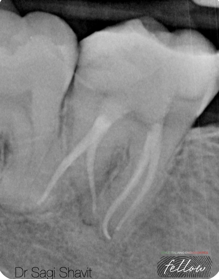
Fig. 1
The patient's primary concern was dental tenderness to pressure on the UR5(15). Initial diagnostic imaging, specifically pre-operative periapical radiographs, demonstrated a distinct periapical lesion contiguous with the root. It was also noted an unusual root morphology.
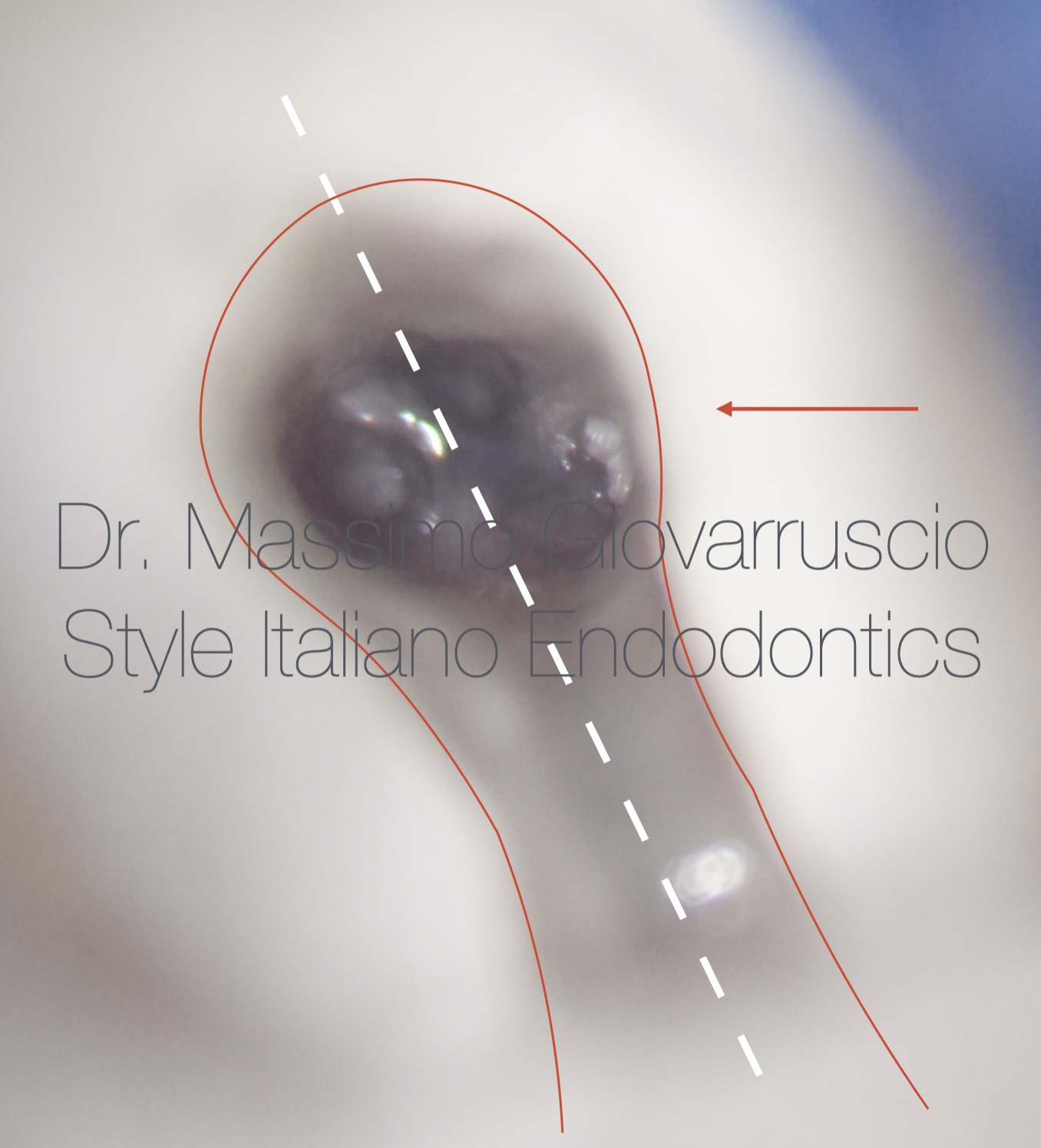
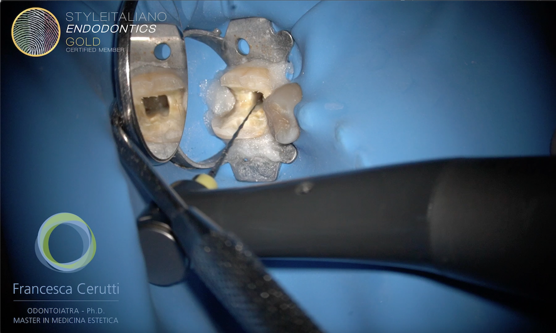
Fig. 2
A critical aspect of treating three-rooted maxillary first premolars involves overcoming the inherent difficulty in locating and accurately preparing the mesiobuccal (MB) and distobuccal (DB) canals. This complexity is often exacerbated when these canals present with a confluent coronal orifice that subsequently bifurcates into distinct canal systems more apically. Such variations necessitate meticulous exploration to prevent missed anatomy and ensure comprehensive disinfection.
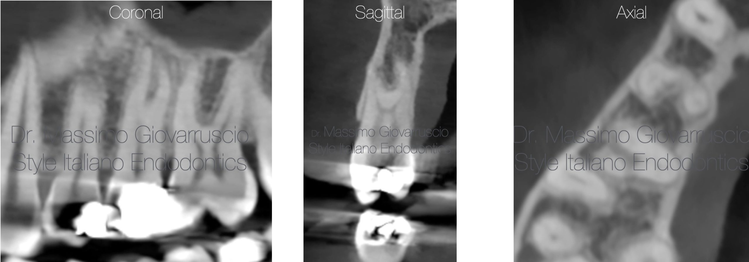
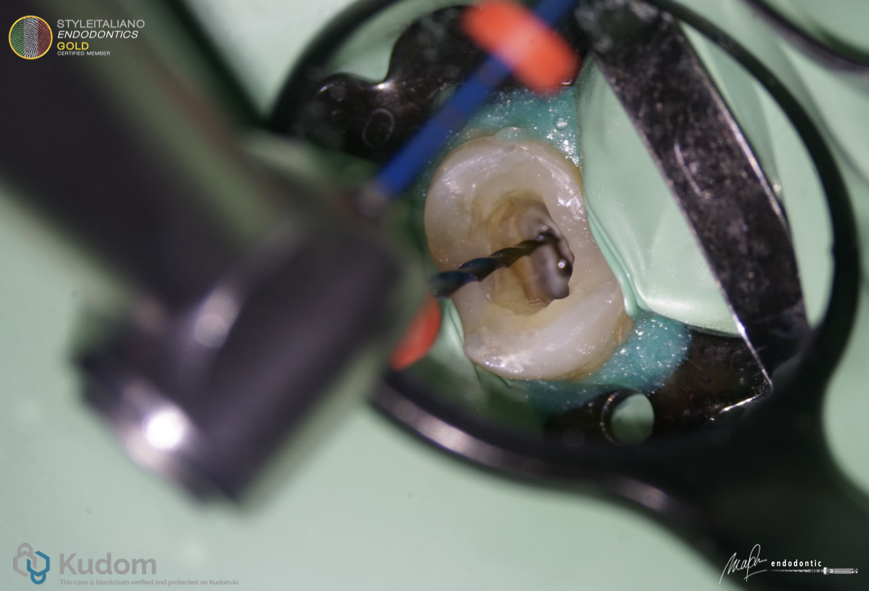
Fig. 3
The acquisition of cone-beam computed tomography (CBCT) images offers a distinct advantage in the precise delineation and comprehensive understanding of the root canal morphology, specifically highlighting the anatomical variations of the mesiobuccal (MB), distobuccal (DB), and palatal (P) canals. Given the observed anatomical complexities, a CBCT scan was acquired.
The resultant images, analyzed across axial, coronal, and sagittal planes, confirmed the presence of a significant anatomical variation. Specifically, the mesiobuccal (MB), distobuccal (DB), and palatal (P) roots were distinctly separated. Furthermore, a notably deep divergence was observed between the orifices of the mesiobuccal and distobuccal canals
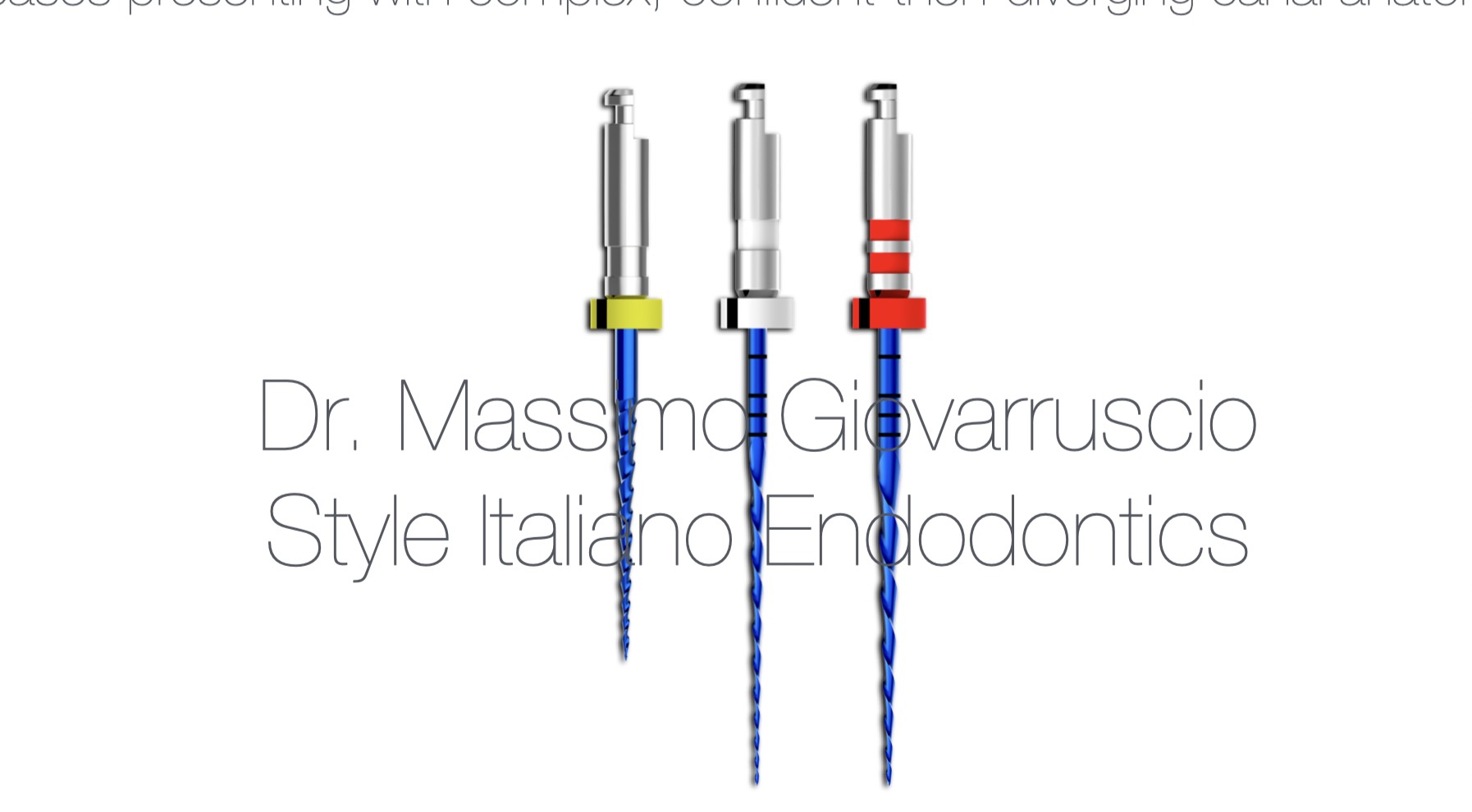
Fig. 4
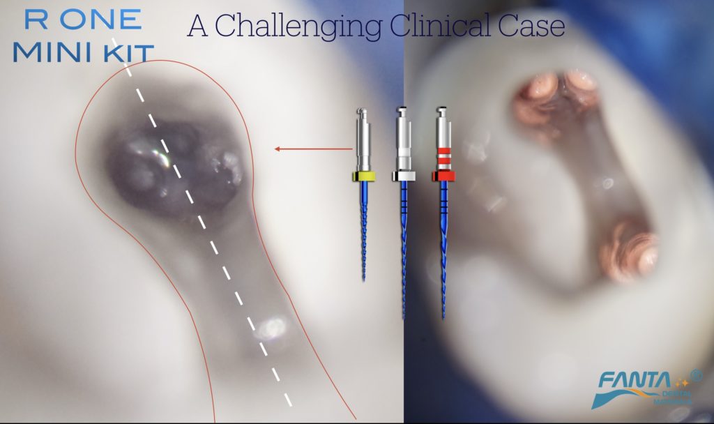
Focus on the Technique
The implementation of contemporary reciprocating file systems, such as the R-One Mini kit, offers a viable solution for navigating these challenging anatomical configurations. Specifically, the inherent flexibility of these files, enhanced by their advanced heat treatment, permits pre-bending of the file tip. This allows for precise manual negotiation and insertion into the separately originating mesiobuccal (MB) or distobuccal (DB) canals. Once the file is seated within the canal, the endodontic motor can be engaged to initiate the reciprocating motion. This approach provides a safe and predictable methodology for achieving comprehensive canal instrumentation in cases presenting with complex, confluent-then-diverging canal anatomy.
Video of the prebending and shaping with the R1 mini kit
Simulation in plastic model
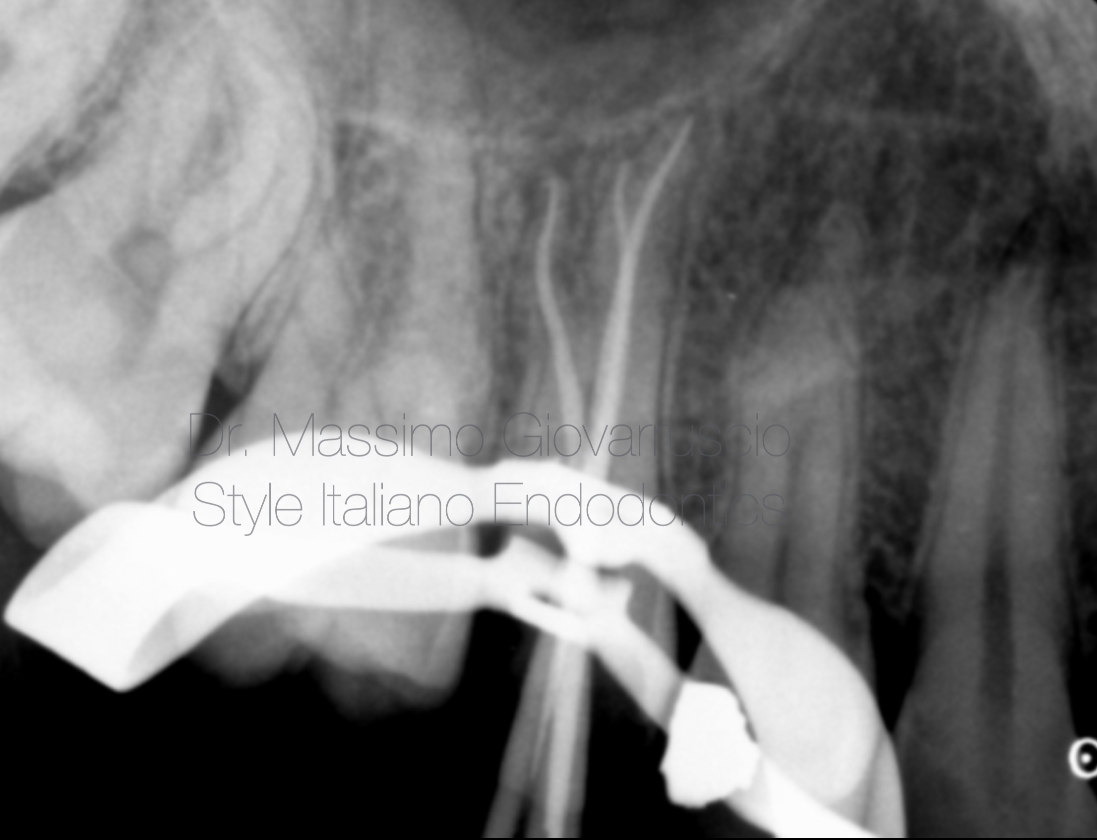
Fig. 5
X- Ray with guttapercha cones
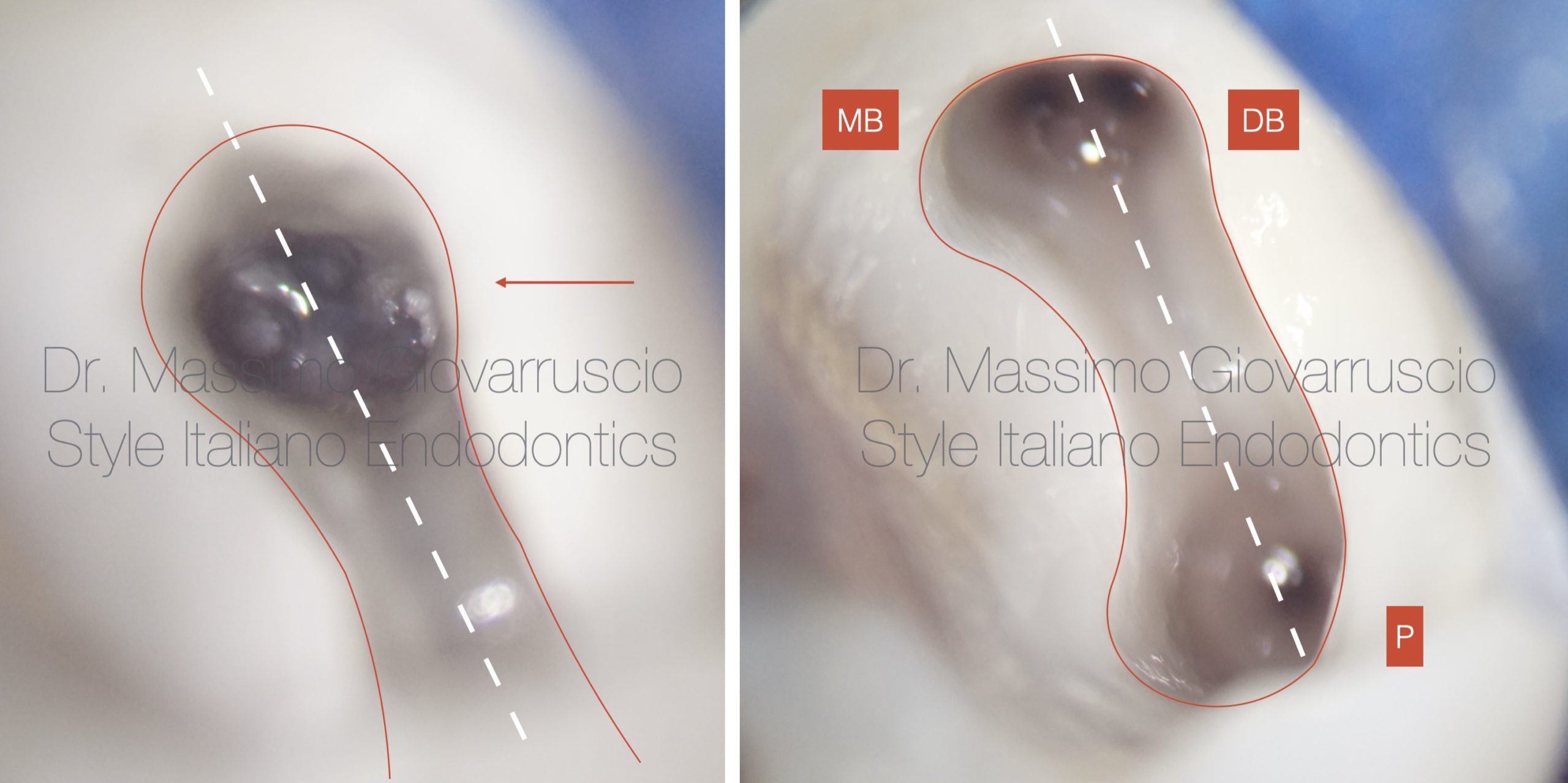
Fig. 6
Clinical image of the orifices
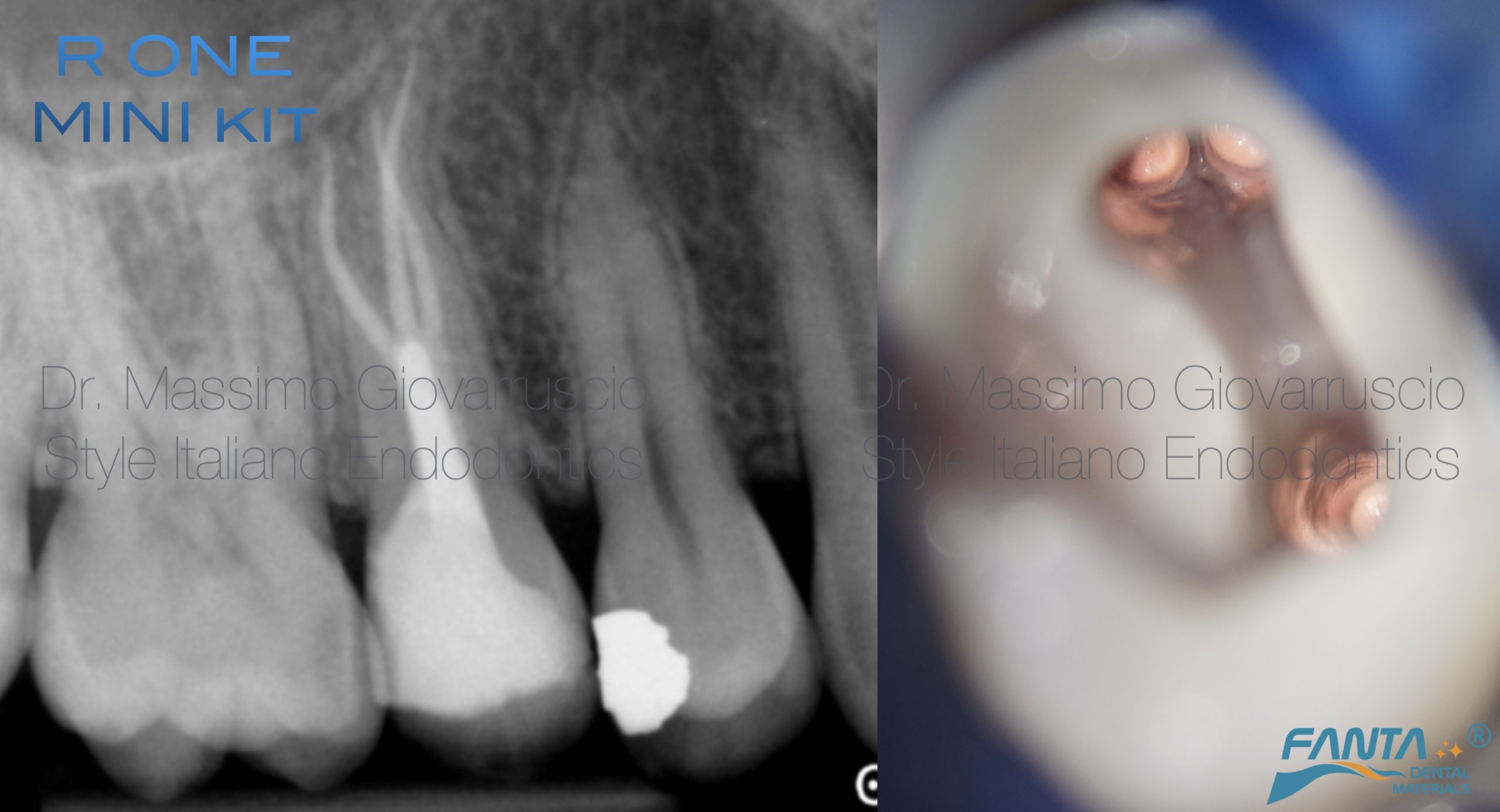
Fig. 7
Final X-ray and image of the pulp chamber prior to composite restoration.
Conclusions
Conclusion
The use of the Reciprocating R-One Mini Kit (Fanta Dental) facilitates the management of complex anatomical variations in endodontic treatment. Owing to advancements in metallurgy, the files are highly flexible, allowing the clinician to pre-bend the tip for improved canal negotiation. This flexibility enables manual insertion of the file into challenging canals prior to connection with the Endodontic motor.
This technique enhances both the predictability and simplicity of the procedure, contributing to more consistent and successful clinical outcomes.
Bibliography
1. Vertucci FJ. Root canal anatomy of the human permanent teeth. Oral Surg Oral Med Oral Pathol. 1984;58:589–99.
2. Vertucci FJ, Gegauff A. Root canal morphology of the maxillary first premolar. J Am Dent Assoc. 1979;99:194–8.
3. Walker RT. Root form and canal anatomy of maxillary first premolars in a southern Chinese population. Dent Traumatol. 1987;3:130–4.
4. Senan, E.M., Alhadainy, H.A., Genaid, T.M. et al. Root form and canal morphology of maxillary first premolars of a Yemeni population. BMC Oral Health 18, 94 (2018). https://doi. org/10.1186/s12903-018-0555-x
5. B. Dadresanfar, Z. Khalilak, and S. Shahmirzadi, “Endodontic treatment of a maxillary first premolar with type IV buccal root canal: a case report,” Iranian Endodontic Journal, vol. 4, no. 1, pp. 35–37, 2009.
6. Sieraski SM, Taylor GN, Kohn RA. Identification and endodontic management of three-canalled maxillary premolars. J Endod 1989;15(1):29–32.
7. Ingle J, Bakland L, Baumgartner JC. Ingle’s Endodontics, ed 6. BC Decker Inc, Hamilton; 2008.