Endodontic microsurgery sometimes is the only way
03/01/2024
Giuseppe Carrieri
Warning: Undefined variable $post in /var/www/vhosts/styleitaliano-endodontics.org/endodontics.styleitaliano.org/wp-content/plugins/oxygen/component-framework/components/classes/code-block.class.php(133) : eval()'d code on line 2
Warning: Attempt to read property "ID" on null in /var/www/vhosts/styleitaliano-endodontics.org/endodontics.styleitaliano.org/wp-content/plugins/oxygen/component-framework/components/classes/code-block.class.php(133) : eval()'d code on line 2
Endodontic microsurgery is a powerful tool for the endodontist because it can help us solving cases that could not be managed in any other way.
When there is a persistent infection despite the presence of a good root canal therapy there is no sense in trying to retreat, because the percentage of success of a retreatment are lower than those of an initial treatment.
In these situation a good pre operative analysis can help us deciding which approach is more appropriate.

Fig. 1
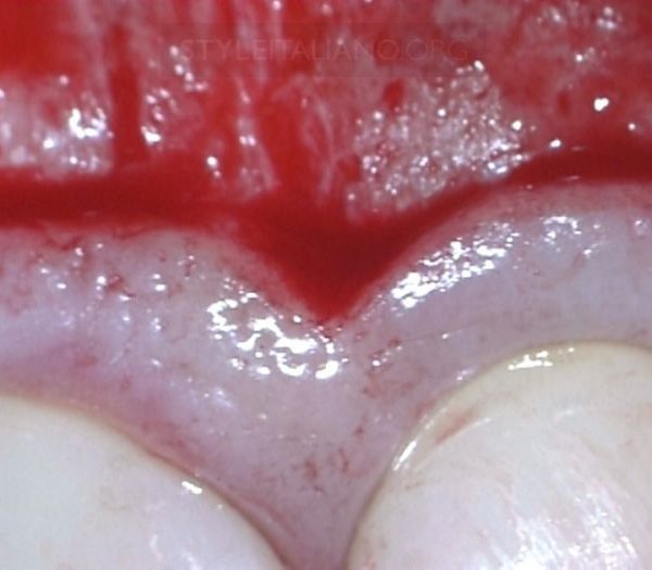
A 28 yers old patient was referred to me due to repeated abscesses on tooth 2.2

Fig. 2
The patient had a sinus tract on 2.2.
The referring dentist asked to make a root canal treatment on tooth 2.1 (negative response to pulp sensitivity test, negative response to electric test) and specification of tooth 2.2

Fig. 3
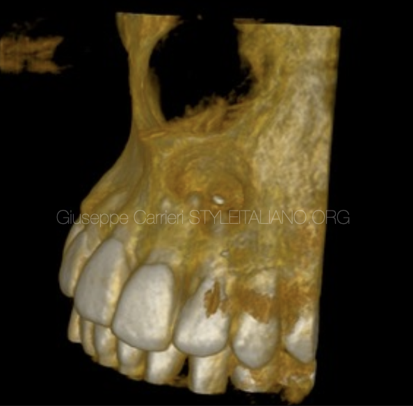
The pre operative CBCT showed the width of the lesion and the size of the apex
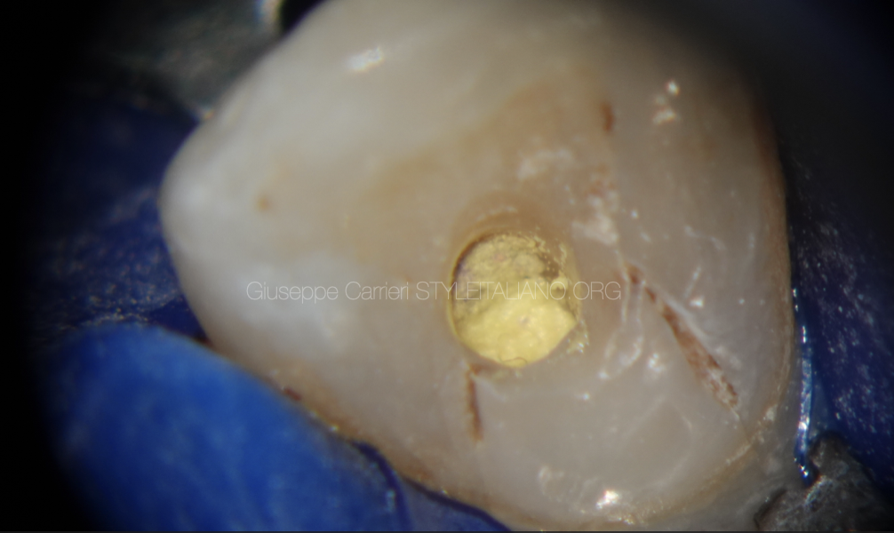
Fig. 4
The access cavity on tooth 2.2 was corrected
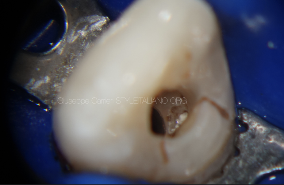
Fig. 5
The temporary root canal filling material was removed
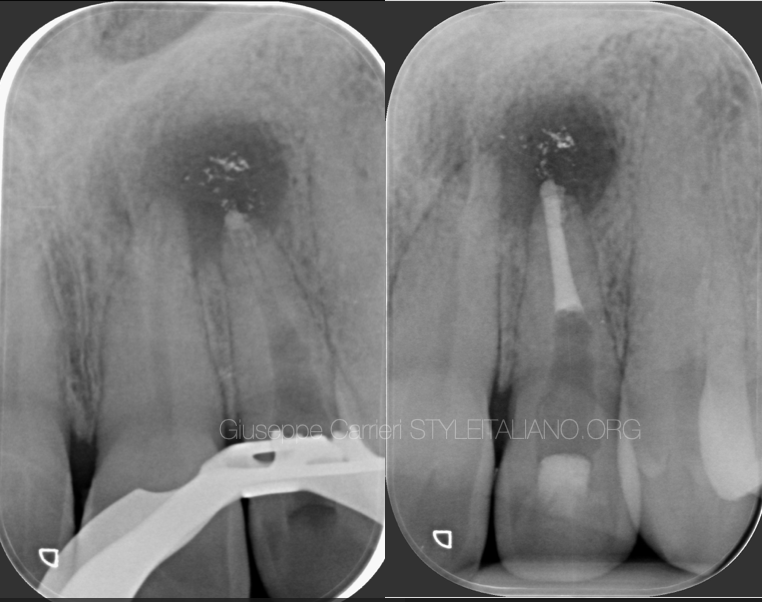
Fig. 6
The intra and post operative X-ray show the removal of the canal dressing and the apical plug with MTA.

Fig. 7
72 hours later the MTA hardening was checked and the root canal therapy was finished.

Fig. 8
In a following appointment the root canal treatment of tooth 2.1 was done
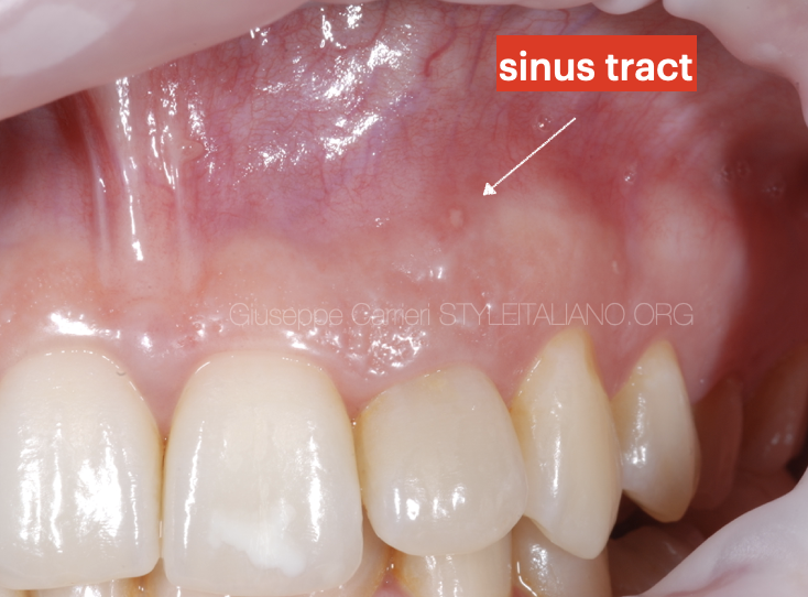
Fig. 9
1 month later the patient still had a sinus tract on tooth 2.2 and in the time between the initial therapy and the recall she had multiple abscessed

Fig. 10
It was decided to treat both incisors with a surgical approach. After raising a flap and removing the lesion, methylene bue helped identifying the anatomy.

Fig. 11
Both apices were resected

Fig. 12
After the retro-preparation, a root end filling material was positioned.

Fig. 13
The material set was checked before finishing the therapy
Video of the procedure
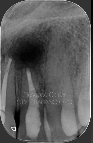
Fig. 14
Post operative X-ray
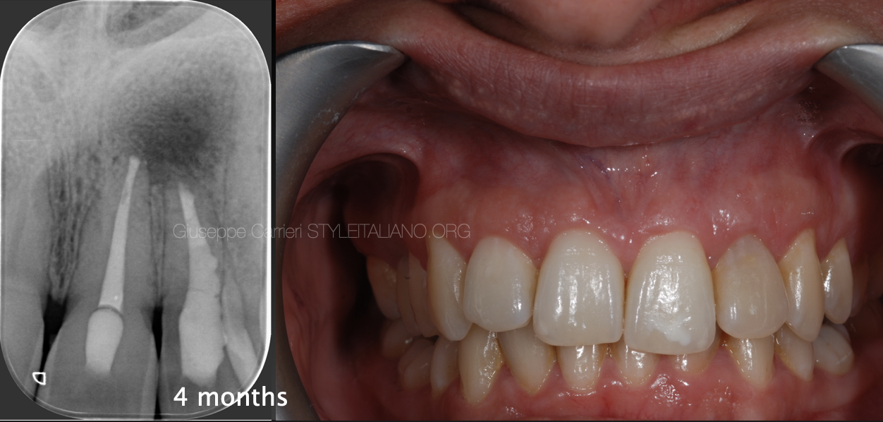
Fig. 15
4 months follow up
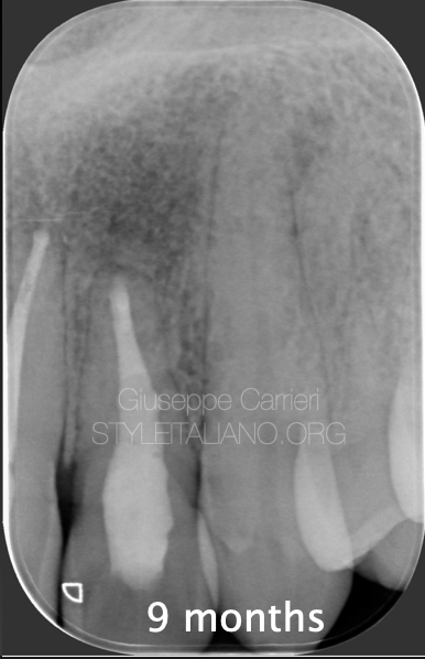
Fig. 16
9 months follow up
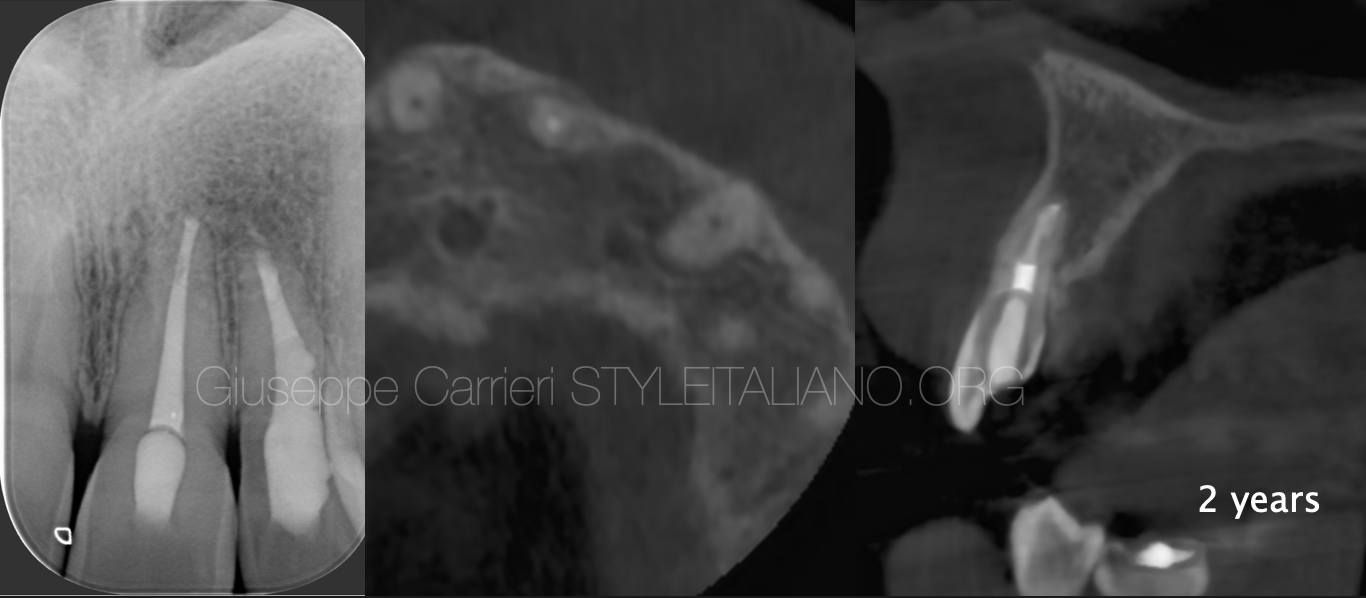
Fig. 17
2 years follow up
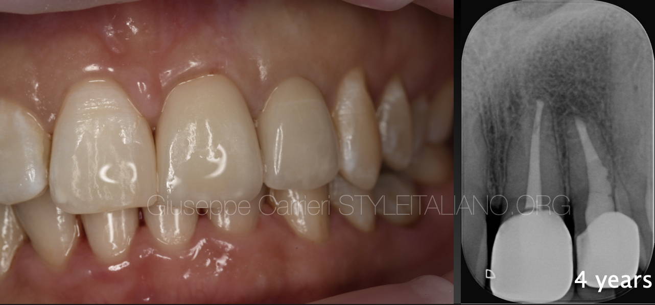
Fig. 18
4 years follow up
Conclusions
A careful analysis of the reasons for failure of the initial treatment help the clinician picking the correct solution for each endodontic case.
Bibliography
Azim AA, Albanyan H, Azim KA, Piasecki L. The Buffalo study: Outcome and associated predictors in endodontic microsurgery- a cohort study. Int Endod J. 2021;54(3):301-18.
Curtis DM, VanderWeele RA, Ray JJ, Wealleans JA. Clinician-centered Outcomes Assessment of Retreatment and Endodontic Microsurgery Using Cone-beam Computed Tomographic Volumetric Analysis. J Endod. 2018;44(8):1251-6.
Eskandar RF, Al-Habib MA, Barayan MA, Edrees HY. Outcomes of endodontic microsurgery using different calcium silicate–based retrograde filling materials: a cohort retrospective cone-beam computed tomographic analysis. BMC Oral Health. 2023;23(1):70.


