Management of C-shaped canal system
21/08/2023
Fellow
Warning: Undefined variable $post in /var/www/vhosts/styleitaliano-endodontics.org/endodontics.styleitaliano.org/wp-content/plugins/oxygen/component-framework/components/classes/code-block.class.php(133) : eval()'d code on line 2
Warning: Attempt to read property "ID" on null in /var/www/vhosts/styleitaliano-endodontics.org/endodontics.styleitaliano.org/wp-content/plugins/oxygen/component-framework/components/classes/code-block.class.php(133) : eval()'d code on line 2
A thorough knowledge of the root canal anatomy and its variations is required for achieving success in root canal therapy, along with diagnosis, treatment planning and clinical expertise, One such variation of the root canal system is the C-shaped canal configuration.
It is termed so because of the C-shaped cross-sectional anatomical configuration of the root and root canal, This condition was described for the first time in literature by Cooke and Cox in 1979, Though most commonly found in mandibular second molars
the C-shaped canal configuration may also occur in mandibular premolars , maxillary molars and mandibular third molars
The C-shaped canal configuration presents with variations in both the number and location of the canal(s), as the canal(s) courses from the coronal to the apical third
The complexity of this canal configuration proves to be a challenge with respect to debridement and obturation and possibly the prognosis during root canal therapy.
Recognition of a C-shaped canal configuration before treatment can facilitate effective management, which will prevent irreparable damage that may put the tooth in severe jeopardy.
The main cause of a C-shaped root is due to the failure of the Hertwig’s epithelial root sheath to fuse on the lingual or buccal root surface .
The roots of molars with C-shaped canals may be conical and fused. For these characteristics, studies suggested that C-shaped root canals could be recognized based on preoperative radiographs .
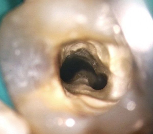
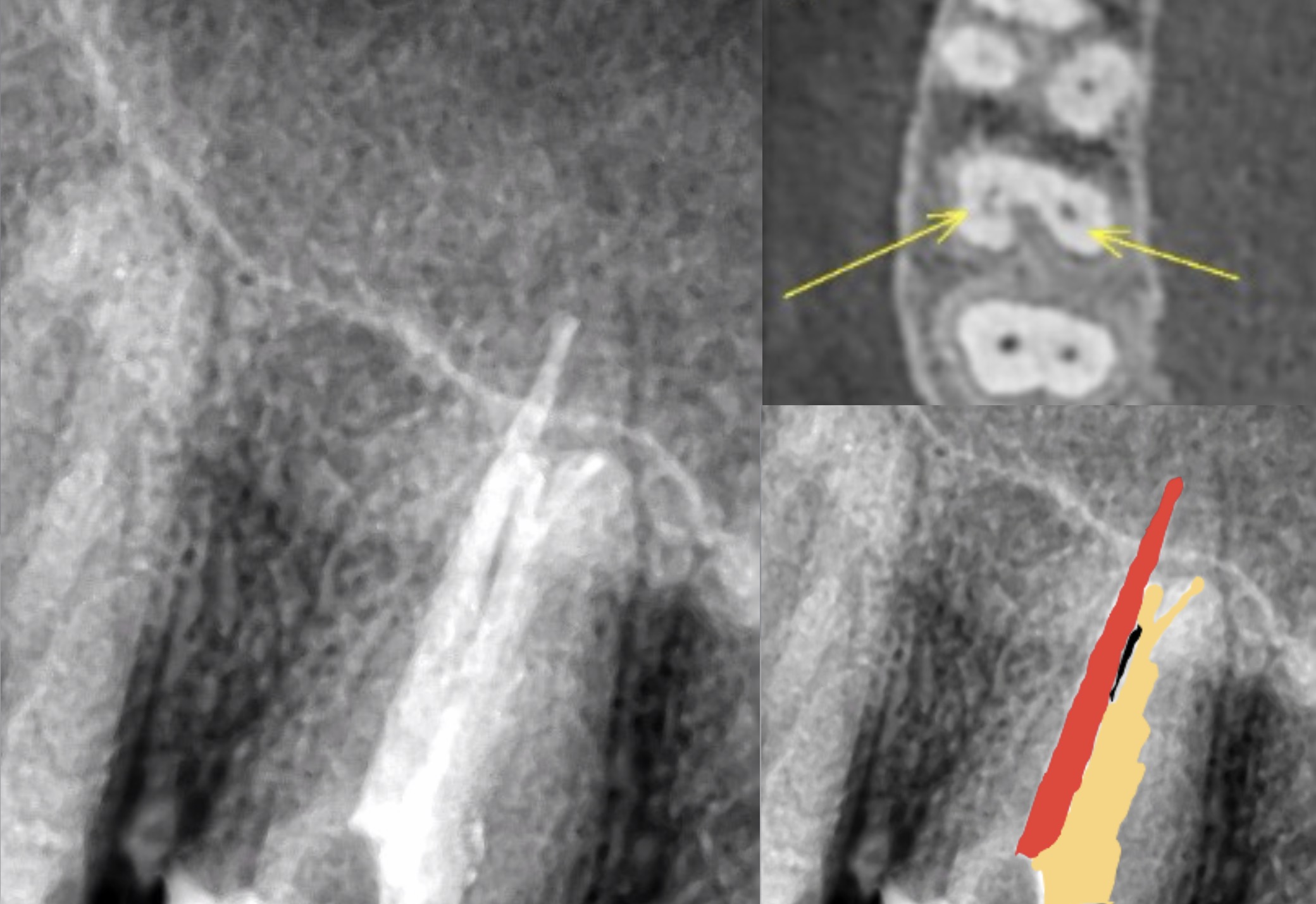
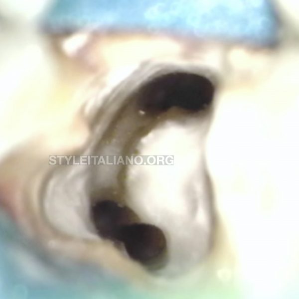
C-shaped canals normally have three canals. The MB and distal canals are interconnected. The ML canal usually is separate. Therefore, we refer to this shape as a C-shaped canal. Sometimes, all three canals are connected in a "horseshoe" type ring. This is called a true C-shaped canal.
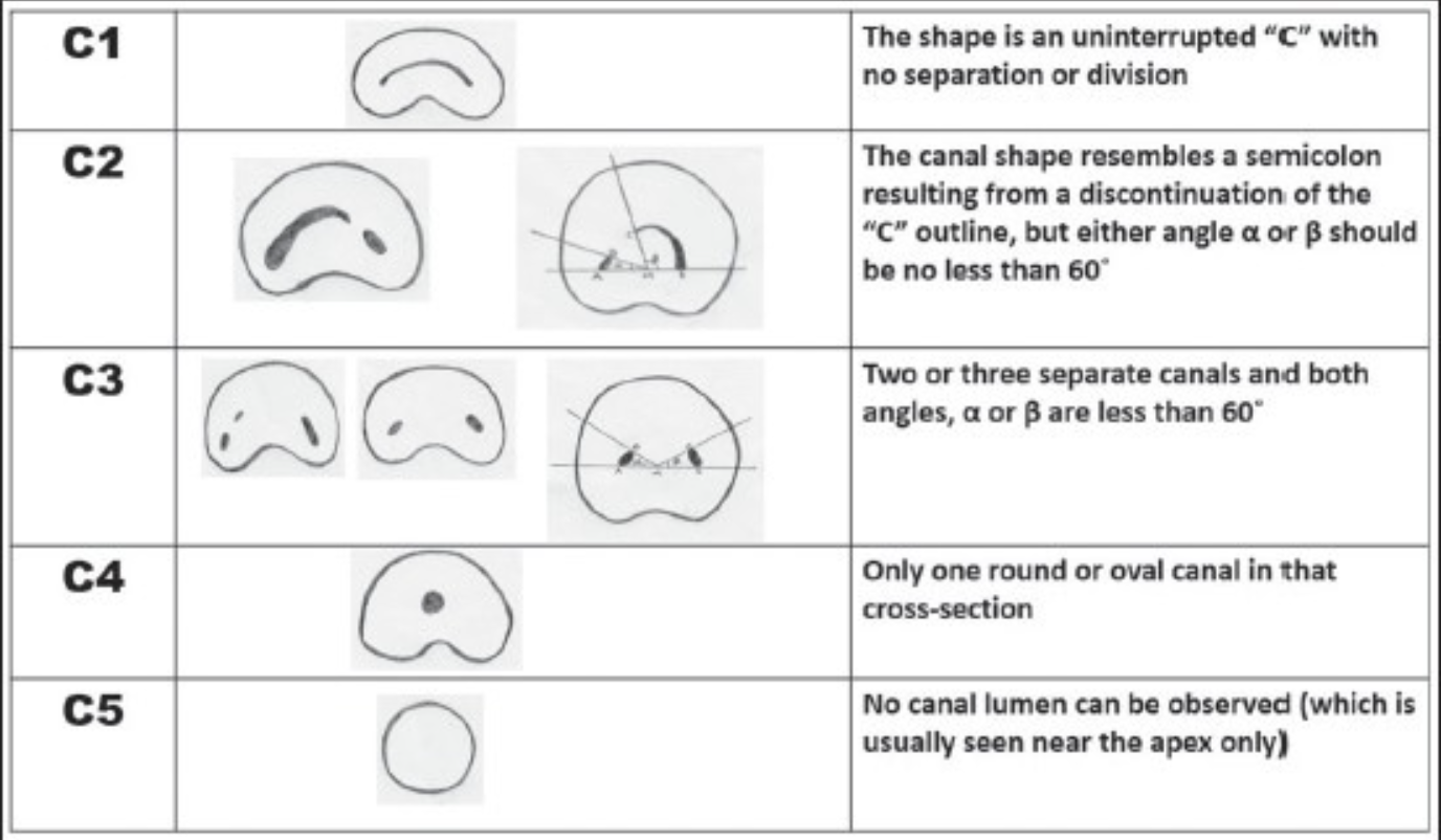
Fig. 1
Classification of C shaped canals
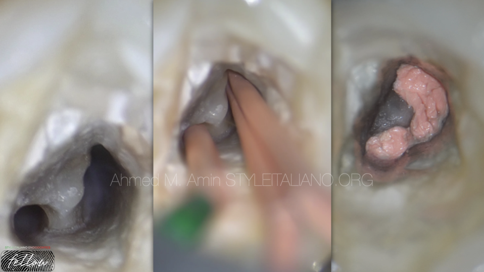
Fig. 2
Case 1
C1, Type 1
A- Access Cavity
B- Cone fitting
C- Final Obturation Resembles C- Shape
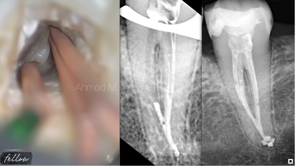
Fig. 3
Down packing and back filling
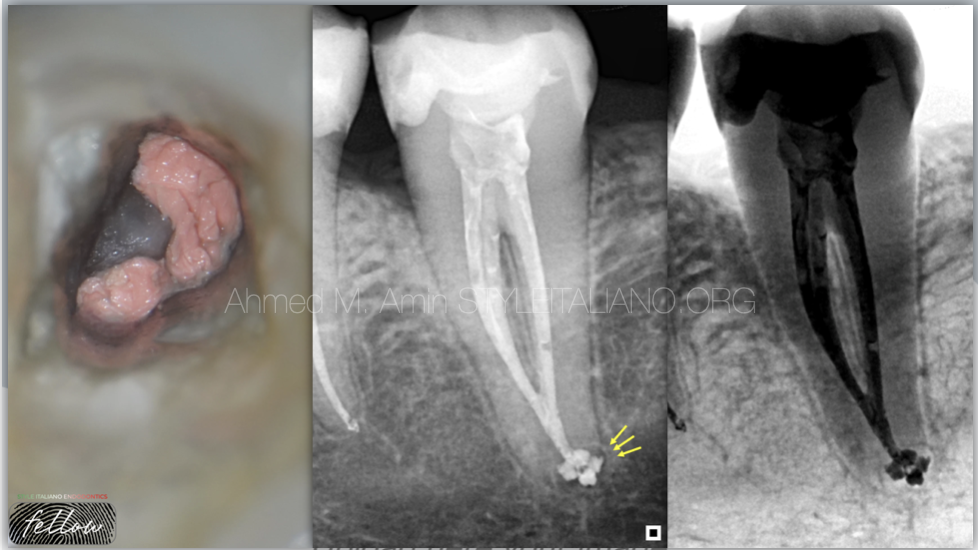
Fig. 4
Notice apical delta after obturation
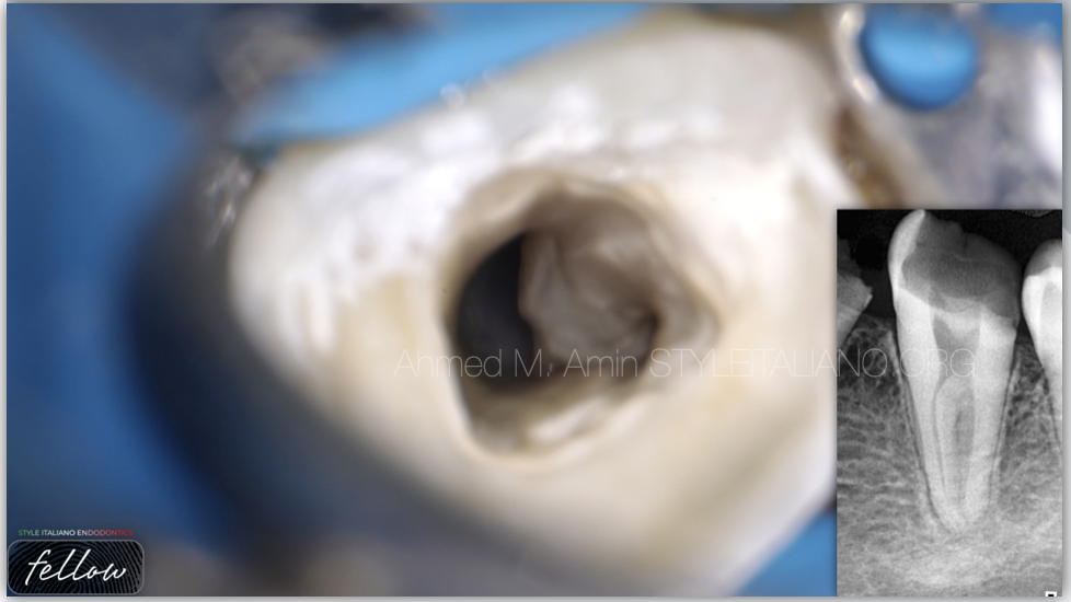
Fig. 5
CASE 2
C2 , Type III
Access Cavity & Pre-OP Xray
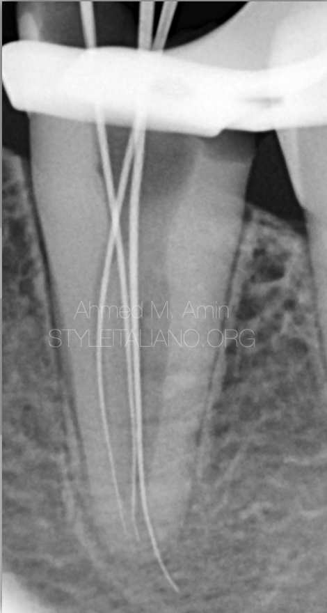
Fig. 6
Working length determination
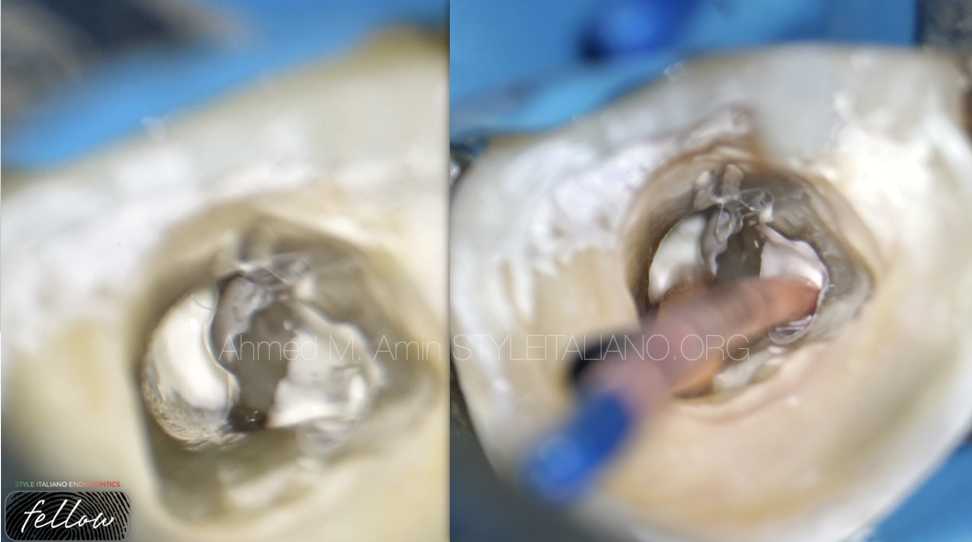
Fig. 7
A- Injecting BC Sealer
B- Cone Fitting
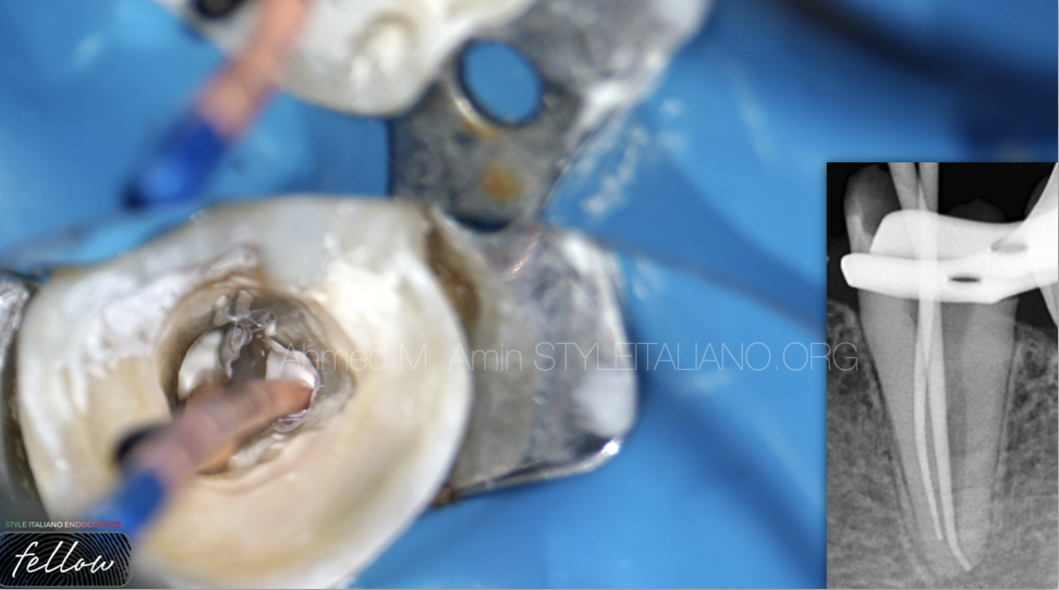
Fig. 8
Cone fitting and clinical X-ray
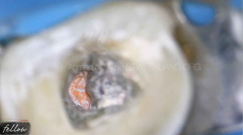
Fig. 9
Down packing
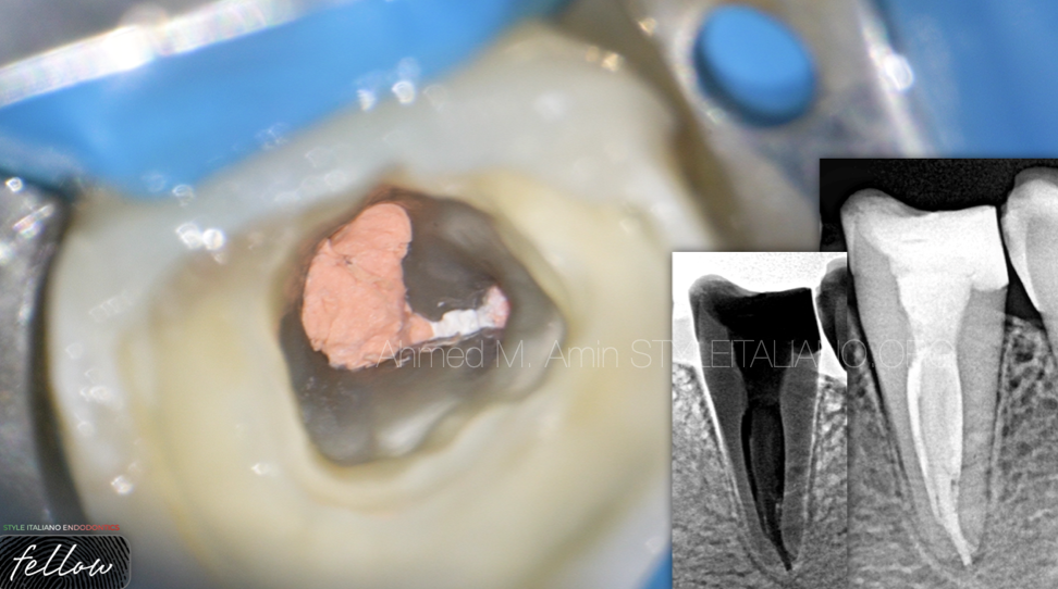
Fig. 10
Final obturation and X-ray
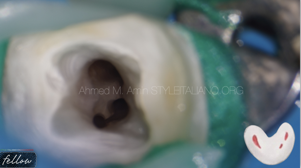
Fig. 11
CASE 3
C3 , Type I
Access Cavity “ with 3 main canal orifices & we can see the isthmus between them clearly “
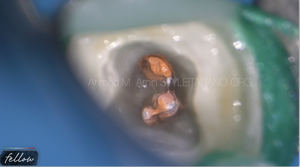
Fig. 12
Down packing
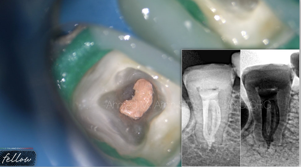
Fig. 13
Final obturation
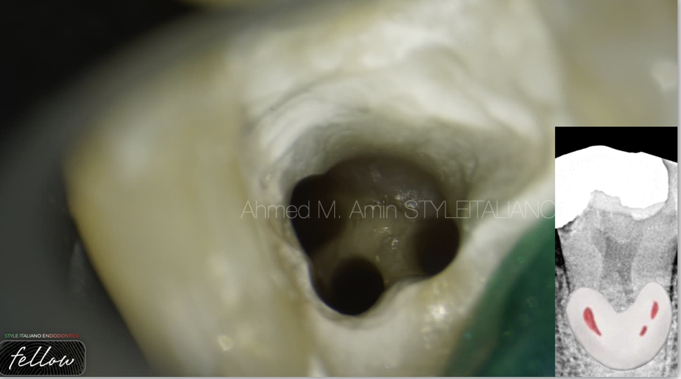
Fig. 14
CASE 4
C3, Type I
Access Cavity
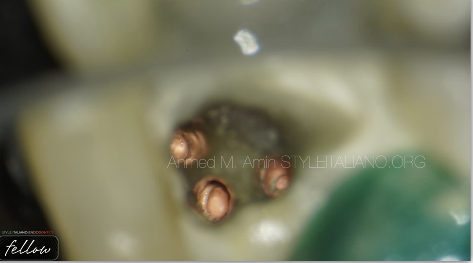
Fig. 15
Down packing
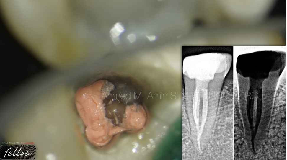
Fig. 16
Final obturation
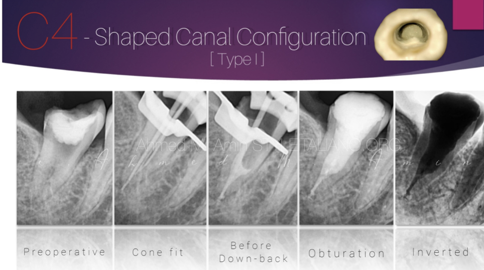
Fig. 17
CASE 5
C4
A- Pre-OP Xray
B- Cone Fitting
C- Down-Packing
D- Obturation
E- Negative
Conclusions
- The C-shaped root canal configuration has an ethnic predilection and a high prevalence rate in mandibular second molars.
-The C-shaped anatomy represents an important and challenging anatomical variation during negotiation, debridement and obturation, Understanding the anatomical presentations of this variation will enable the clinician to manage these cases effectively.
Bibliography
Cooke HG, Cox FL. C-shaped canal configurations in mandibular molars. J Am Dent Assoc 1979; 99: 836-839
Yang ZP, Yang SF, Lin YL. C-shaped root canals in mandibular second molars in Chinese population. Endod Dent Traumatol 1988; 4: 160-163
Manning S. Root canal anatomy of mandibular second molars. Part II C-shaped canals. Int. Endod. J 1990; 23: 40-45
Melton DC, Krell KV, Fuller MW. Histological Anatomical and features of C-shaped canals in mandibular second molars. J Endod 1991; 17: 384-388.
Sabala CL, Benenati FW, Neas BR. Bilateral root or root canal aberra- tions in a dental school patient population. J Endod 1994; 20: 38-42
Haddad GY, Nehme WB, Ounsi HF. Diagnosis, classification, and frequency of C-shaped canals in mandibular ?second molars in the Lebanese population. J Endod 1999; 25: 268-271.
Sidow SJ, West LA, Liewehr FR, Loushine RJ. Root canal morphology of human maxillary and mandibular third molars. J Endod 2000; 26: 675-678
Gulabivala K, Aung TH, Alavi A, Ng YL. Root and canal morphology of Burmese mandibular molars. Int Endod J 2001; 34: 359-370.
Gulabivala K, Opasanon A, Ng YL, Alavi A. Root and canal morphology of Thai mandibular molars. Int Endod J 2002; 35: 56-62.
Fan B, Cheung GS, Fan M, Gutmann JL, Bian Z. C-shaped canal system in mandibular second molars: Part I: Anatomical features. J Endod 2004; 30: 899-903.


