Endo Resto With Dentoclic Fiber Post Build Up
30/06/2022
Marc Habib
Warning: Undefined variable $post in /home/styleendo/htdocs/styleitaliano-endodontics.org/wp-content/plugins/oxygen/component-framework/components/classes/code-block.class.php(133) : eval()'d code on line 2
Warning: Attempt to read property "ID" on null in /home/styleendo/htdocs/styleitaliano-endodontics.org/wp-content/plugins/oxygen/component-framework/components/classes/code-block.class.php(133) : eval()'d code on line 2
In the following clinical case, a maxillary first premolar was presented with invading decay reaching the pulp chamber. The tooth was diagnosed with irreversible pulpitis needing root canal treatment. The pre-operative X-ray revealed a 3 rooted premolar, a special configuration that can be found in 5% of the cases as cited by Vertucci.
Knowing that the anatomy is the ultimate challenge during root canal treatment, special care was taken to preserve tooth structure as much as possible. This was done in order to maximize a conservative approach and use the adhesive technique to core build up and restore this tooth.
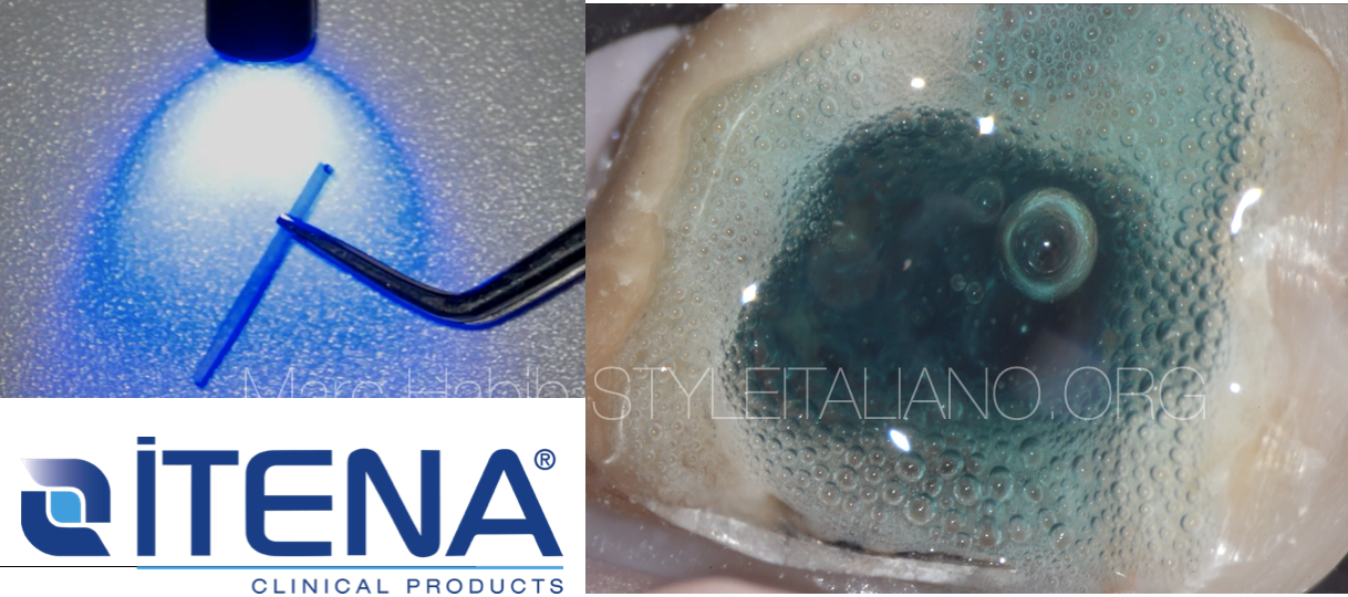
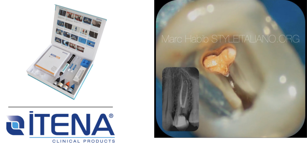
The purpose of this clinical Video is to highlight the possible management of core build up of endodontically treated premolar teeth with ITENA Clinical’s Corono-Radicular Restoration Kit.
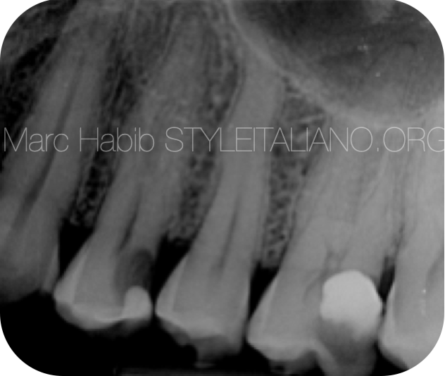
Fig. 1
Pre Operative X ray
The patient presented to the clinic with deep caries on tooth 24
Primary root canal treatment is scheduled
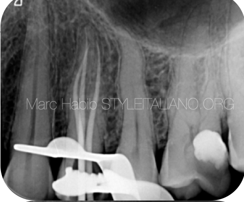
Fig. 2
Treatment planning to restore this maxillary first premolar includes first a root canal retreatment.
The X-ray taken during the root canal treatment shows the Master cones check
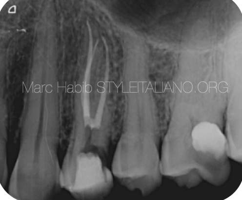
Fig. 3
Post Operative X-ray taken the day of the treatment.
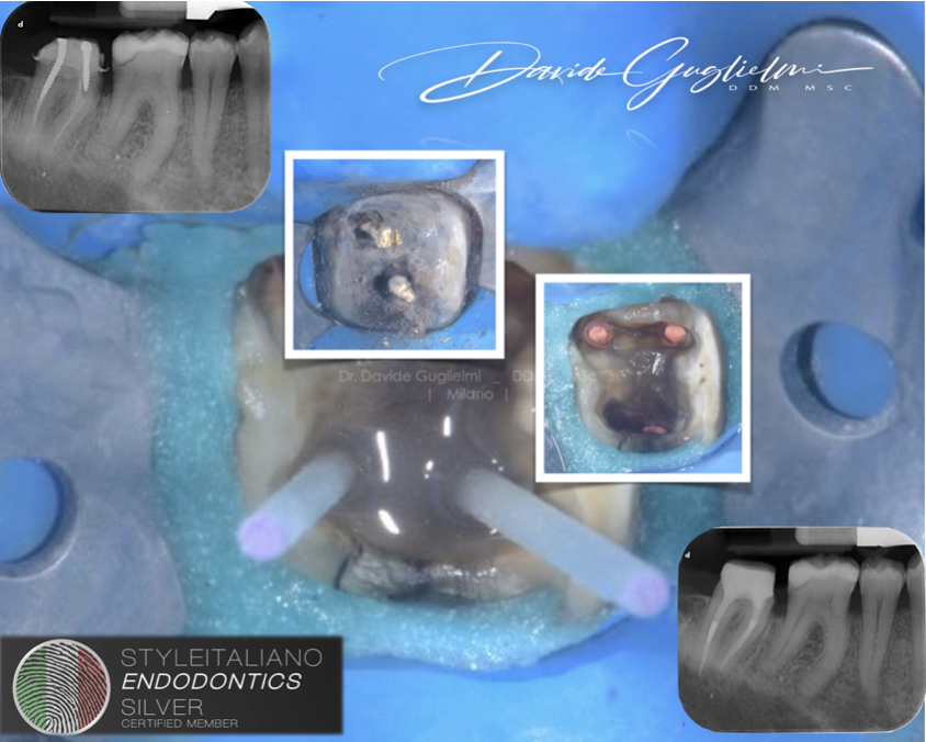
Step by step post endo restoration
Conclusions
In summary, Itena Clinical's resin core build up material and fiber posts can provide excellent restoration results if managed properly. Adequate steps for dentine or enamel preparation before and during bonding should be carried out for optimum result. Moreover, the clinician should respect all stages of the adhesive techniques taking special care for the time frame during etching and bonding of the tooth structures.
Bibliography
- Bhuva B, Giovarruscio M, Rahim N, Bitter K, Mannocci F. The restoration of root filled teeth: a review of the clinical literature. Int Endod J. 2021 Apr;54(4):509-535. doi: 10.1111/iej.13438. Epub 2021 Jan 5. PMID:33128279.
- Figueiredo FE, Martins-Filho PR, Faria-E-Silva AL. Do metal post-retained restorations result in more root fractures than fiber post-retained restorations? A systematic review and meta-analysis. J Endod. 2015 Mar;41(3):309-16. doi: 10.1016/j.joen.2014.10.006. Epub 2014 Nov 11. PMID: 25459568.
- Tay, F. R., & Pashley, D. H. (2007). Monoblocks in Root Canals: A Hypothetical or a Tangible Goal. Journal of Endodontics, 33(4), 391–398. doi:10.1016/j.joen.2006.10.00
- Scotti, N., Forniglia, A., Bergantin, E., Paolino, D. S., Pasqualini, D., & Berutti, E. (2013). Fibre post adaptation and bond strength in oval canals. International Endodontic Journal, 47(4), 366–372. doi:10.1111/iej.12156
- Dietschi D, Duc O, Krejci I, Sadan A. Biomechanical considerations for the restoration of endodontically treated teeth: a systematic review of the literature. Part II: evaluation of fatigue behavior, interfaces, andin vivo studies. Quintessence Int 2008;39:117—29.


