Deep Margin Elevation in an endodontically treated premolar: a conservative restorative strategy
09/11/2026
Warning: Undefined variable $post in /home/styleendo/htdocs/styleitaliano-endodontics.org/wp-content/plugins/oxygen/component-framework/components/classes/code-block.class.php(133) : eval()'d code on line 2
Warning: Attempt to read property "ID" on null in /home/styleendo/htdocs/styleitaliano-endodontics.org/wp-content/plugins/oxygen/component-framework/components/classes/code-block.class.php(133) : eval()'d code on line 2
Deep cervical margins remain among the most demanding situations in adhesive restorative dentistry. When a proximal lesion extends below the gingival margin, access, rubber dam isolation, matrix adaptation, bonding procedures and removal of restorative excesses become increasingly difficult. Deep margin elevation (DME), also described as cervical margin relocation, addresses this problem by using a bonded restorative material to move the operative margin coronally in carefully selected cases [1,2].
The rationale is not to make a deep defect disappear, but to transform an inaccessible adhesive interface into one that can be visualized, finished and maintained more predictably. This may be particularly relevant when endodontic treatment and substantial coronal reconstruction must be coordinated within the same restorative plan.
DME is nevertheless not a substitute for periodontal diagnosis. The position of the cervical margin relative to the supracrestal tissue attachment, the ability to obtain a reliable matrix seal and complete moisture control, and the quality of the final emergence profile all determine whether the technique is appropriate [3,4].
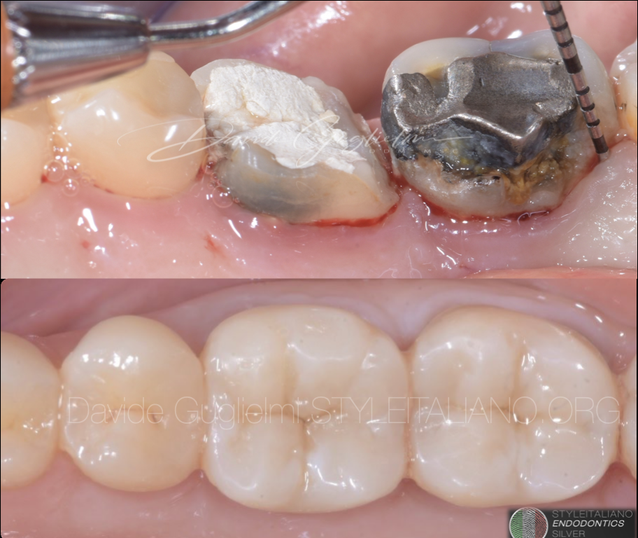
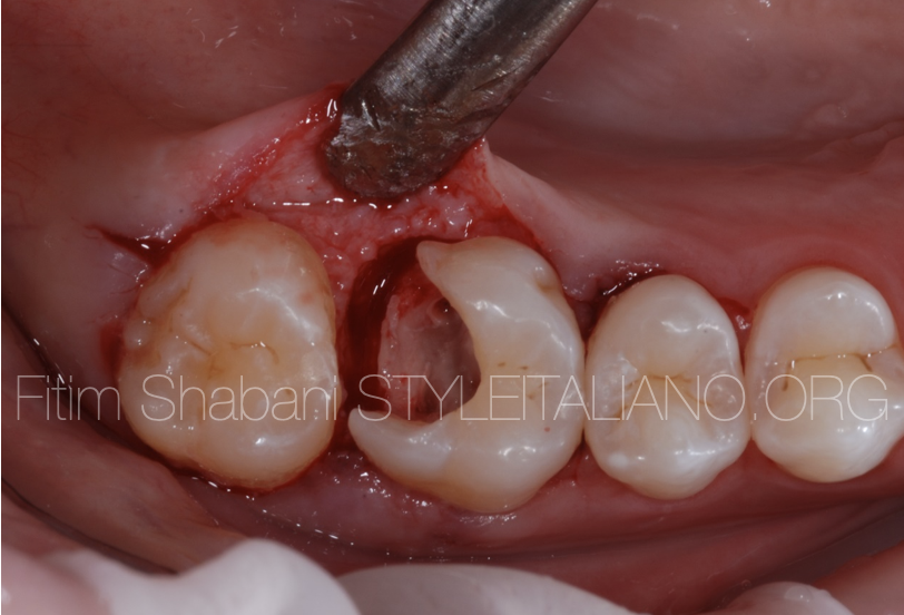
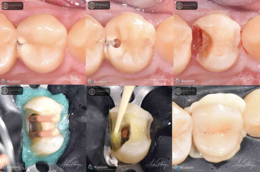
The photographic sequence illustrates the management of a premolar with a deep proximal defect in the context of endodontic treatment and subsequent adhesive reconstruction. The initial occlusal image is followed by the radiographic assessment and by clinical views showing the considerable loss of proximal tooth structure after access and preparation.
Once the defect is exposed, restorability must be assessed in both technical and biological terms. The fact that a margin can be reached with an instrument is not sufficient: it must be possible to isolate it, adapt a stable matrix to the entire cervical perimeter and respect the supracrestal tissues. Where those conditions are absent, surgical crown lengthening, orthodontic extrusion or an alternative treatment plan may be more appropriate.
The following images document preparation and endodontic stages under rubber dam isolation. Endodontic and restorative objectives are closely related: an adequate field supports treatment quality, while a coronal seal and thoughtful reconstruction contribute to the overall prognosis of the tooth.
When case selection supports DME, the first priority is complete removal of the diseased or defective substrate and verification of the actual cervical boundary. The matrix system must engage and seal the deepest margin without trapping gingival tissue or rubber dam between the band and the tooth. Matrix geometry, support from the remaining walls, and careful management of wedges or adjunctive sealing techniques are more important than the choice of a particular commercial band [2,3].
After adhesive pretreatment according to the selected system, a resin composite is used to relocate the margin coronally in controlled increments. The operator should avoid overhangs, voids, and excessively convex emergence profiles; finishing, polishing and removal of excess material are integral to the technique rather than optional cosmetic steps. A highly filled flowable composite may be convenient in suitable situations, but current clinical evidence does not establish one resin-composite category as universally superior for DME [7].
The supplied operative photographs show an adhesive reconstruction phase and the resulting occlusal morphology. The dedicated matrix-adaptation and cervical relocation step is not separately documented in the high-resolution images provided; its precise material and sequence should be confirmed in the final editorial version of this case report
Relocating a cervical margin does not relocate the periodontal attachment. The original tooth–restoration interface remains deep and must be smooth, well sealed and biologically acceptable. Appropriate patient selection, plaque control and periodontal maintenance are therefore fundamental. A 2024 prospective controlled study found increased bleeding on probing at DME sites compared with control surfaces after one year, despite no significant change in probing depths [5].
A 2025 scoping review reported encouraging restorative outcomes while also highlighting inconsistent periodontal findings and limited long-term evidence [6]. More recent pooled clinical data support DME as a conservative option in selected posterior restorations, without demonstrating a clear clinical advantage for a specific polymer-based elevation material [7]. These findings argue for careful execution and monitoring rather than unqualified claims of biological neutrality.
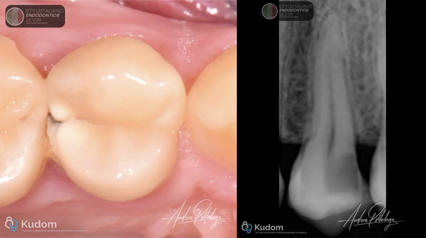
Fig. 1
Pre Op Xray and clinical situation
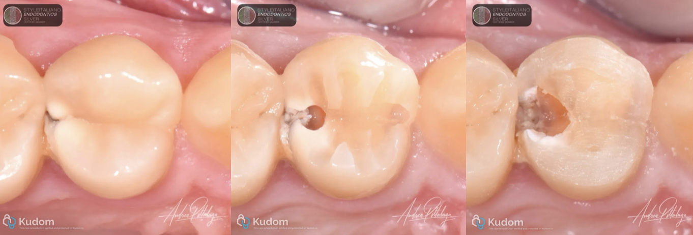
Fig. 2
Cavity Prep
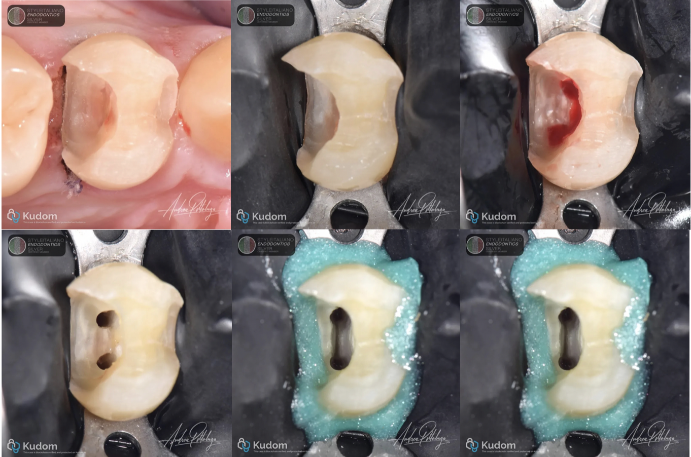
Fig. 3
Isolation and access cavity
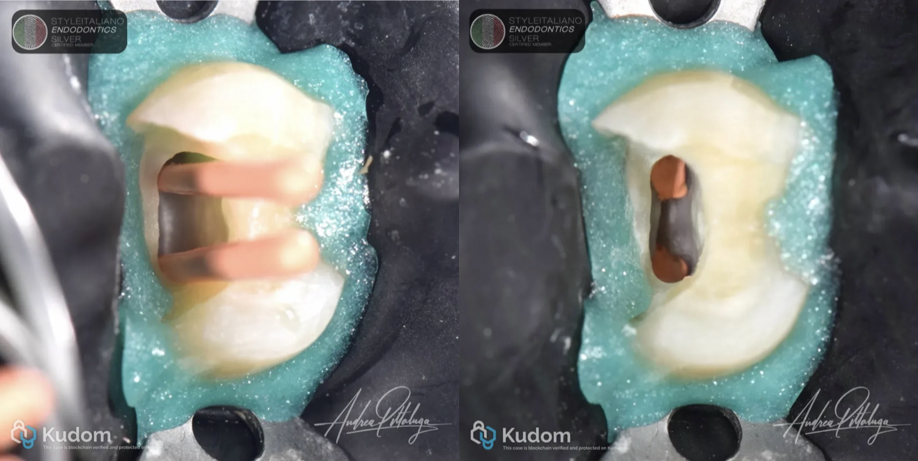
Fig. 4
Cone fit and 3d obturation
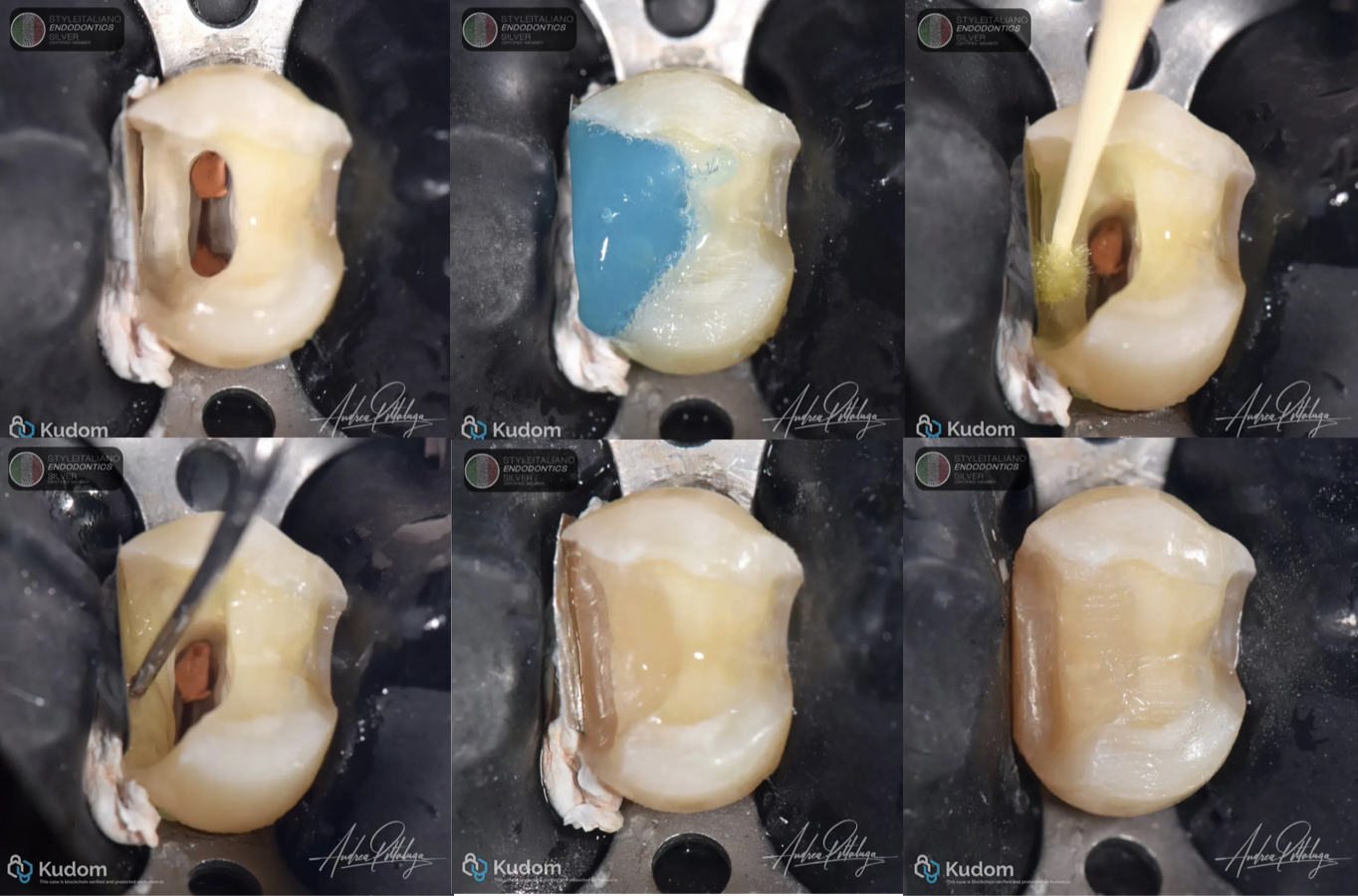
Fig. 5
Adhesion steps
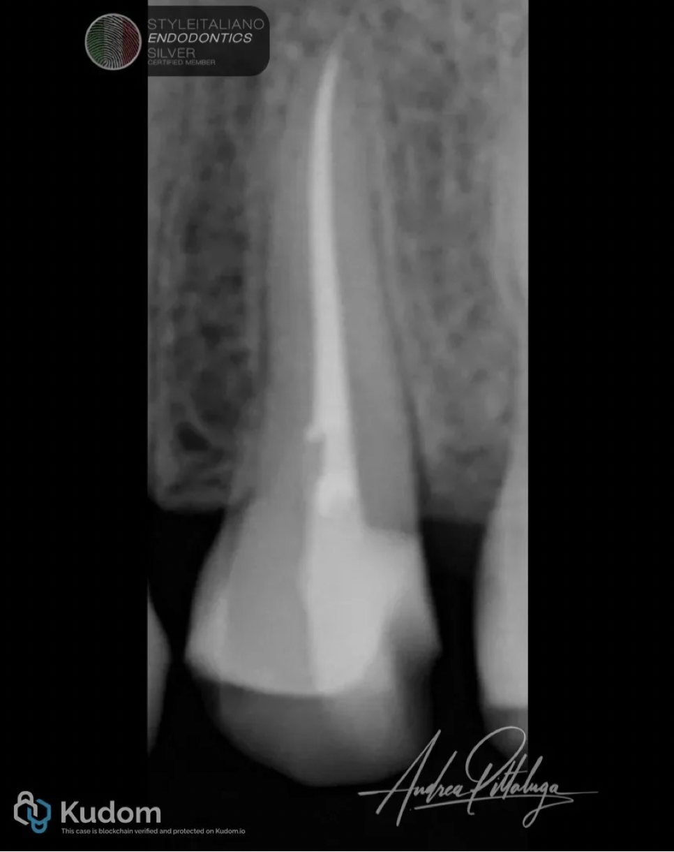
Fig. 6
Final x ray
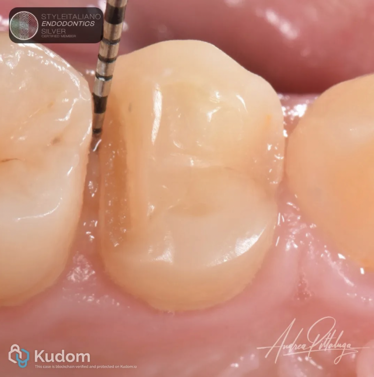
Fig. 7
Two weeks probing control
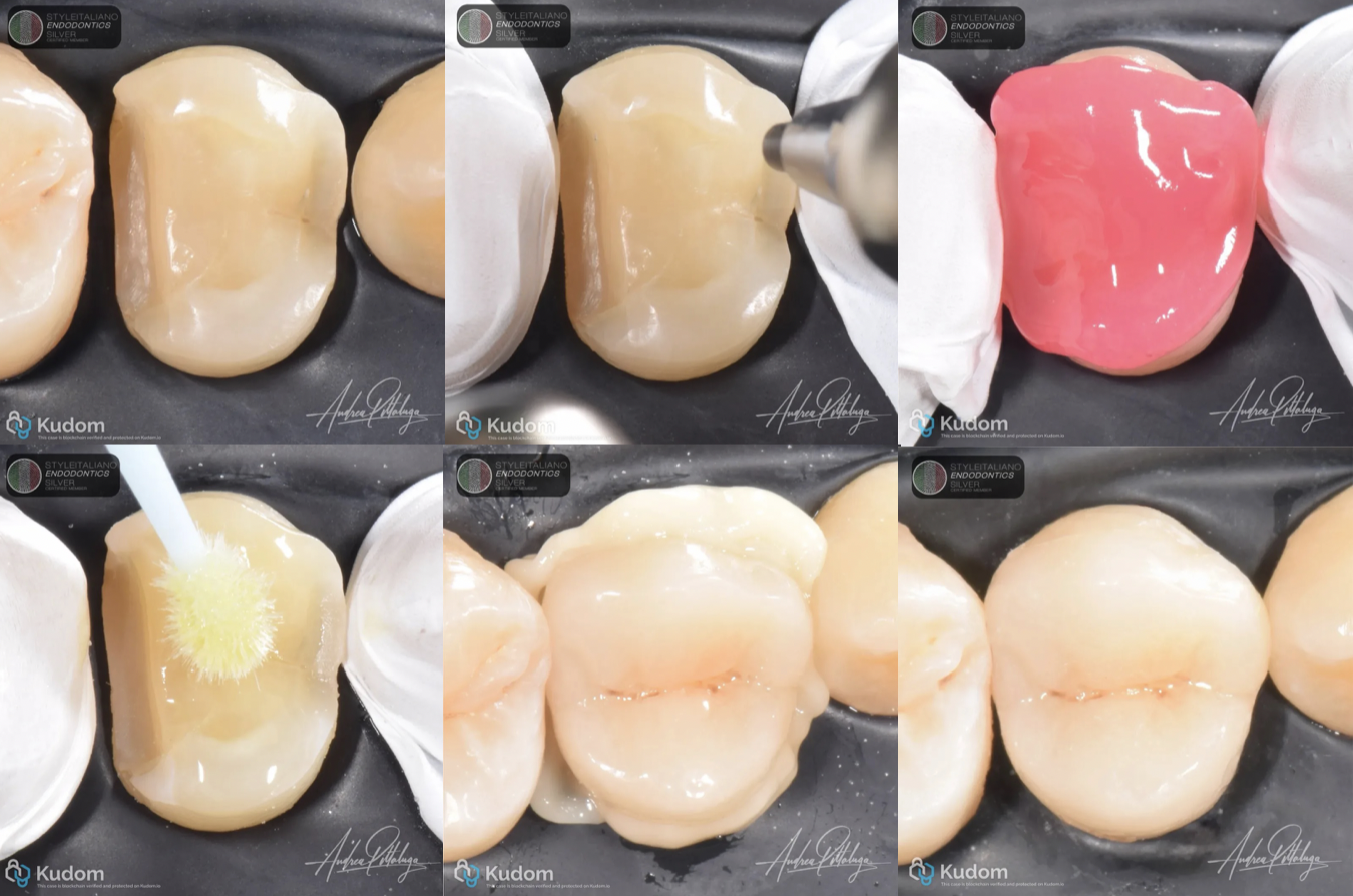
Fig. 8
Bonding steps
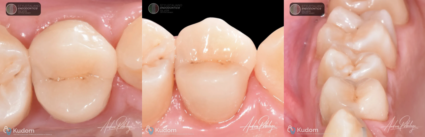
Fig. 9
Final result
Conclusions
Deep margin elevation is best regarded as a case-dependent restorative strategy, not a universal solution for every subgingival lesion. Its value lies in creating a more accessible adhesive margin while preserving dental tissues and enabling the endodontic and restorative phases to be planned as a coherent whole.
Three decisions are decisive: whether the margin can be isolated and perfectly adapted by a matrix; whether its relationship with the supracrestal attachment is biologically acceptable; and whether the relocated interface can be finished, cleaned and maintained over time. The photographically documented reconstruction illustrates the restorative challenge, while a complete report of the DME-specific procedure and periodontal recall is needed to establish the clinical outcome of this individual case.
Bibliography
[1] Dietschi D, Spreafico R. Current clinical concepts for adhesive cementation of tooth-colored posterior restorations. Pract Periodontics Aesthet Dent. 1998;10(1):47–54. PMID: 9582662.
[2] Magne P, Spreafico RC. Deep margin elevation: a paradigm shift. Am J Esthet Dent. 2012;2:86–96.
[3] Magne P. M-i-M for DME: matrix-in-a-matrix technique for deep margin elevation. J Prosthet Dent. 2023;130(4):434–438. doi:10.1016/j.prosdent.2021.11.021.
[4] Eggmann F, Ayub JM, Conejo J, Blatz MB. Deep margin elevation—present status and future directions. J Esthet Restor Dent. 2023;35(1):26–47. doi:10.1111/jerd.13008.
[5] Hausdörfer T, Lechte C, Kanzow P, Rödig T, Wiegand A. Periodontal health in teeth treated with deep-margin-elevation and CAD/CAM partial lithium disilicate restorations—a prospective controlled trial. Clin Oral Investig. 2024;28:670. doi:10.1007/s00784-024-06053-y.
[6] do Nascimento Santos JV, et al. Deep margin elevation and its influence on periodontal health and the longevity of indirect restorations—a scoping review. J Esthet Restor Dent. 2025;37(11):2379–2399. doi:10.1111/jerd.13510.
[7] Bourgi R, et al. Polymer-based restorative outcomes following deep margin elevation: a systematic review and meta-analysis. Front Dent Med. 2026;7:1854285. doi:10.3389/fdmed.2026.1854285.