Expect the unexpected : Four canals in mandubular premolar
10/09/2026
Warning: Undefined variable $post in /home/styleendo/htdocs/styleitaliano-endodontics.org/wp-content/plugins/oxygen/component-framework/components/classes/code-block.class.php(133) : eval()'d code on line 2
Warning: Attempt to read property "ID" on null in /home/styleendo/htdocs/styleitaliano-endodontics.org/wp-content/plugins/oxygen/component-framework/components/classes/code-block.class.php(133) : eval()'d code on line 2
According to Vertucci, mandibular premolars have one single canal in approximately 74% of the cases. On occasion, mandibular premolars have two canals, in around 25.5%, but higher numbers are very uncommon.
Finding four separate canals is extremely rare anatomical variation. The canals are classified as mesiobuccal, distobuccal, mesiolingual and distolingual canal. This significantly increases the complexity of endodontic treatment. Use of magnification, such as dental operating microscope, is highly recommended. Since untreated canals are the main reason of root canal treatment failure, when there is uncertainty regarding the number of canals clinicians should consider using CBCT in order to predictably locate all the canals. Once the canals are located, shaping, cleaning and obturation of the canals should be straighforward.
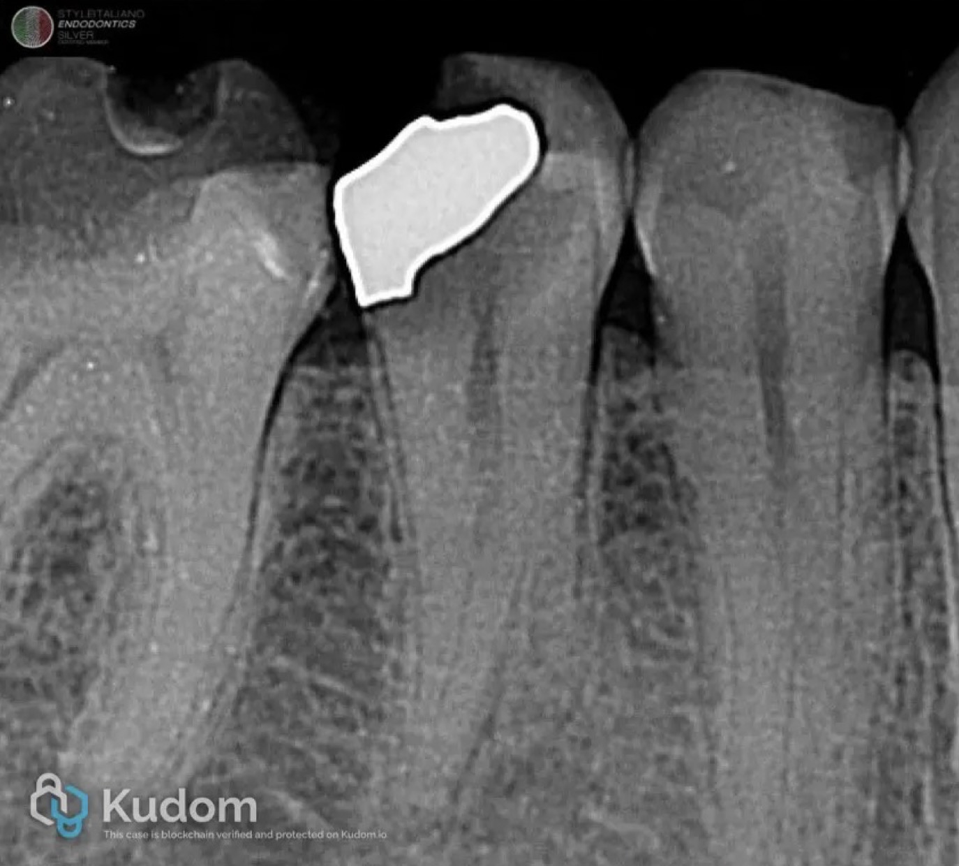
Fig. 1
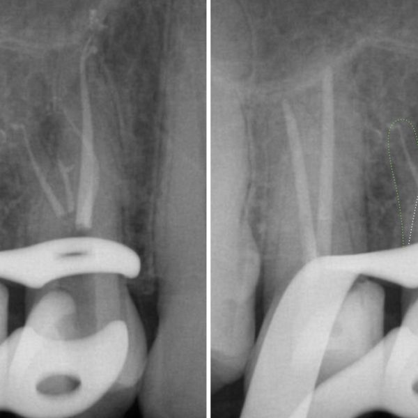
Pre operative radiograph of mandibular second premolar. Note the two separate roots and potentially complex canal anatomy.
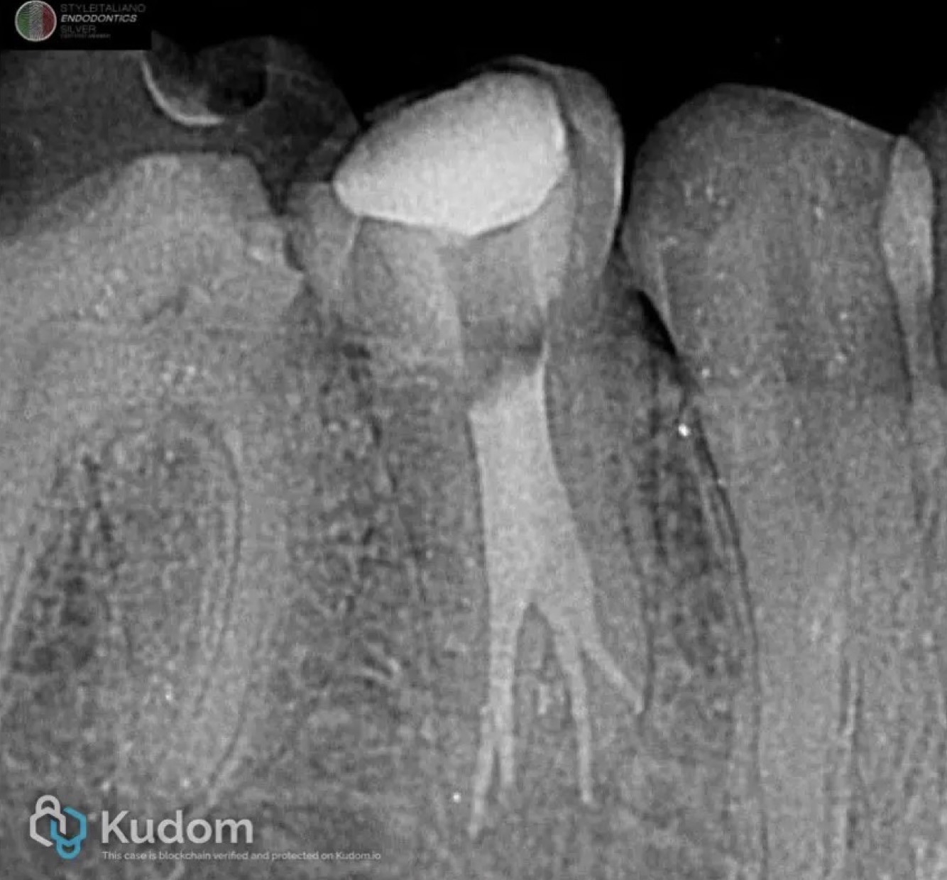
Fig. 2
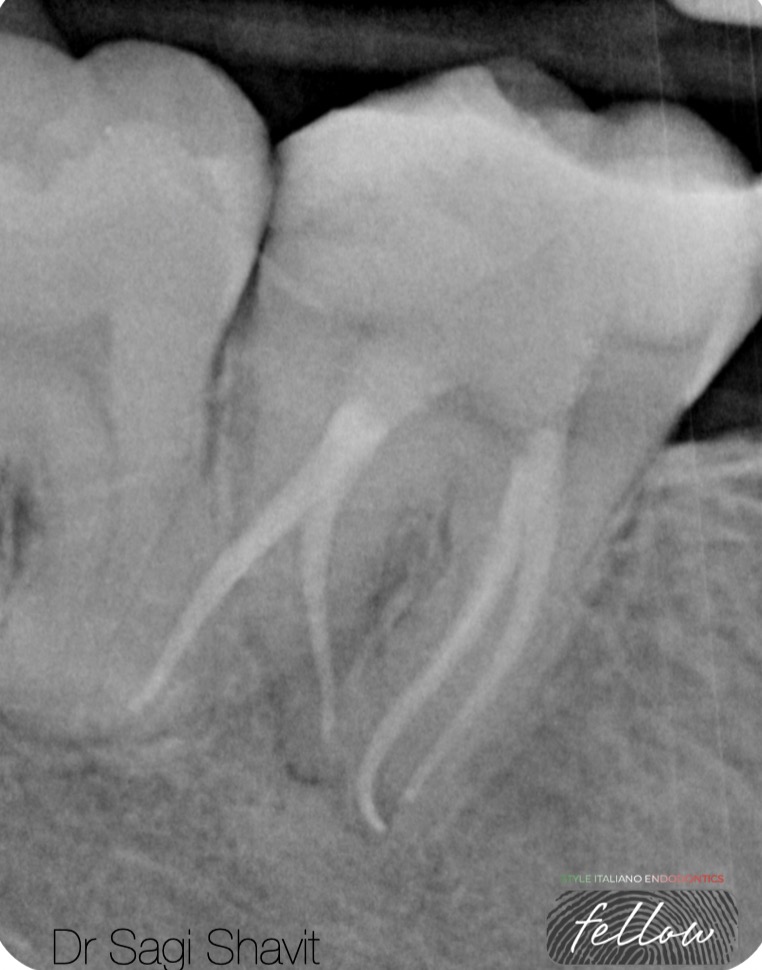
Post operative radiograph demonstrates four separate canals, two buccal and two lingual canals.
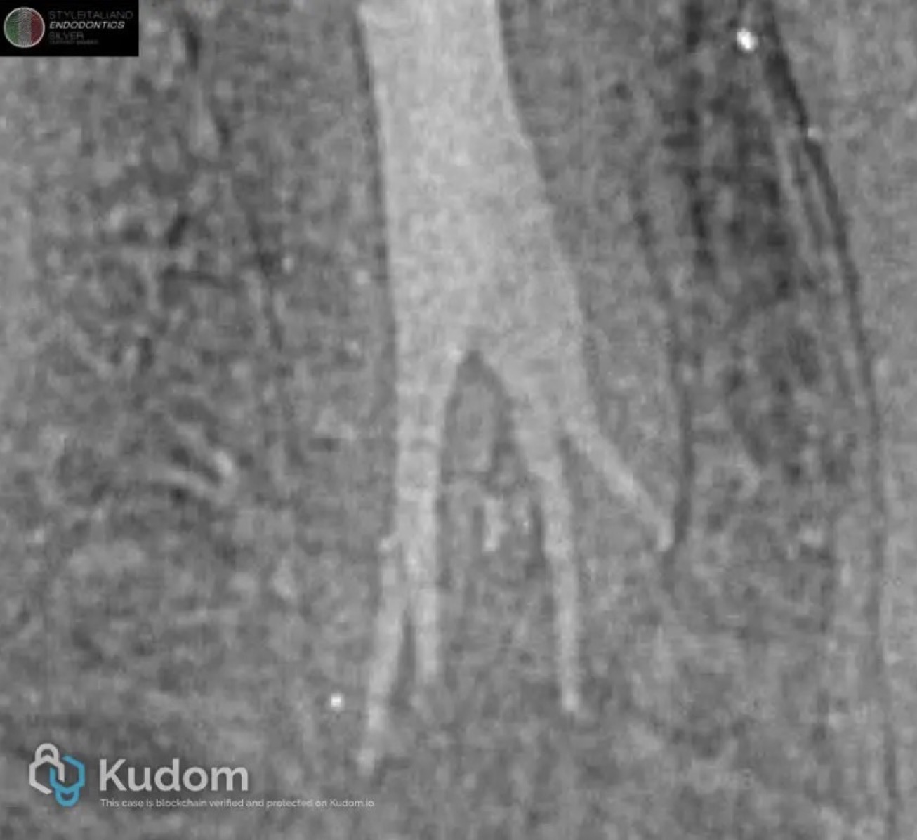
Fig. 3
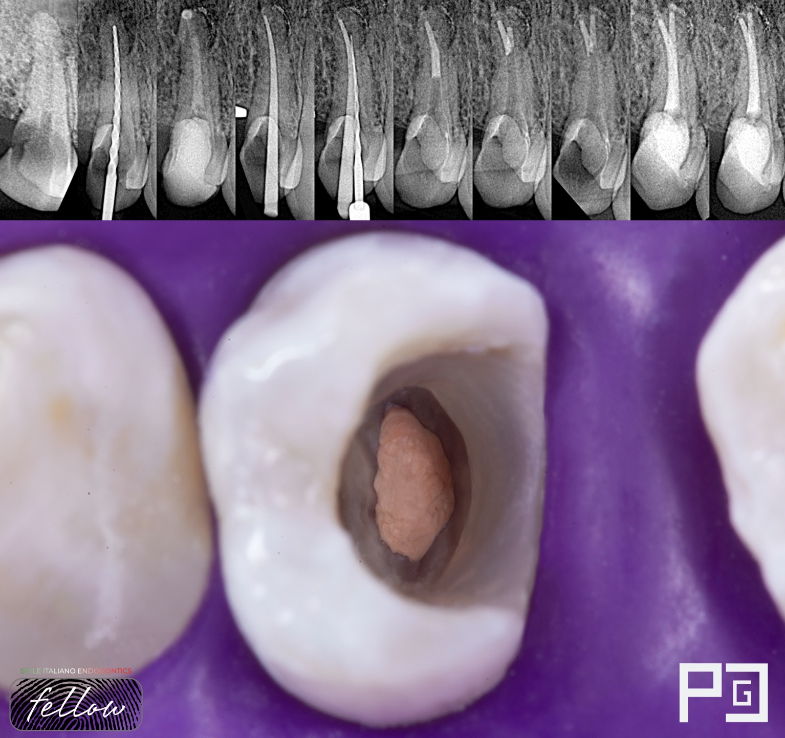
Close up view of post operative radiograph. Canals were filled using warm vertical compaction technique.
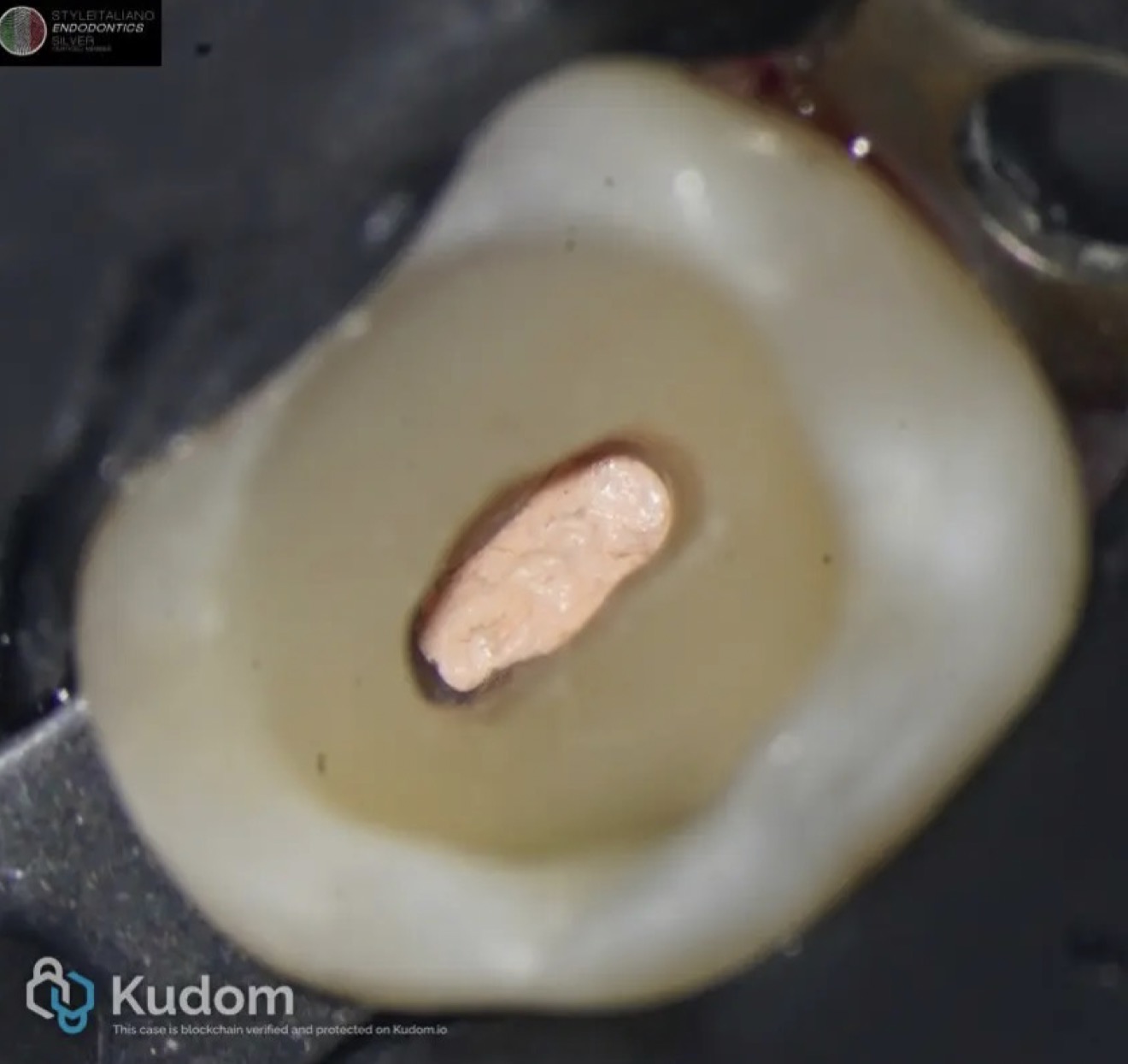
Fig. 4
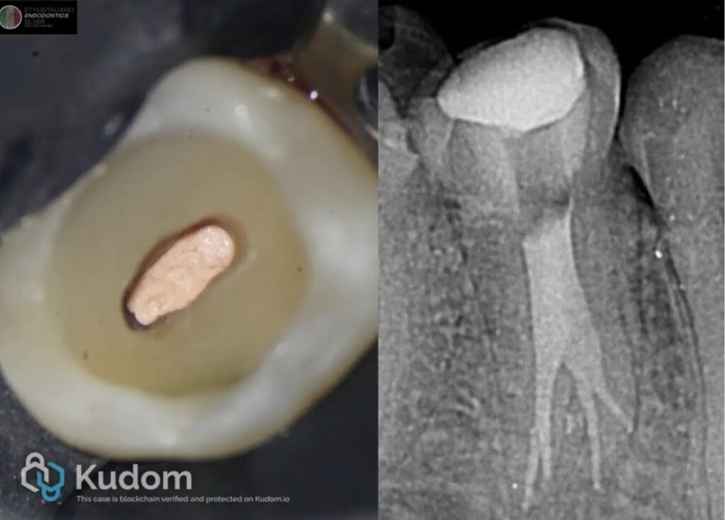
Post obturation clinical photo. Note the conservative access cavity. Tooth will be restored with ceramic overlay.
Conclusions
Treatment of lower premolars could present significant challenge for clinicians. Uniqueness of every single tooth should be always kept in mind. But afterall this is what makes the game of endodontics so fun.
Bibliography
- Bertani P, Gagliani M, Gorni F: Retreatments. Milan: Edra S.p.A; 2020
- Berutti E, Castellucci A: Cleaning and shaping the root canal system. In Castellucci A, editor. Endodontics, Vol. II. Florence: Tridente S.r.l; 2006. p. 396-437
- Schilder H. Filling root canals in three dimensions, Dent Clin North Am; 723-744, November 1967.