Radix entomolaris
26/10/2026
Warning: Undefined variable $post in /home/styleendo/htdocs/styleitaliano-endodontics.org/wp-content/plugins/oxygen/component-framework/components/classes/code-block.class.php(133) : eval()'d code on line 2
Warning: Attempt to read property "ID" on null in /home/styleendo/htdocs/styleitaliano-endodontics.org/wp-content/plugins/oxygen/component-framework/components/classes/code-block.class.php(133) : eval()'d code on line 2
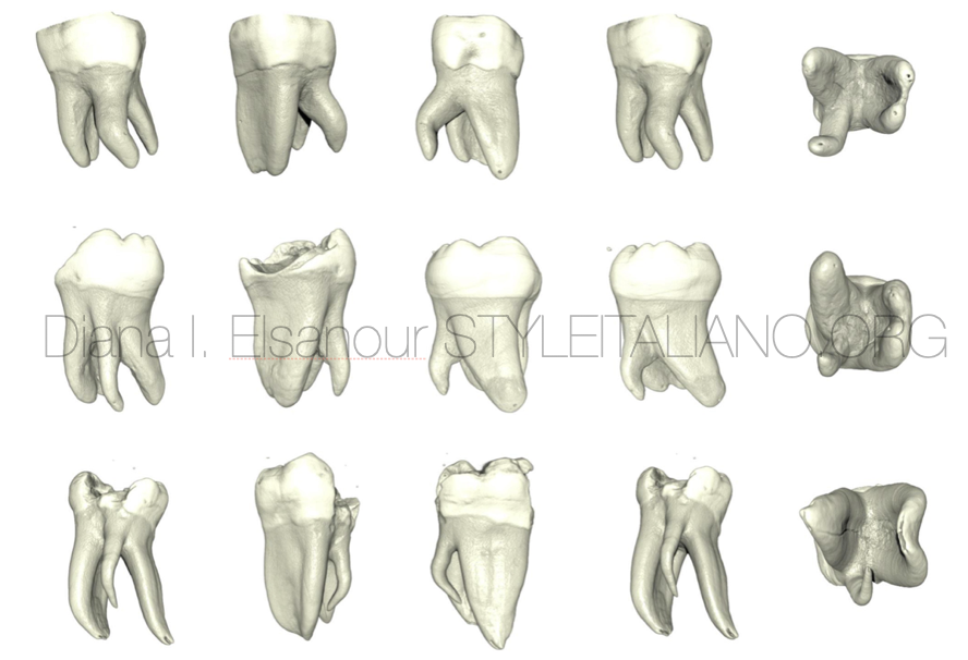
Endodontic treatment is always challenging when anatomy differs from its normal form. The majority of mandibular second molars have two roots (71%), but teeth with one root (27%) and teeth with three roots (2%) are also seen. When these teeth have an extra third root on the distolingual side, it's called "radix entomolaris," and when an extra root located distobuccal side it's called as "radix paramolaris".
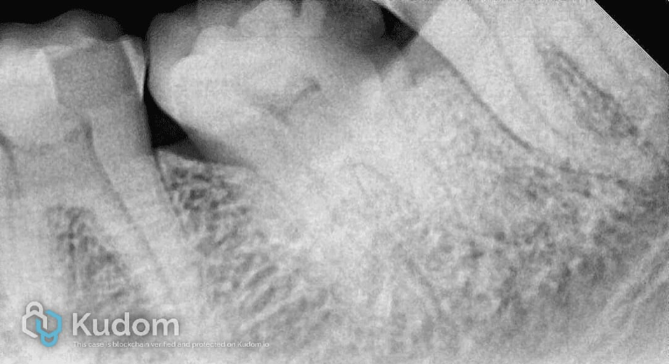
Fig. 1
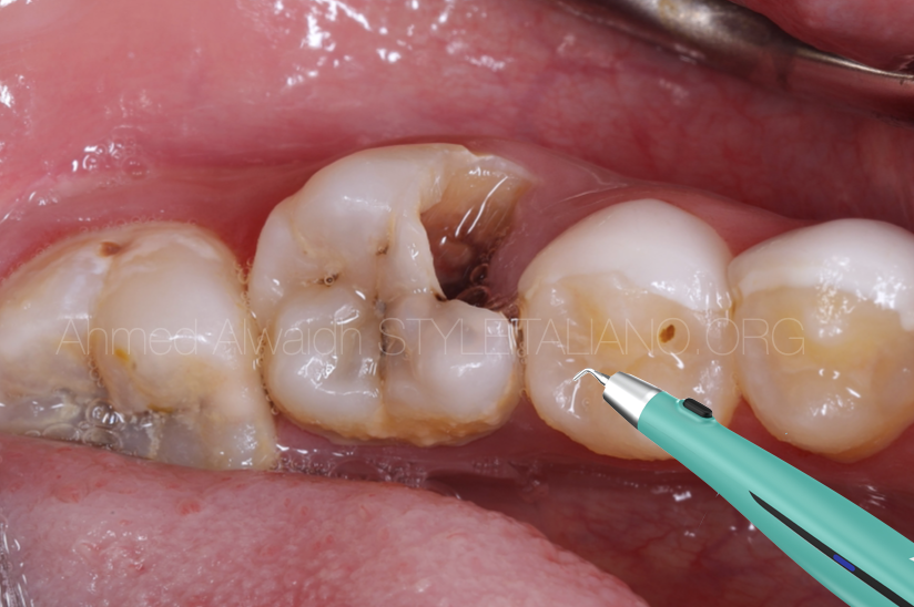
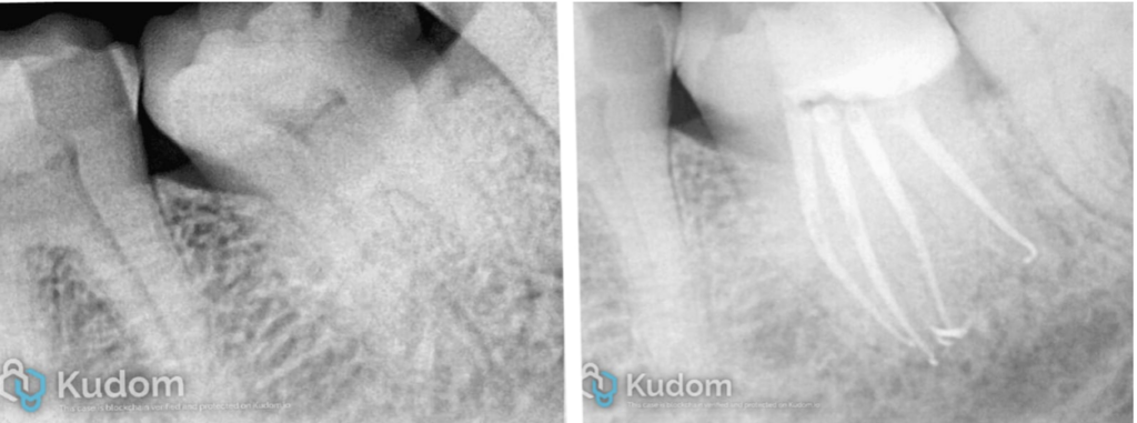
In this case, the patient reported to our office with a complaint of pain and sensitivity when taking in cold in the lower left back tooth region. During the oral examination, proximal caries were found in the lower second molar, and a fractured cusp was seen in the first molar. During the cold test, the second molar had lingering pain, and the first molar gave a normal response when compared with the opposite side's lower molar. The radiograph shows proximal caries extended to the pulp in the lower second molar. Symptoms and cold test suggest symptomatic irreversible pulpitis in lower second molar.
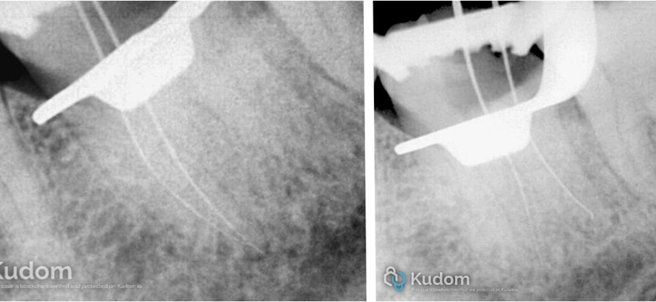
Fig. 2
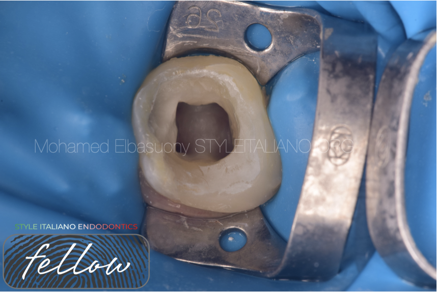
After rubber dam isolation, caries was removed and deroofing was done. Initial scouting was done with 10 number K-files, and working length was measured. This fig. shows mesial and distal canals’ working lengths are being establised. Protaper Gold rotary files were used to enlarge the canal space. Recapitulation was done after every rotary file and in the presence of sodium hypochlorite. Irrigation activation was done with air sonic device. EDTA liquid was use before the final rinsing with sodium hypochlorite.
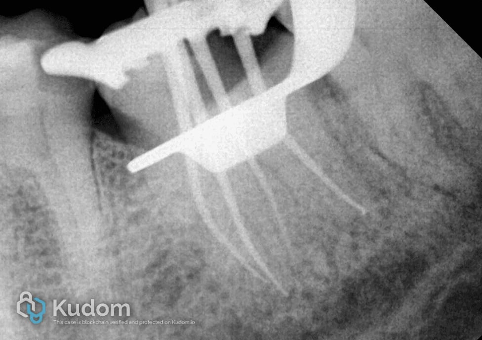
Fig. 3
This figure shows master cone x-ray in mesiobuccal, mesiolingual, distobuccal and distolingual canal. All the canals have separate portal of exits.
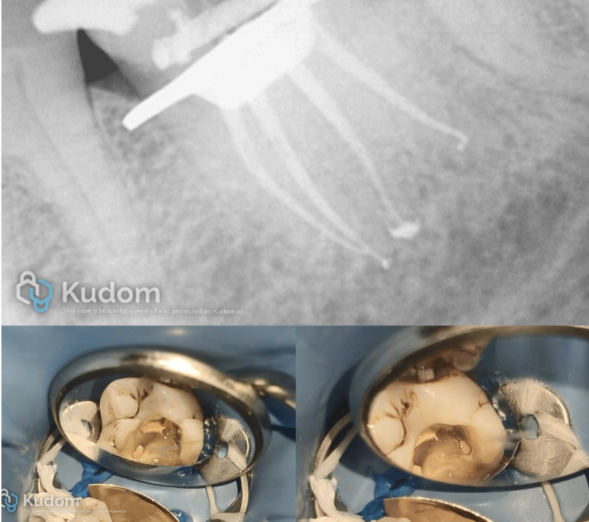
Fig. 4
Obturation was done with continuous wave of compaction and AH Plus sealer.
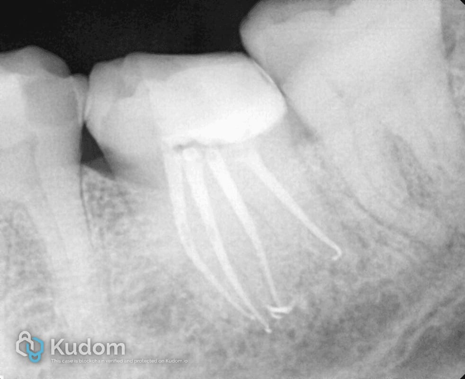
Fig. 5
Post operative film with post endodontic restoration.
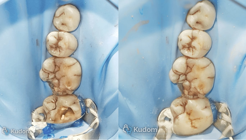
Fig. 6
Tooth was restored with direct composite restoration with the help of fiber-reinforced composite.

Fig. 7
About the author
Dr Parth Padhya
Conclusions
The goal of endodontic is to clean the root canal systems by mechanical and more with chemical. To achieve this first we have to identify the entrance of the canals. Like in this case most of the time clinician missed the extra root canal because of difference in the anatomy which is not everyday we see. From the diagnostic radiograph we can identify variation in root anatomy. After access, visual inspection with laws of orifices location help us to identify orifices, even in this type of cases. Having knowledge of anatomy and its variation help clinician to perform better endodontic treatment and give the successful outcome of root canal treatment.
Bibliography
- Grossman’s endodontic practice- 14th edition, chapter 13, page 259.
- Sarah Abrami : The radix entomolaris : management of the distolingual root canal, Giornale Italiano di Endodonzia,volume 30, November 2016, page 120-123.
- Ajinkya Mansingh Pawar, Anda Kfir, Shishir Singh, Prasanna Neelakantan: Root canal morphology and variations in mandibular second molar teeth of an Indian population: an in vivo cone-beam computed tomography analysis, Clinical oral investigations, march 2017, volume 21, pages 2801-2809.
- Filip L. Calberson, Roeland J. De Moor, Christophe A. Deroose: The radix entomolaris and paramolaris: clinical approach in endodontics, Journal of endodontics, volume 33, January 2007, pages 58-63.