Apexification of an immature mandibular premolar: a case report
03/09/2026
Warning: Undefined variable $post in /home/styleendo/htdocs/styleitaliano-endodontics.org/wp-content/plugins/oxygen/component-framework/components/classes/code-block.class.php(133) : eval()'d code on line 2
Warning: Attempt to read property "ID" on null in /home/styleendo/htdocs/styleitaliano-endodontics.org/wp-content/plugins/oxygen/component-framework/components/classes/code-block.class.php(133) : eval()'d code on line 2
The endodontic treatment of immature permanent teeth with an open apex represents a clinical challenge due to the absence of apical constriction, which makes working-length control, canal obturation and the achievement of a predictable apical seal more difficult.
In cases involving persistent intraradicular infection and apical periodontitis, treatment should promote effective disinfection of the root canal system while creating an adequate apical barrier. Apexification with mineral trioxide aggregate (MTA) allows the formation of an immediate apical plug and facilitates subsequent root canal obturation.
Due to its biocompatibility, sealing ability and potential to promote periapical tissue repair, MTA provides a conservative and predictable treatment option that supports the preservation and long-term function of immature teeth.
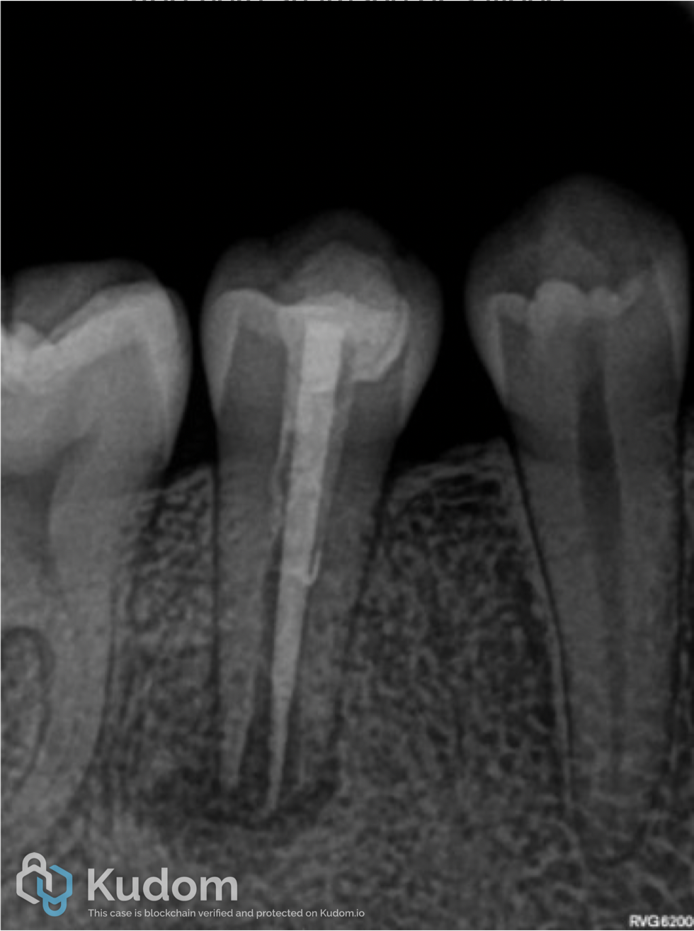
Fig. 1
A 26-year-old male patient was referred in May 2024 for evaluation of the mandibular right second premolar (tooth 45). The tooth had previously undergone endodontic treatment and the patient reported persistent discomfort that was exacerbated during mastication.
Clinical examination revealed periodontal probing within physiological limits, no swelling or sinus tract, normal tooth mobility and tenderness to vertical percussion.
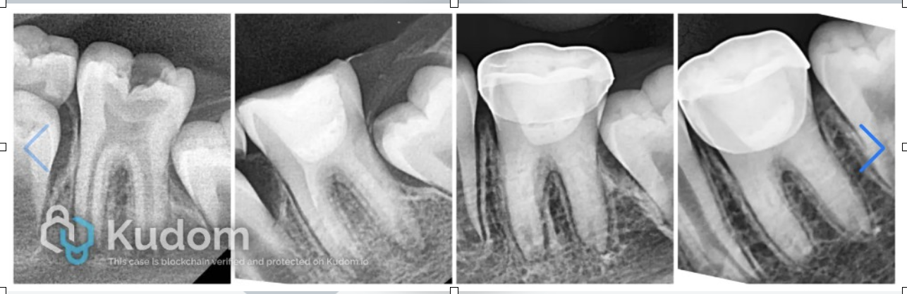
The initial periapical radiograph showed incomplete root development with an open apex, an inadequate previous root canal filling and the absence of a predictable apical seal.
A periapical radiolucency associated with the root apex was also observed.
Non-surgical endodontic retreatment was proposed and accepted by the patient.
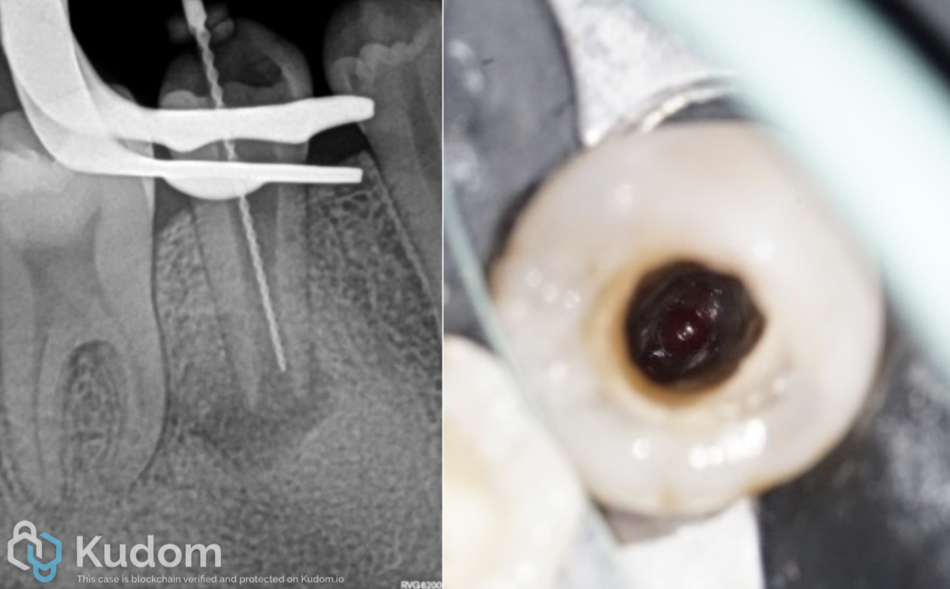
Fig. 2
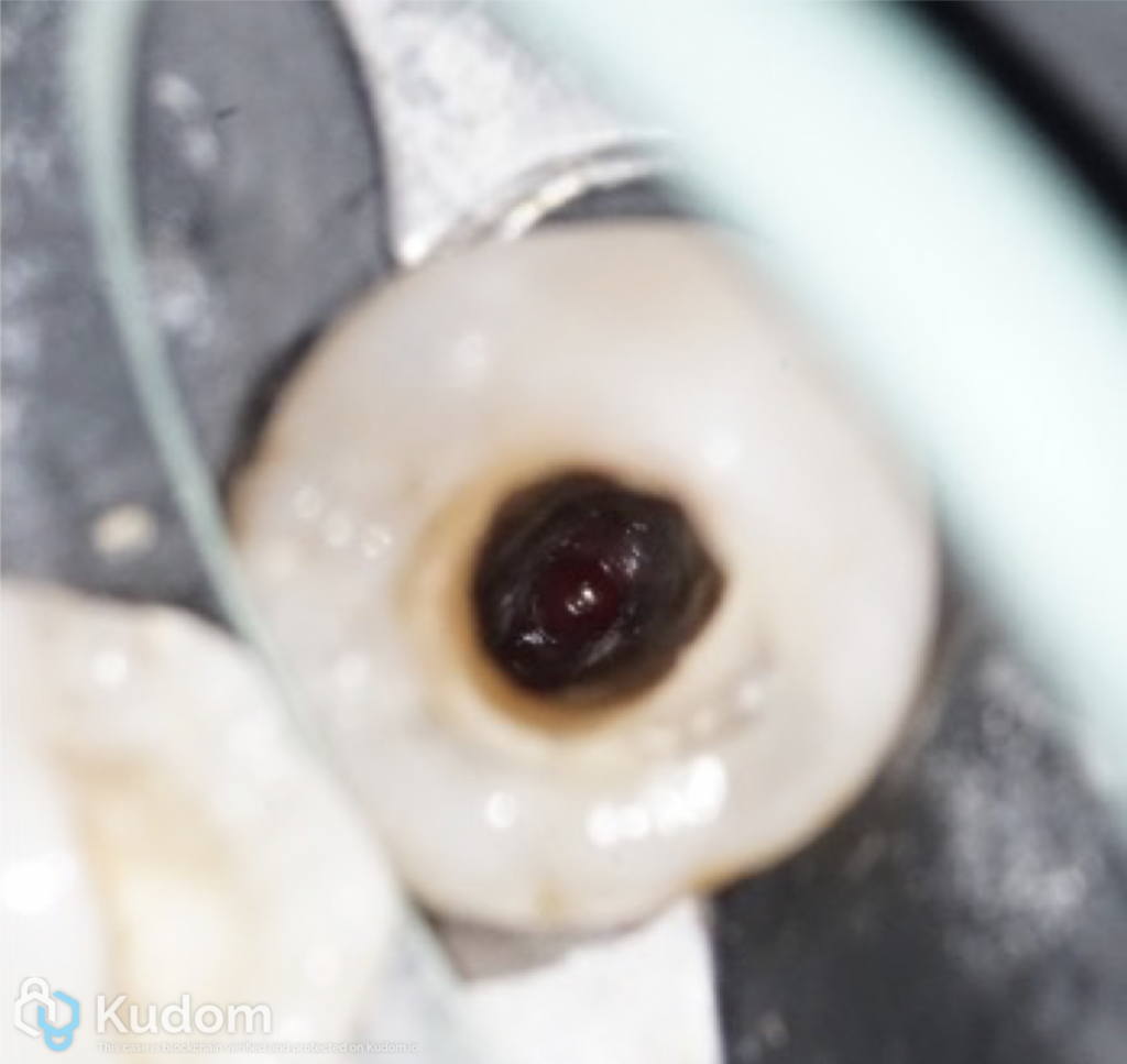
Following the administration of local anaesthesia, the tooth was isolated with a rubber dam to ensure an aseptic operating field.
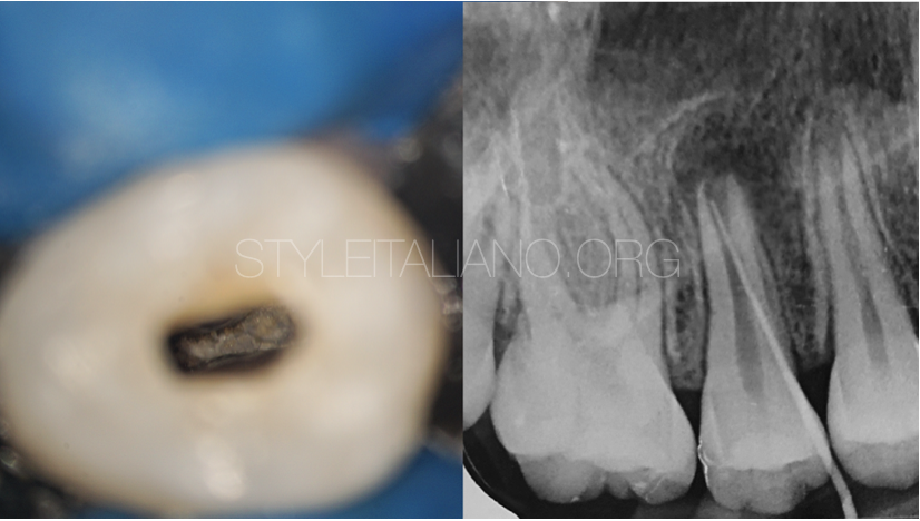
The pre-existing root canal filling material was completely removed using Reciproc R25, R40 and R50 instruments (VDW, Germany). The working length was subsequently determined, and the entire root canal system was thoroughly disinfected using 5.25% sodium hypochlorite and 17% EDTA.
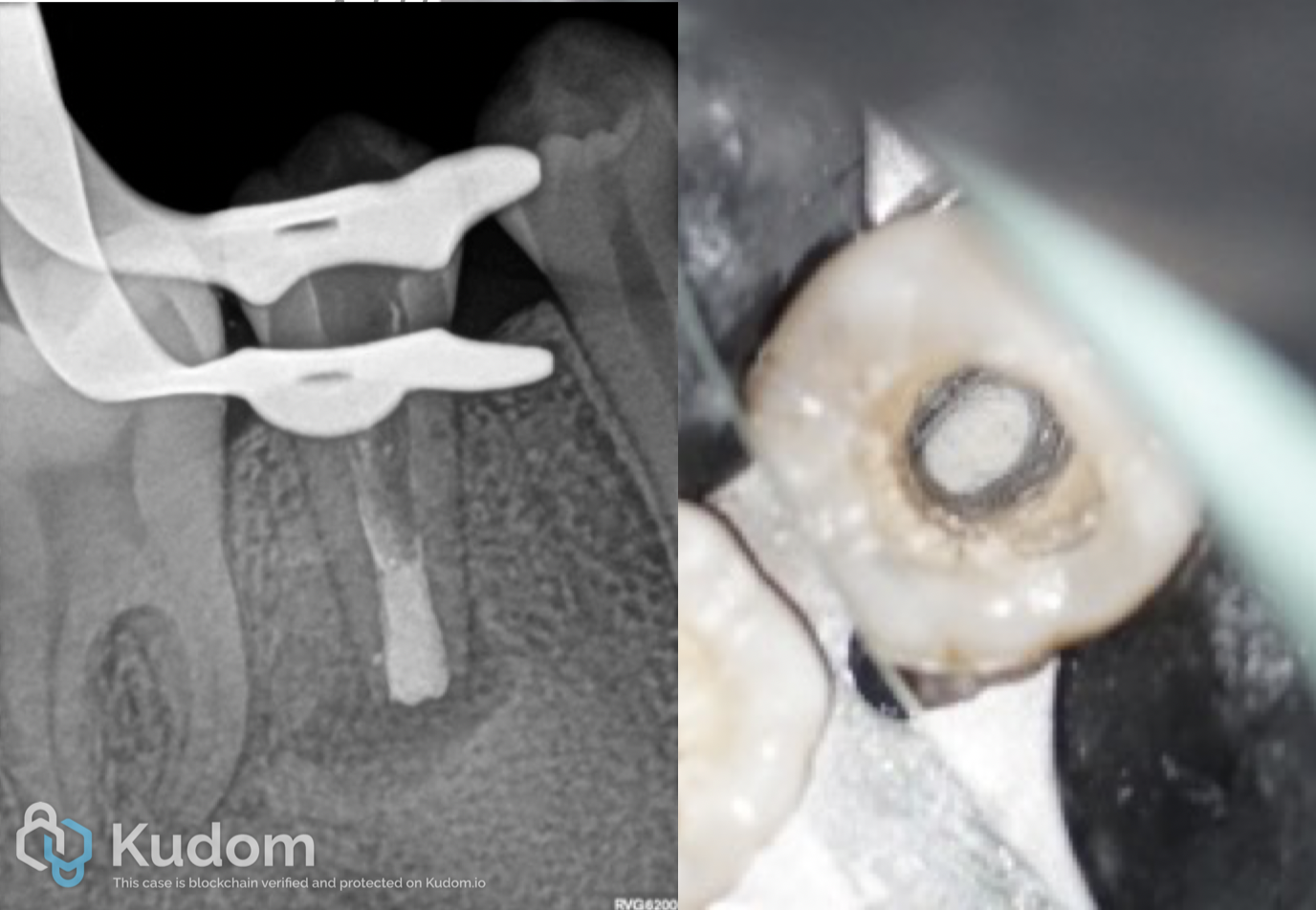
Fig. 3
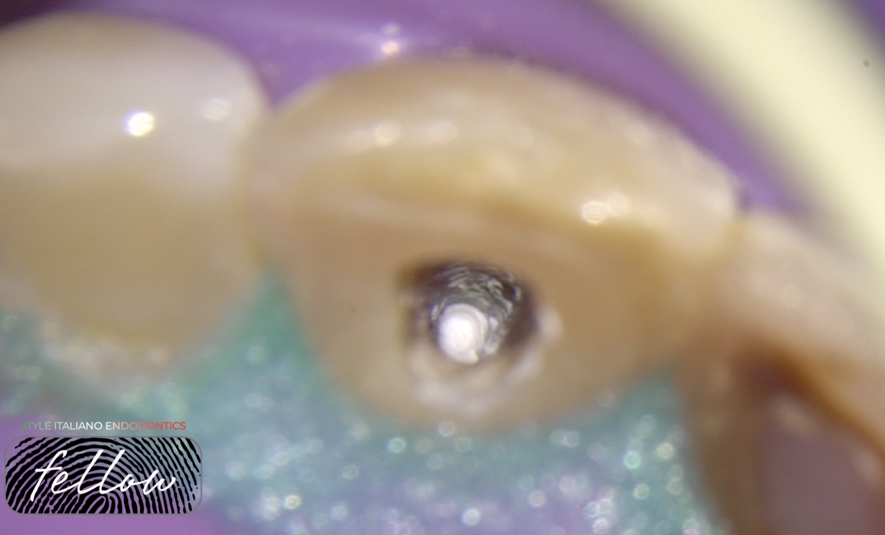
A manual plugger was selected and tested inside the canal. It was positioned 1 mm short of the established working length to ensure adequate control during placement and condensation of the apical material. The root canal was then carefully dried.
A 4–5 mm apical barrier was created using MTA+ (Cerkamed). The material was positioned and condensed in the apical portion of the canal, ensuring appropriate apical control and the formation of a predictable apical seal.
Following placement of the MTA, a moist cotton pellet was positioned inside the tooth and a provisional restoration was placed.
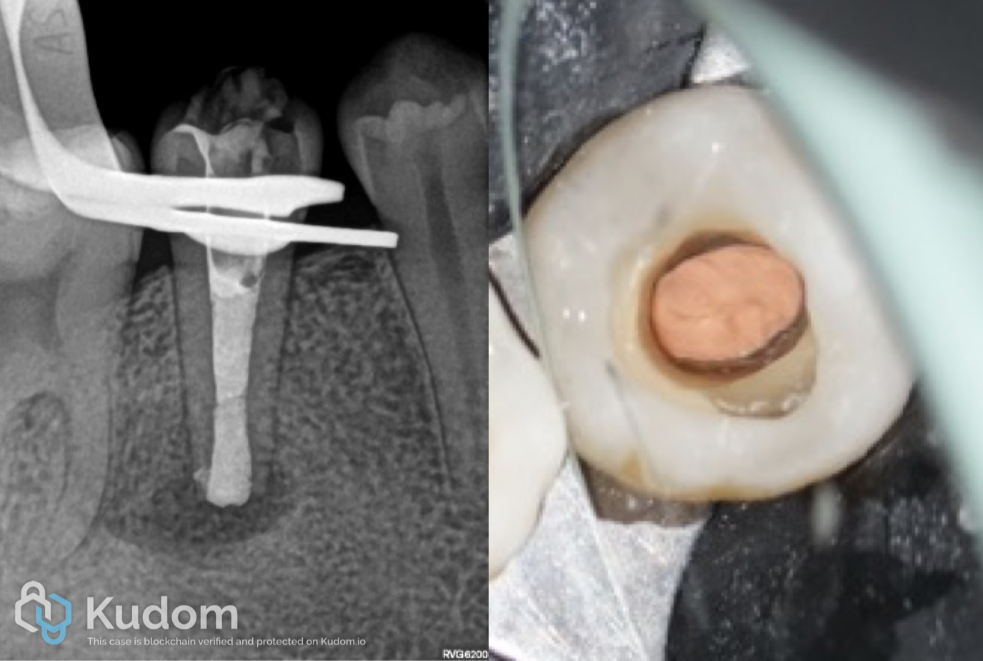
Fig. 4
After the MTA had set, the remaining root canal space was backfilled with injected gutta-percha in association with AH Plus Jet resin-based sealer (Dentsply Sirona). The canal was filled up to the cementoenamel junction.
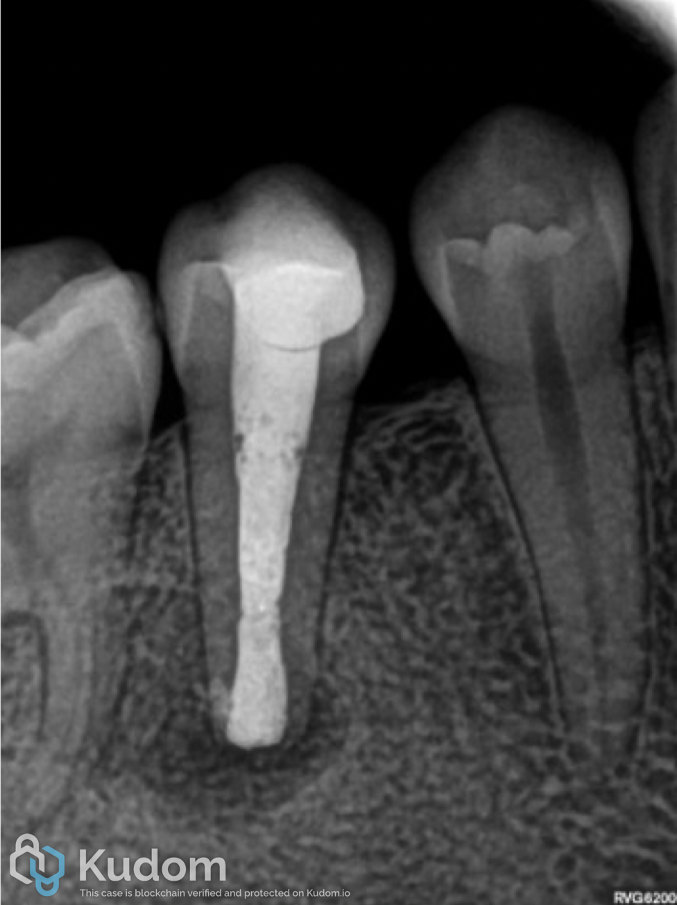
Fig. 5
An intracoronal seal was subsequently performed, followed by placement of a new provisional restoration.
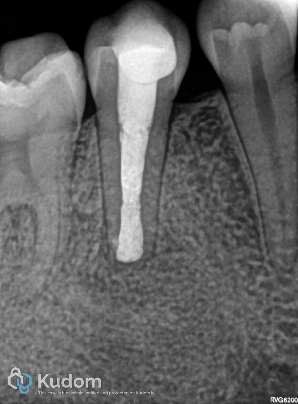
Fig. 6
At the follow-up examination performed in September 2025, approximately 16 months after treatment, the patient was asymptomatic.
The follow-up periapical radiograph demonstrated a favourable evolution of the periapical tissues and evidence of periapical healing, confirming the clinical and radiographic success of the treatment.
Conclusions
This clinical case demonstrates that non-surgical endodontic retreatment combined with MTA apexification can provide a predictable and biologically favourable treatment option for a previously treated immature permanent tooth with an open apex and symptomatic apical periodontitis.
The successful outcome depended on the complete removal of the pre-existing root canal filling, effective disinfection of the root canal system, creation of a 4–5 mm MTA apical barrier and adequate root canal obturation and intracoronal sealing.
At the 16-month follow-up, the patient remained asymptomatic and the periapical radiograph showed favourable tissue healing. This conservative approach allowed the resolution of the periapical pathology while preserving the tooth and maintaining its function.
Bibliography
- Mente J, Leo M, Panagidis D, Ohle M, Schneider S, Lorenzo Bermejo J, Pfefferle T. Treatment outcome of mineral trioxide aggregate in open apex teeth. J Endod. 2013 Jan;39(1):20-6
- Pace R, Giuliani V, Nieri M, Di Nasso L, Pagavino G. Mineral trioxide aggregate as apical plug in teeth with necrotic pulp and immature apices: a 10-year case series. J Endod. 2014 Aug;40(8):1250-4
- Ree MH, Schwartz RS. Long-term Success of Nonvital, Immature Permanent Incisors Treated With a Mineral Trioxide Aggregate Plug and Adhesive Restorations: A Case Series from a Private Endodontic Practice. J Endod. 2017 Aug;43(8):1370-1377
- Torabinejad M, Nosrat A, Verma P, Udochukwu O. Regenerative Endodontic Treatment or Mineral Trioxide Aggregate Apical Plug in Teeth with Necrotic Pulps and Open Apices: A Systematic Review and Meta-analysis. J Endod. 2017 Nov;43(11):1806-1820