Internal bleaching of endodontically treated teeth
04/08/2026
Warning: Undefined variable $post in /home/styleendo/htdocs/styleitaliano-endodontics.org/wp-content/plugins/oxygen/component-framework/components/classes/code-block.class.php(133) : eval()'d code on line 2
Warning: Attempt to read property "ID" on null in /home/styleendo/htdocs/styleitaliano-endodontics.org/wp-content/plugins/oxygen/component-framework/components/classes/code-block.class.php(133) : eval()'d code on line 2
Teeth discolorations can be extrinsic or intrinsic. The first type is usually caused by food, drinks, smoking and other substances that lead to the deposit of stains on the enamel. The second type are the discolorations that lie within the enamel and/or dentin. Correct diagnosis leads to correct management. Internal bleaching treats only intrinsic discolorations.
These can have many causes:
- Local causes:
- Dental trauma, intra pulpal hemorrhage and pulp necrosis: After a dental trauma, pulp blood vessels rupture, and erythrocytes go into dentinal tubules causing discoloration. Some bacteria can contaminate the necrotic pulp space and interact with the iron released from the hemolysis and lead to the formation of black ferric sulphide and tooth discoloration.
- Pulp calcification: Calcifications of the pulp chamber lead to a reduction in translucency, shown as a yellowish crown.
- Incomplete access cavity: Following endodontic treatment when necrotic tissue remnants in the root canal system (especially the pulp horns of maxillary anterior teeth) are not completely eliminated usually due to improper access cavity preparation.
- Endodontics solutions and sealers: Chlorhexidine when combined with sodium hypochlorite produces a dark brown precipitate that stains the enamel and dentine. Also when combined with EDTA, it can produce a pink precipitate. Some intracanal medicaments like the triple antibiotic paste can lead to a greyish discoloration. Sealer remnants in the access cavity and MTA containing bismuth oxide could be associated with tooth discoloration, that’s why cleaning the access cavity from these remnants is of utmost importance.
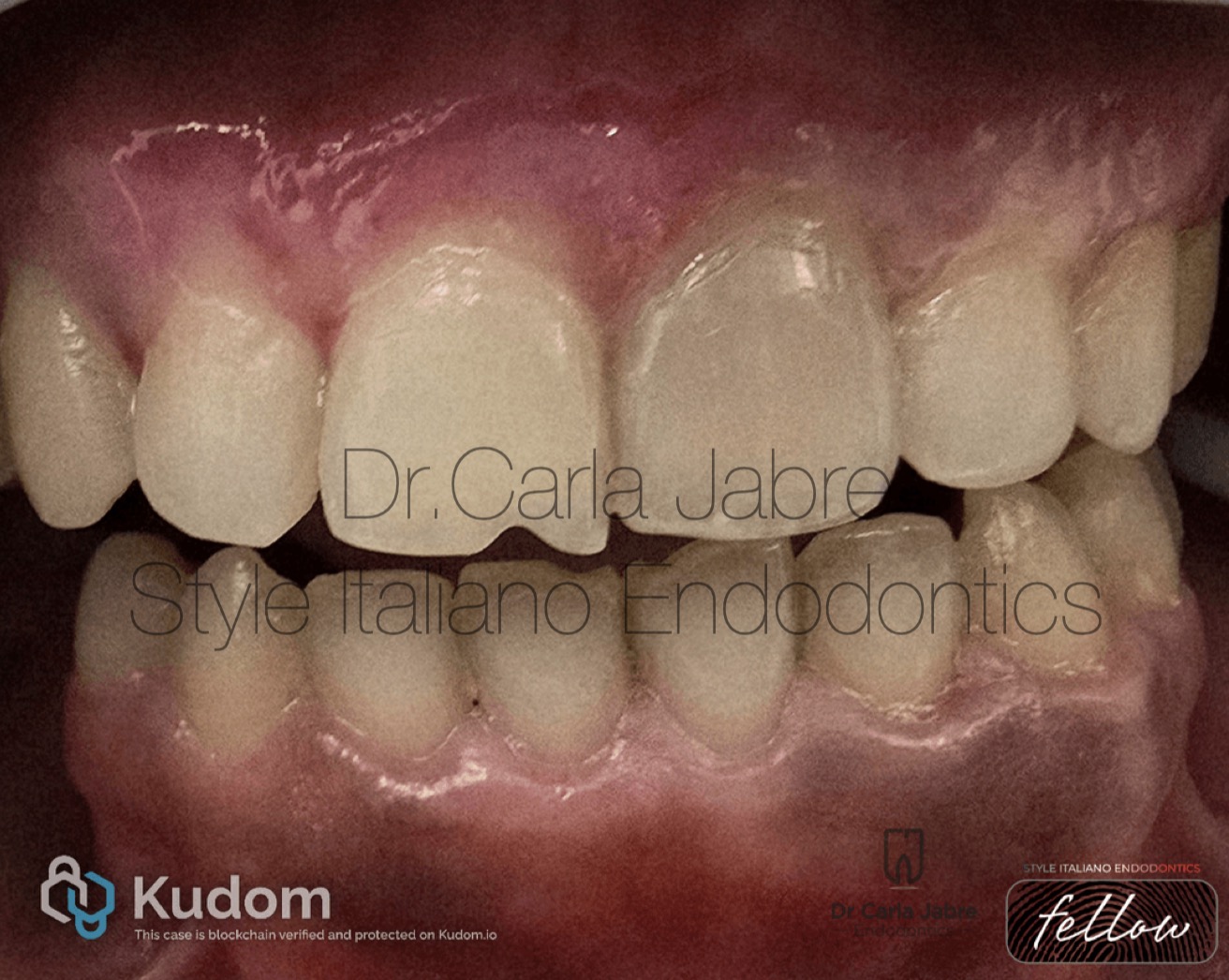
Fig. 1
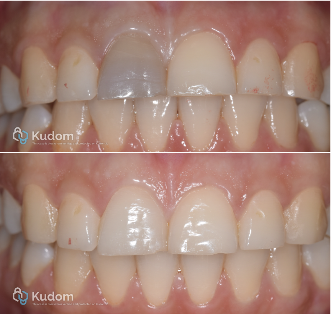
A 25-year-old woman presented to the clinic concerned about a discolored left central incisor that she noticed after the removal of her braces.
The tooth was asymptomatic, with no history of symptoms. The patient reported a history of trauma during her childhood.
Clinical examination revealed no response to cold and heat, no pain to percussion and palpation and no mobility.
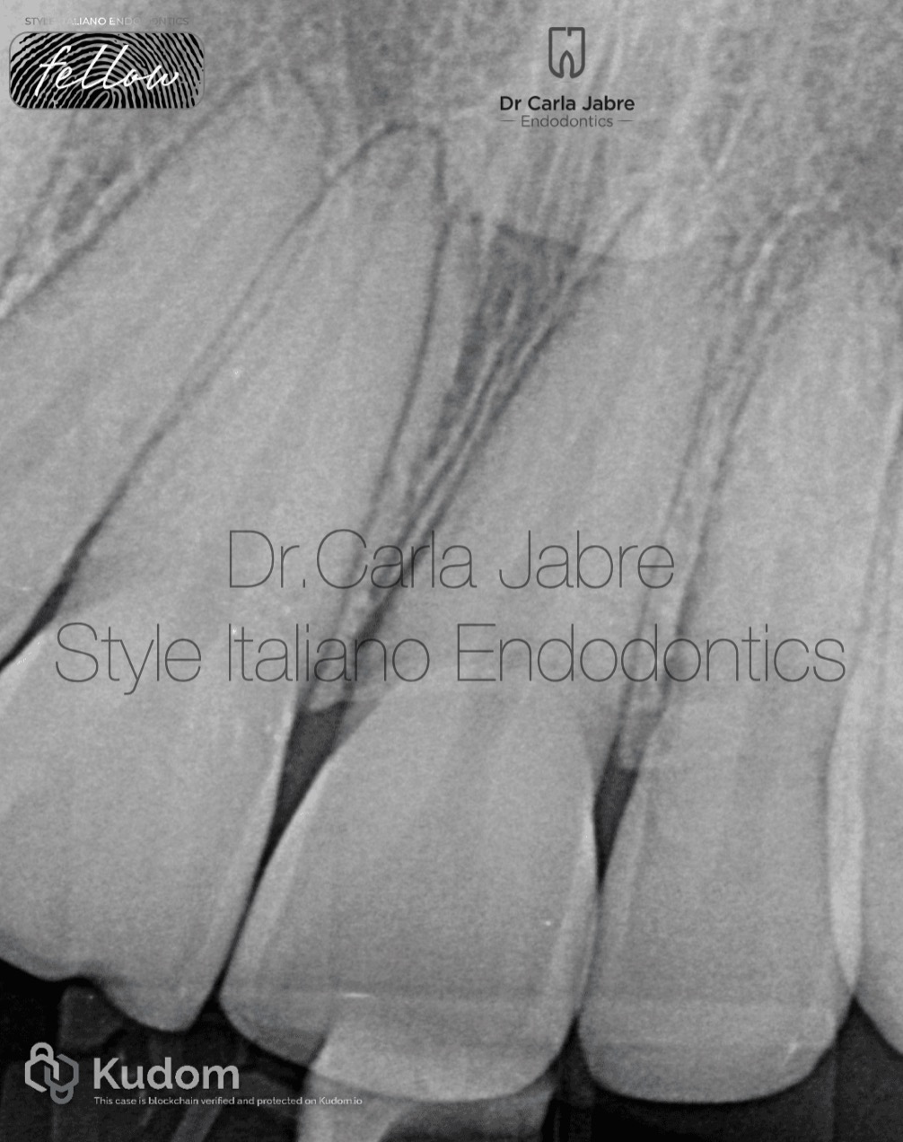
Fig. 2
Radiographic examination showed no apical radiolucency and no pulpal calcifications.
The diagnosis established was pulp necrosis and normal periapical tissues.
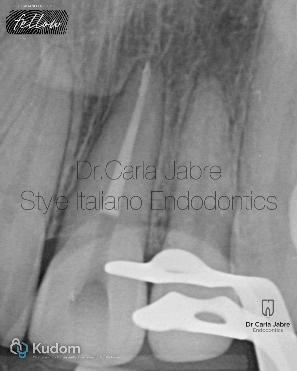
Fig. 3
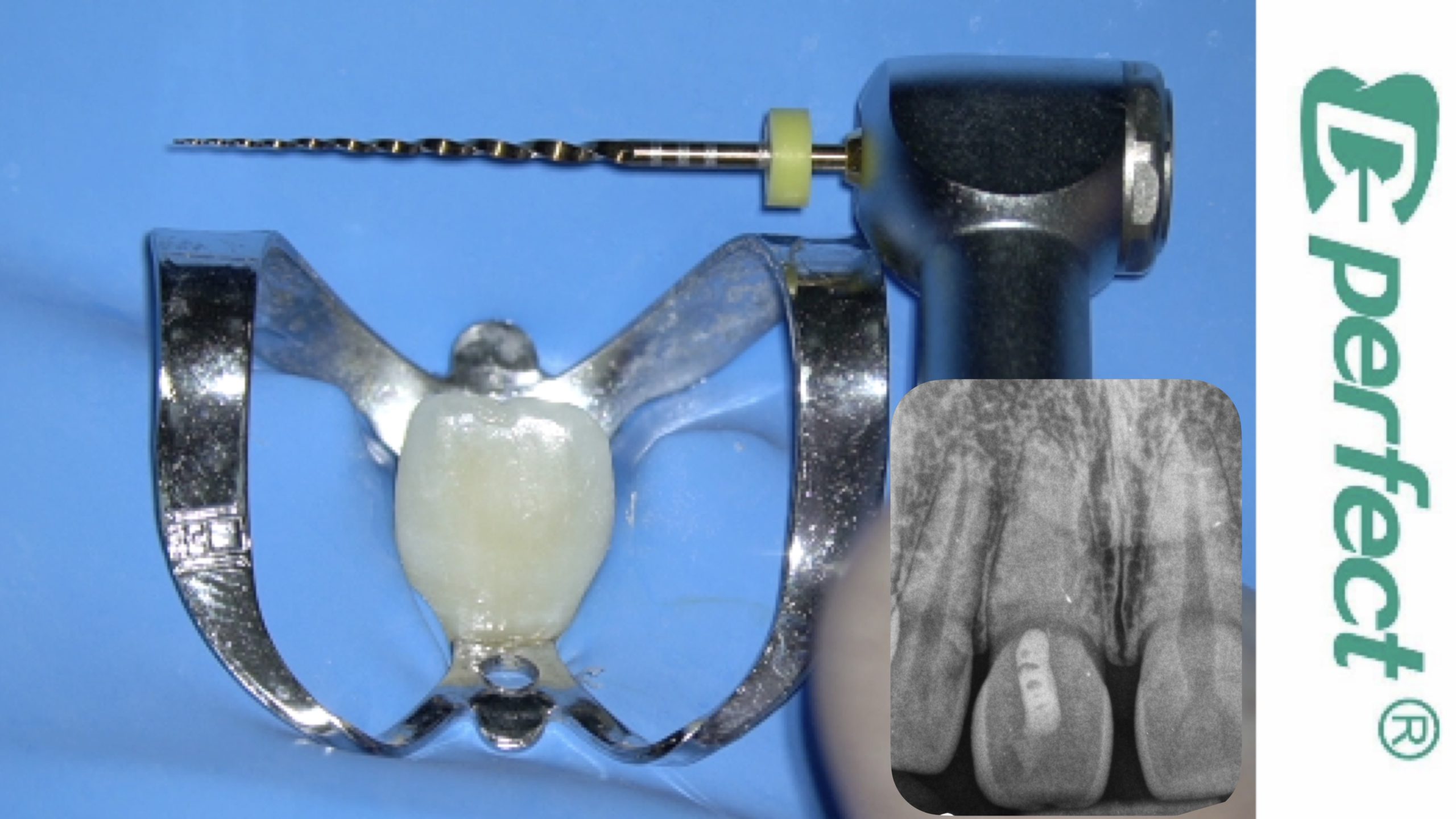
After anesthesia and rubber dam placement, the root canal treatment was done in one session:
- Shaping to the size 30/.06
- Copious irrigation using 5,25% NaOCl with sonic activation.
- Warm vertical condensation technique for obturation. The level of the backfill was stopped at 2mm below the CEJ.
The access cavity was carefully cleaned from any remaining sealer using an ethanol saturated micro brush followed by ultrasonic scaler with water. This is an important step to remove any sealer remnants that could lead to future discolorations.
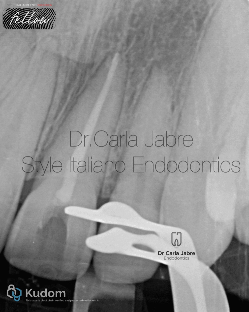
Fig. 4
GIC was used to fill the 2mm between the obturation and the CEJ. To do that, a periodontal probe is used to measure the distance between the incisal edge and the CEJ from the outside, and then transferred to the pulp chamber.
This layer prevents the penetration of the whitening product into the root space. External cervical invasive root resorption has been reported as an adverse effect of internal bleaching, especially when no barrier was used between the bleaching agent and the root canal obturation.
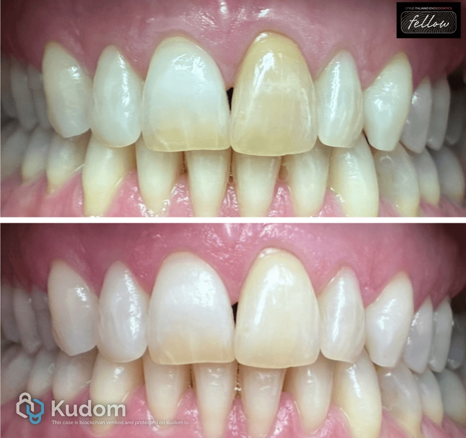
Opalescence endo, a 35% hydrogen peroxide, was used for the whitening process. It was injected into the access cavity and left there for 4 days. The access cavity was sealed with a white temporary filling.
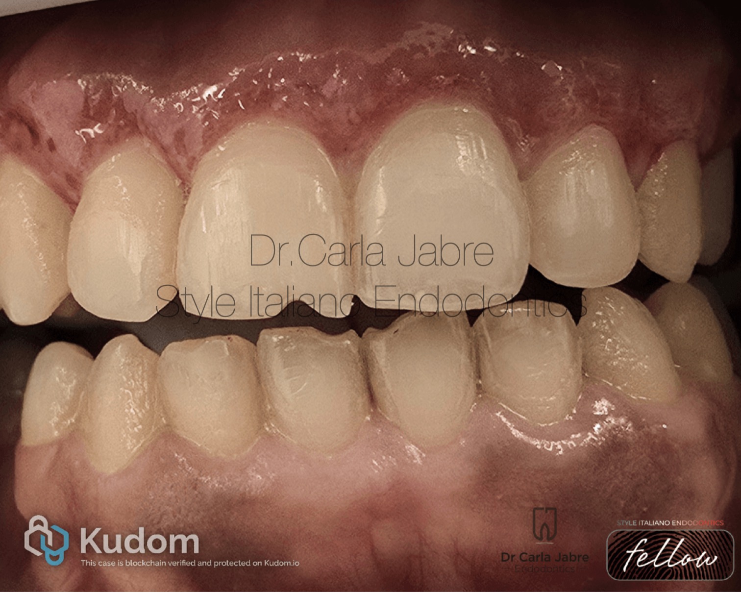
Fig. 5
After 4 days, the patient was satisfied with the result. The product was washed away with normal saline.
Some in vitro studies show that hydrogen peroxide compounds can affect bonding to enamel and dentine up to an average of 7 to 14 days. Therefore, a temporary filling was used for 2 weeks before the final restoration of the access cavity to prevent interference of the whitening product with bonding agents.
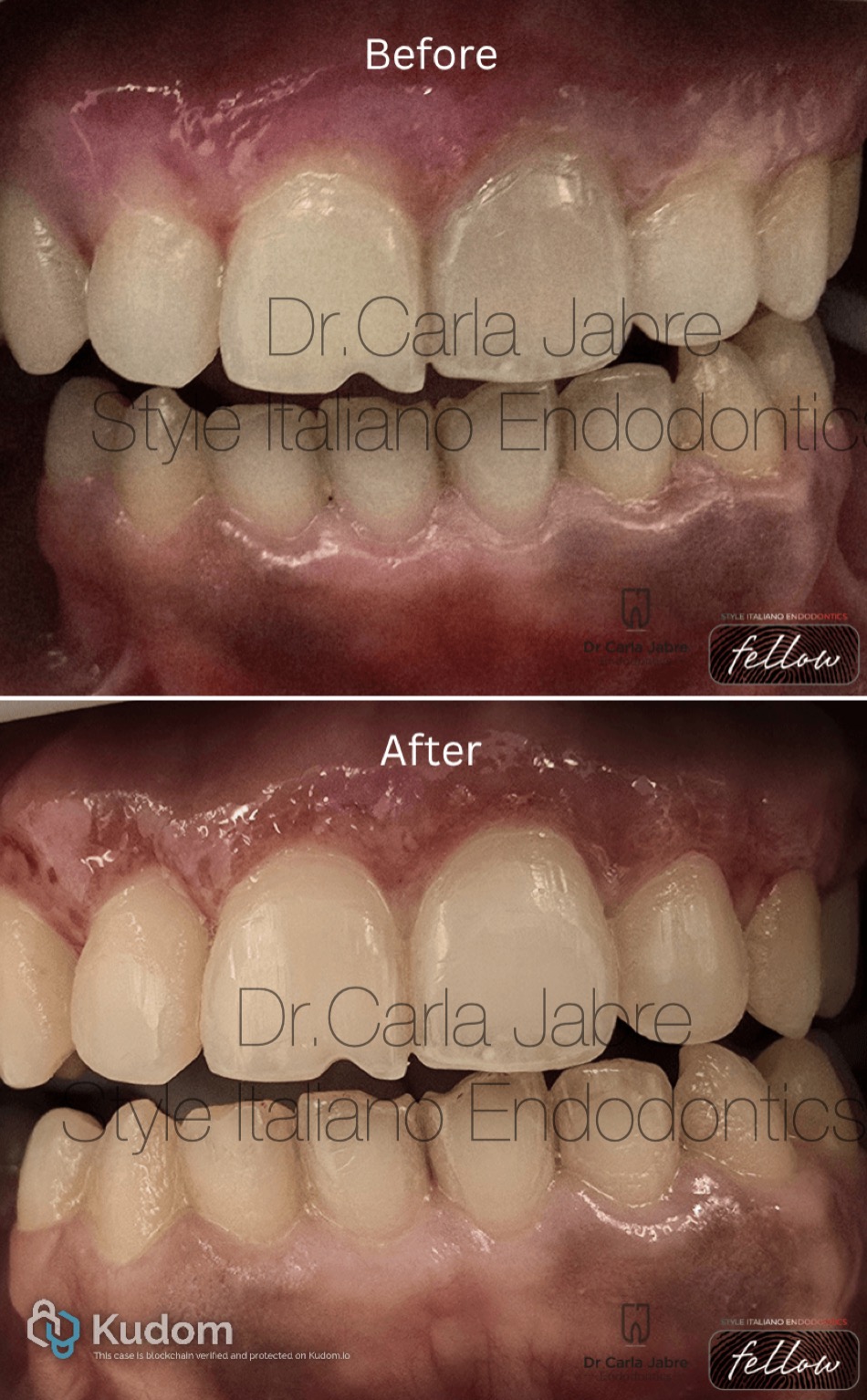
Fig. 6
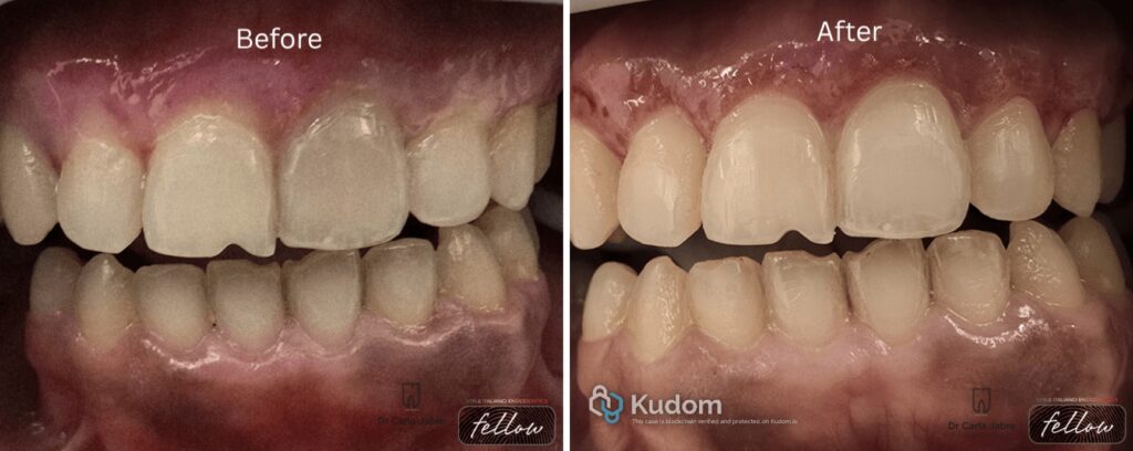
Before and after

Fig. 7
About the author:
Dr Carla Jabre
2008-2013: Undergrad studies at the faculty of Dentistry at Saint Joseph University, Beirut, Lebanon.
2013-2016: Masters in Endodontics at the faculty of Dentistry at Saint Joseph University, Beirut, Lebanon.
2018-2019: Masters in Biomaterials at Saint Joseph University, Beirut, Lebanon.
Dr Jabre has been a clinical instructor, guiding undergraduate students, in the department of Endodontics at Saint Joseph University, since 2016.
She is a member of the Lebanese Society of Endodontics, and a fellow in the Style Italiano Endodontics group.
Her private practice is limited to endodontics.
Conclusions
The internal bleaching is an easy and conservative solution to discolored and endodontically treated teeth.
Satisfactory results rely on the diagnosis of the cause of the discoloration and the use of safe protocols.
Bibliography
- Amer M. Intracoronal tooth bleaching - A review and treatment guidelines. Aust Dent J. 2023 Jun;68 Suppl 1:S141-S152. doi: 10.1111/adj.13000. Epub 2023 Nov 17. PMID: 37975331.
- Frank AC, Kanzow P, Rödig T, Wiegand A. Comparison of the Bleaching Efficacy of Different Agents Used for Internal Bleaching: A Systematic Review and Meta-Analysis. J Endod. 2022 Feb;48(2):171-178. doi: 10.1016/j.joen.2021.10.011. Epub 2021 Nov 9. PMID: 34762968.
- Jin Y, Paranhos KS, Salamone A, Bongiorno W, Brizuela M. Internal Tooth Whitening. 2024 May 7. In: StatPearls [Internet]. Treasure Island (FL): StatPearls Publishing; 2026 Jan–. PMID: 38753915.