Clinical and Anatomical Challenges in Mandibular Molars: A Comparative Analysis of Two-Canal versus Six-Canal Systems with Apical Pathosis
19/01/2026
Fellow
Warning: Undefined variable $post in /home/styleendo/htdocs/styleitaliano-endodontics.org/wp-content/plugins/oxygen/component-framework/components/classes/code-block.class.php(133) : eval()'d code on line 2
Warning: Attempt to read property "ID" on null in /home/styleendo/htdocs/styleitaliano-endodontics.org/wp-content/plugins/oxygen/component-framework/components/classes/code-block.class.php(133) : eval()'d code on line 2
Mandibular molars exhibit one of the highest degrees of anatomical variability in the human dentition. While the classical configuration involves two or three canals, extreme variations exist, ranging from simplified two-canal systems to highly complex six-canal anatomies. When combined with apical pathosis, these anatomical extremes pose significant diagnostic and therapeutic challenges.
Successful endodontic therapy is based on three fundamental principles: complete debridement, effective disinfection, and three-dimensional obturation of the root canal system. However, these principles are strongly influenced by root canal anatomy. Mandibular molars demonstrate exceptional anatomical variability, particularly in the mesial and distal roots, where accessory canals, isthmuses, and middle mesial canals are frequently present.
While some mandibular molars present a simplified anatomy of two large canals, others may exhibit up to six independent canals, dramatically increasing the biological and mechanical difficulty of treatment.
When apical periodontitis is present, untreated anatomical complexity becomes the dominant factor leading to persistent infection and treatment failure.
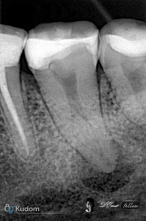
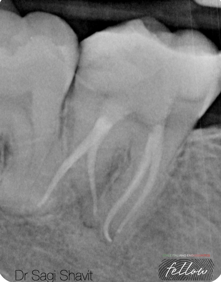
Fig. 1
Two-Canal Mandibular Molars
Two-canal mandibular molars typically consist of one mesial and one distal canal, often with round or oval cross-sections. These canals are usually wide, relatively straight, and accessible, allowing efficient mechanical shaping and irrigant penetration.
Radiographic examination showed a well-defined periapical radiolucency
Diagnosis
Necrotic pulp
Chronic apical periodontitis
Anatomy
Access revealed:
One mesial canal
One distal canal in one apical foramen
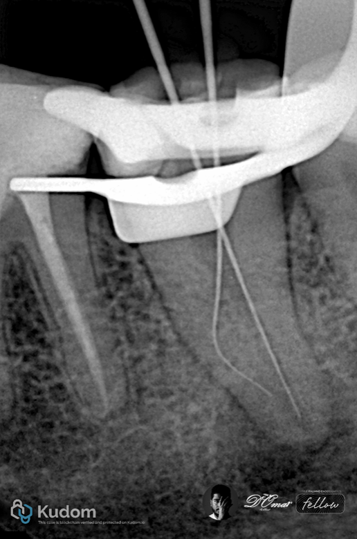
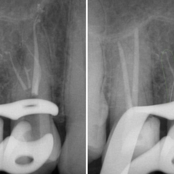
Fig. 2
Working length established using apex locator and radiographs
Rotary NiTi instrumentation to full working length
Irrigation using NaOCI with ultrasonic activation
EDTA for smear layer removal
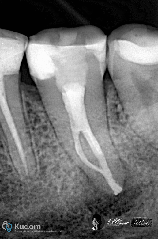
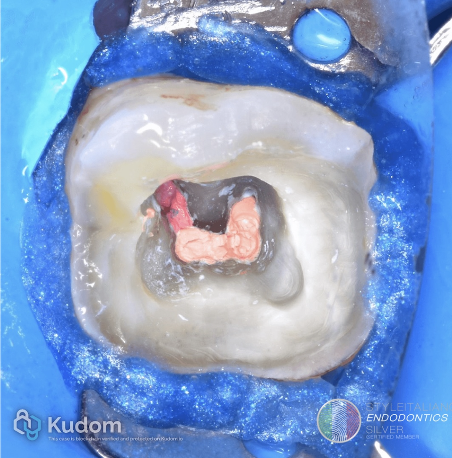
Fig. 3
Obturation done by BC sealer
with warm vertical compaction
Two-canal mandibular molars typically consist of one mesial and one distal canal, often with round or oval cross-sections. These canals are usually wide, relatively straight, and accessible, allowing efficient mechanical shaping and irrigant penetration.
Such anatomy provides:
-Better irrigant flow
-Fewer uninstrumented recesses
-Higher obturation predictability
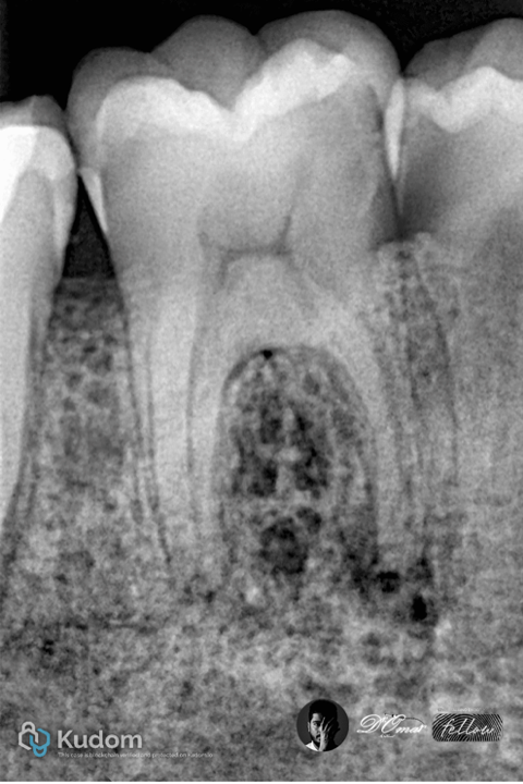
Fig. 4
Case No.2
Six-Canal Mandibular
Molar with Apical Lesion
A 41-year-old patient presented with spontaneous pain and swelling related to a mandibular first molar.
Radiography showed a large apical radiolucency involving mesial roots.
CBCT Findings
Three mesial canals (mesiobuccal, mesiolingual, middle mesial)
Three distal canals (distobuccal, distolingual, middle distal)
Multiple isthmuses were present between canals.
Diagnosis
Necrotic pulp
Symptomatic apical periodontitis
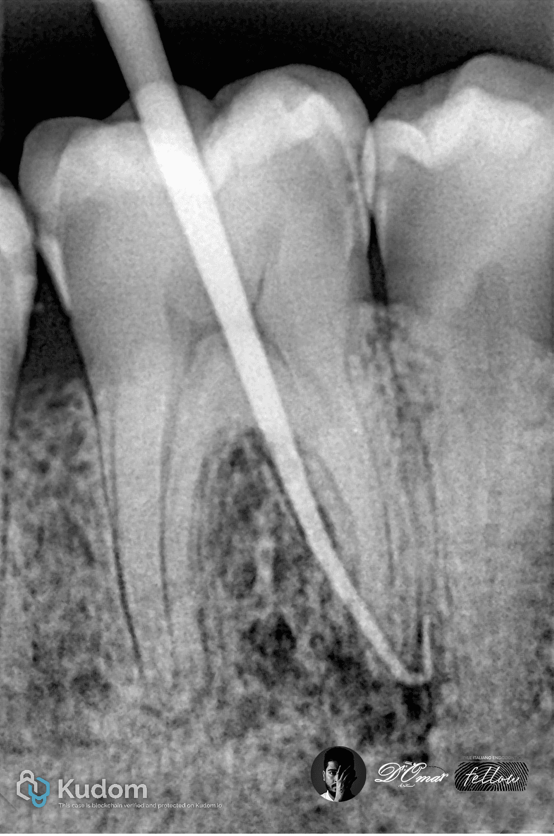
Fig. 5
Radiographic examination showed a well-defined periapical radiolucency associated with the mesial root.
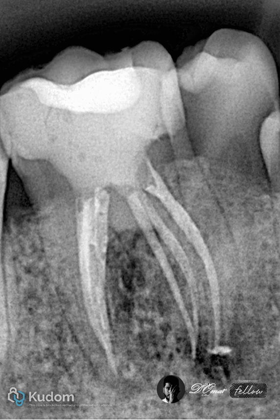
Fig. 6
Canal orifices were extremely narrow and difficult to locate
Complex curvatures increased the risk of file separation
Irrigant penetration was limited by multiple canal interconnections
Despite advanced irrigation and careful shaping, some areas remained mechanically untouched.
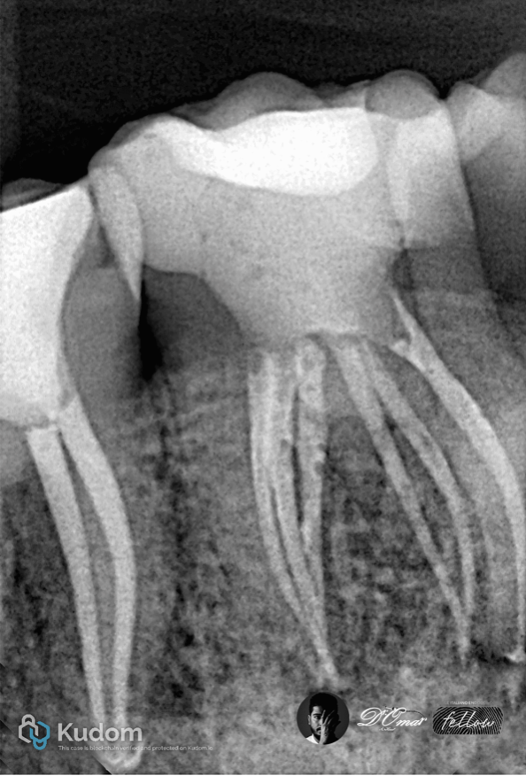
Fig. 7
Mesial shift PA x-ray show the
6 canals ..6 oraface and 3 roots

Fig. 8
About the author
Omar Alsheikhly
BDS , MSc endodontist
SIE fellow member
CEO dental division (UNITED HORIZON COMPANY)
KOL for Dentsplay sirona (VDW)
KOL for Maruchi
KOL for Denjoy
KOL for Illuco
Bachelor of dental surgery (BDS) Ivan Horbachevsky Ternopil State Medical University UKRAIN 2011-2016
Awarded the certificate of equivalency for the diploma Faculty of Dentistry \University of Baghdad IRAQ 2019
MSc master in advanced Endodontics University of Siena \ ITALY 2021\2023
Conclusions
Although both teeth presented with apical pathosis, their biological and mechanical realities were fundamentally different. The two-canal system allowed nearly complete canal wall contact by instruments and irrigants, enabling rapid bacterial elimination and healing.
In contrast, the six-canal system contained multiple untouched areas where bacteria could survive. Even advanced rotary files and activated irrigation cannot fully clean isthmuses and micro-canals. This explains why periapical healing was delayed despite correct technique.
Mandibular molars with identical apical pathology may behave very differently depending on their internal anatomy. While two-canal systems allow predictable healing, six-canal systems represent a biologically resistant environment where persistent infection is more likely. Understanding and respecting canal anatomy is therefore the key to true endodontic success.
Bibliography
1- Anatomical complexity in mandibular second molars
Madfa AA et al. — Anatomical complexity in mandibular second molars: prevalence of C-shaped canals, radicular grooves, taurodontism, and radices molarum in a Saudi population.
Scientific Reports (2025).
2. Root and canal morphology in an Egyptian population
Saber SM et al. — Root and canal morphology of mandibular second molars in an Egyptian subpopulation: a cone-beam computed tomography study.
BMC Oral Health (2023).
3. Root canal anatomy of mandibular first molars
Akhlaghi NM et al. — Root canal anatomy and morphology of mandibular first molars.
PMC article (2017).
4. Anatomical variations overview
Manik K — Anatomical variations in mandibular molars.
PMC article (2024).
5. Endodontic management of complex molars
Hasan A — Endodontic management of a mandibular first molar with four mesial root canals.
Exploration of Medicine (2025)