Root - Crown -Tissue: The Tooth Salvage Concept
08/12/2025
Fellow
Warning: Undefined variable $post in /home/styleendo/htdocs/styleitaliano-endodontics.org/wp-content/plugins/oxygen/component-framework/components/classes/code-block.class.php(133) : eval()'d code on line 2
Warning: Attempt to read property "ID" on null in /home/styleendo/htdocs/styleitaliano-endodontics.org/wp-content/plugins/oxygen/component-framework/components/classes/code-block.class.php(133) : eval()'d code on line 2
An interdisciplinary approach to dental treatment enables successful clinical outcomes and allows for the long-term functionality of seemingly hopeless teeth. A crucial stage in tooth preservation is the development of a treatment plan, which must be scientifically and clinically justified.
When formulating the plan, three key elements involved in the preservation process must be considered: the root, the crown, and the tissue.
The strategy of this concept lies in the fact that each element must function as a unified mechanism, yet be addressed in distinct groups.
Root involves endodontics and build-up for subsequent restorative work.
Crown entails the ability to achieve healthy gums around the restoration through provisional crowns and the restoration of each tooth's chewing function with permanent prosthetics.
Tissue refers not only to the pink aesthetics around the permanent restorations but also to the long-term periodontal status of the tooth post-treatment.

Fig. 1
In 2018, a patient (female) presented with complaints of pain upon pressing on the tooth, a sensation of fullness in the gum, serous fluid discharge from a fistula, and bad breath

Fig. 2
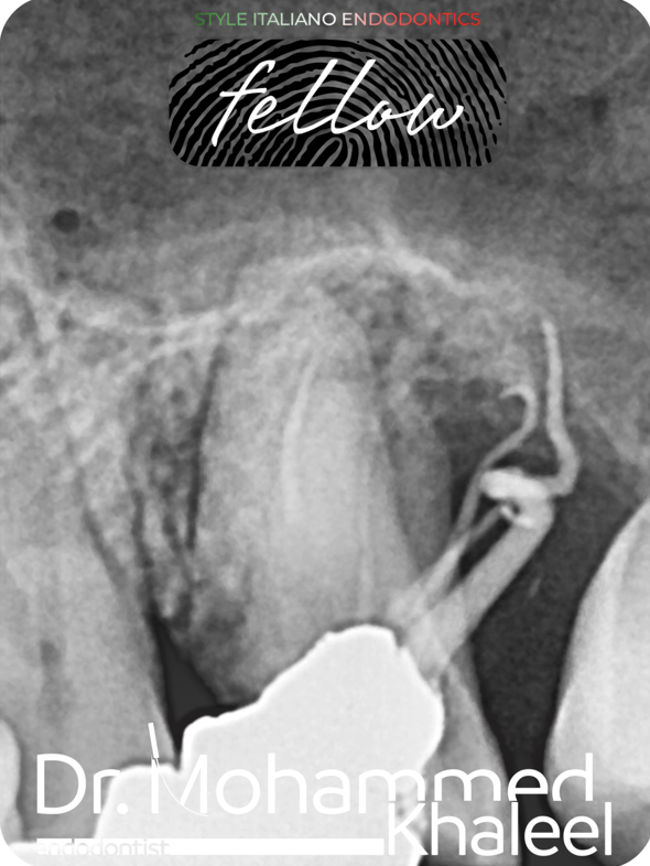
The figure shows incorrect treatment protocol planning and very aggressive root canal preparation for metal intraradicular posts, which subsequently led to a root perforation in the apical portion
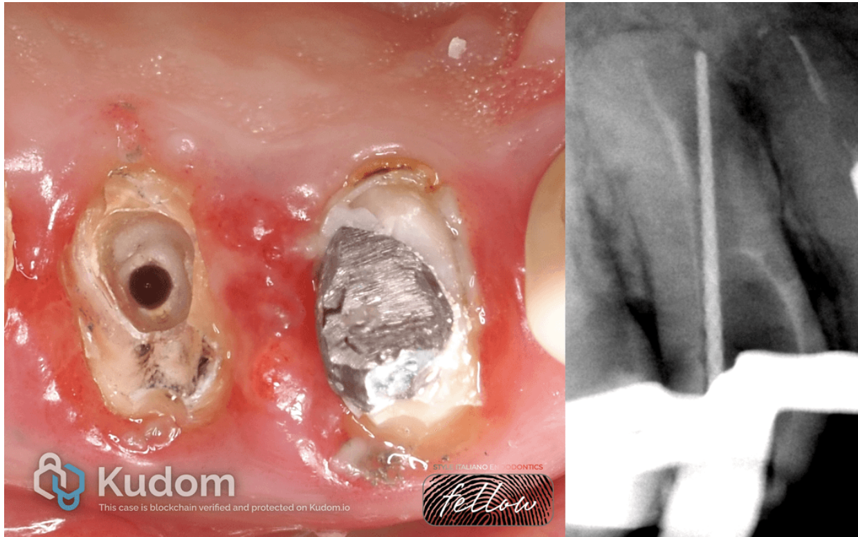
Fig. 3
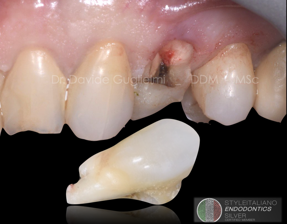
The absence of a ferrule, perforation in the apical portion of the root, and the inability to perform adequate endodontics and prosthodontics on this tooth led to the only option being a plan to replace the tooth with an implant.
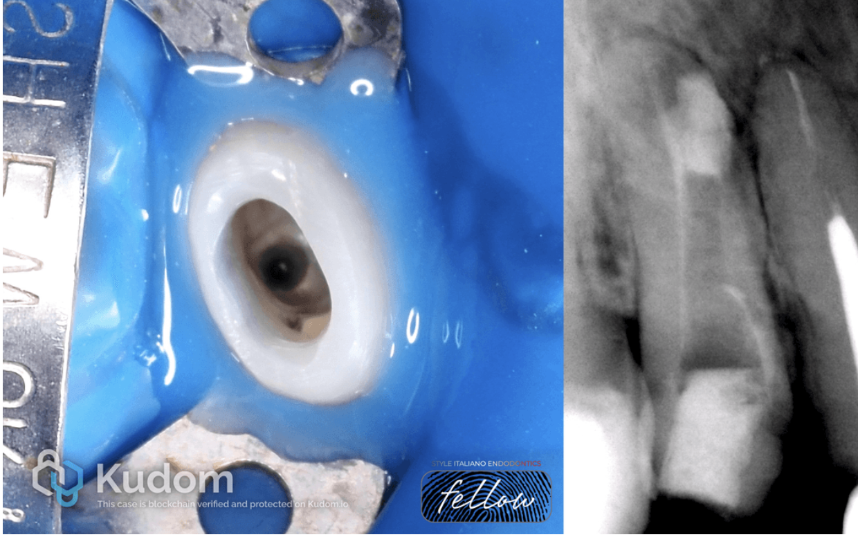
Fig. 4
However, considering the patient's life factors, the decision was made to save this tooth and choose a patient-oriented treatment plan. The program to save this tooth began with sealing the perforation using MTA. The Crown-Tissue aspect became the primary salvage strategy for this clinical case
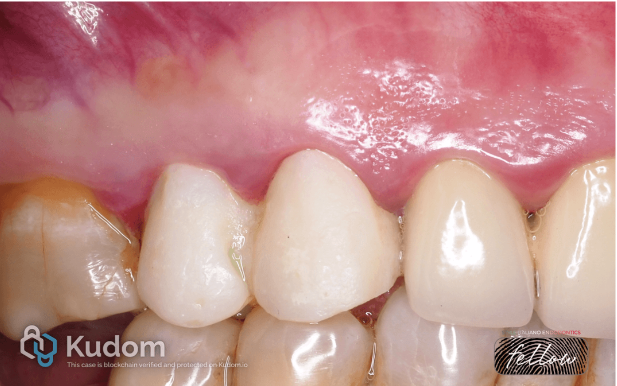
Fig. 5
Crown-Tissue is the main salvage strategy for this clinical case. Immediately after the repeated endodontic treatment, a temporary restoration was fabricated. After 8 days, closure of the fistulous tract, absence of symptoms, and stabilization of the soft tissues are observed

Fig. 6
After 20 days, the crown fabrication process began. Next, a Build-Up was performed using a glass fiber post and composite cement. Utilizing temporary crowns, healthy gums were created around the tooth stumps.
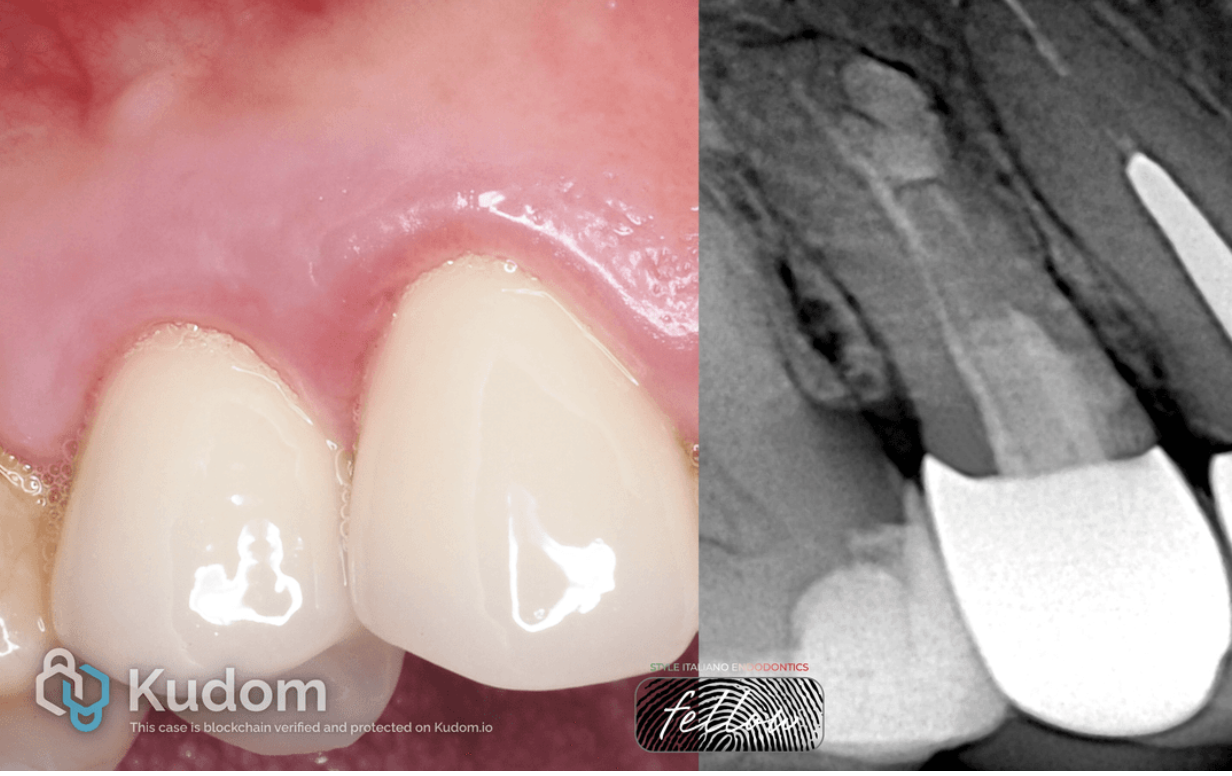
Fig. 7
Subsequently, zirconium dioxide crowns were cemented, and radiographic diagnostics were performed. The photograph shows healthy tissue around the crowns. The vertical preparation technique allows for a more apical placement of the restoration, thereby creating a new cemento-enamel junction.

Fig. 8
Four-year follow-up of this tooth. Healthy tissue around the crowns is noted, with no symptoms of apical periodontitis. The patient has no complaints.
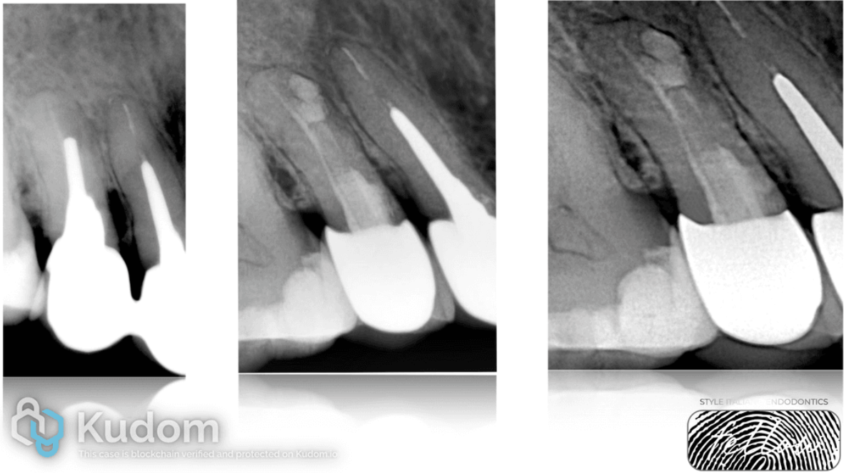
Fig. 9
Dynamic radiographic diagnostics over 4 years also shows excellent results. Healing of the periapical tissues is observed.
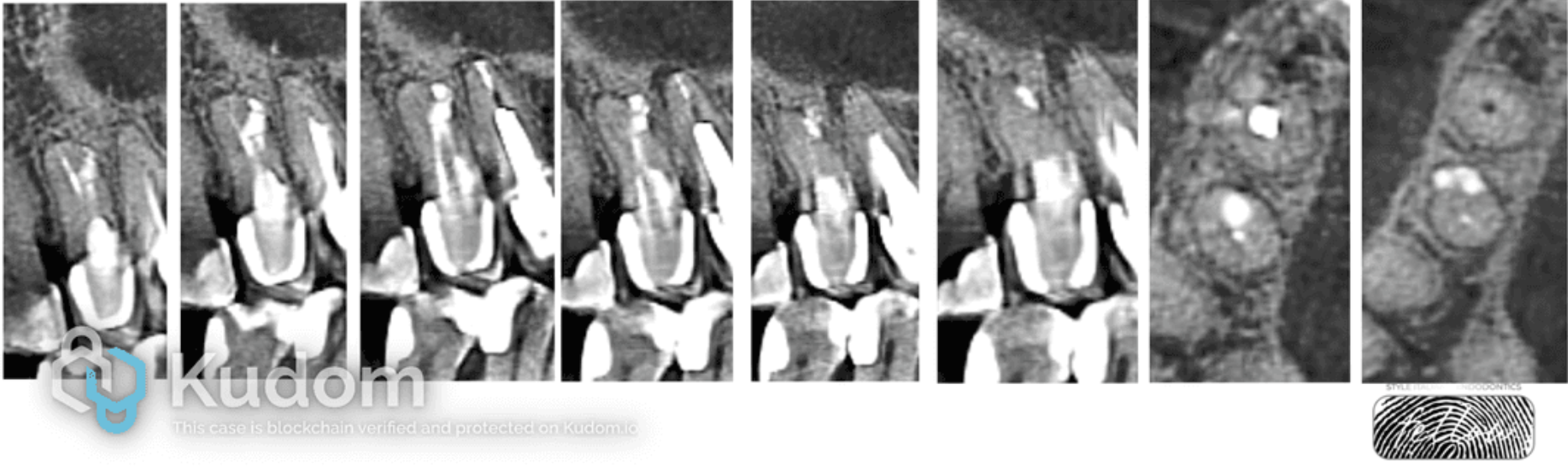
Fig. 10
After 5 years, a CBCT scan was performed, revealing complete elimination of the pathological focus with restoration of the lamina dura and a normal periodontal ligament space around the entire root perimeter. This indicates the successful outcome of the ROOT-CROWN-TISSUE concept.

Fig. 11
About the author:
Nasim Akhatov
Nasim Akhatov was born in 1981 in Dushanbe city, Republic of Tajikistan.
He graduated the Tajik state medical university in 2003.
In 2005 he graduated the clinical residency in the specialty of endodontics.
He has a private practice «Akhatov Dental studio» in Kazan city, Russian Federation.
He works as a Microscopic Endodontist for advanced root canal treatment.
The main direction in practice is saving teeth from root to crown approach.
He has given lectures, hands on courses and training in Endodontics during this time.
He was KOL of DENTSPLY Sirona from 2017.
Conclusions
The healing of a periapical lesion is influenced by many factors. The goal of endodontic treatment is the qualitative cleaning of canals to disinfect infected areas and eliminate root canal defects. And, of course, a crucial factor for the success of endodontic treatment is the fabrication of correct restorations, which not only restore chewing function but also enable the achievement of a dense soft tissue phenotype. The latter is an inseparable part of endodontic treatment
Bibliography
G.M.G.Hommez, C.R.M.Coppens, R.J.G.De Moor : Periapical health related to the quality of coronal restorations and root fillings, International endodontic journal, august 2002/volume 35, issue 8/ pp.680-689.
Zamparini F, Prati C, Taddei P, Spinelli A, Di Foggia M, Gandolfi MG. Chemical-Physical Properties and Bioactivity of New Premixed Calcium Silicate-Bioceramic Root Canal Sealers.
P. Bertani, P. General, F.Gorni, T.Testori Tooth or implant? The recovery of replacement of the severely compromised natural tooth.
Marzadori M, Stefanini M, Sangiorgi M, Mounssif I, Monaco C, Zucchelli G. Crown lengthening and restorative procedures in the esthetic zone. Periodontol 2000. 2018 Jun;77(1):84-92.