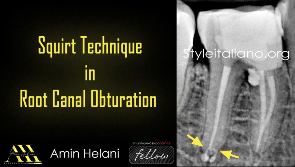
Squirt Technique in Root Canal Obturation
28/02/2025
Fellow
Warning: Undefined variable $post in /home/styleendo/htdocs/styleitaliano-endodontics.org/wp-content/plugins/oxygen/component-framework/components/classes/code-block.class.php(133) : eval()'d code on line 2
Warning: Attempt to read property "ID" on null in /home/styleendo/htdocs/styleitaliano-endodontics.org/wp-content/plugins/oxygen/component-framework/components/classes/code-block.class.php(133) : eval()'d code on line 2
Root canal obturation techniques have developed significantly over the years to improve sealing, reduce treatment times, and ensure more predictable outcomes. The "Squirt Technique," introduced by Hermann and Hülsmann, is a non-master cone method that involves injection of thermoplasticized gutta-percha. This material is introduced via a cartridge tip, often utilizing an electronic obturation unit , and then compacted with cold pressure. When used by experienced hands, with appropriate canal shaping and under a surgical operating microscope , this technique has shown promise for efficient, three-dimensional obturation. However, successful implementation requires maintaining the minor constriction of the apical foramen as a barrier, as canals with transported or resorbed apices are generally unsuitable for this approach.
Despite its potential, the Squirt Technique is not widely documented. Initial reports, such as the 1977 publication by Yee et al., introduced its in vitro applications, and later Marlin's 1981 in vivo study demonstrated clinical success. The author of this case report sought to explore this obturation method in more detail, first using it on a patient with a complex upper wisdom tooth anatomy. This first application yielded favorable results, and the tooth remained stable two years post-treatment. Since then, the author has incorporated the Squirt Technique more frequently into clinical practice, noting various advantages and refining a specific protocol for its use.
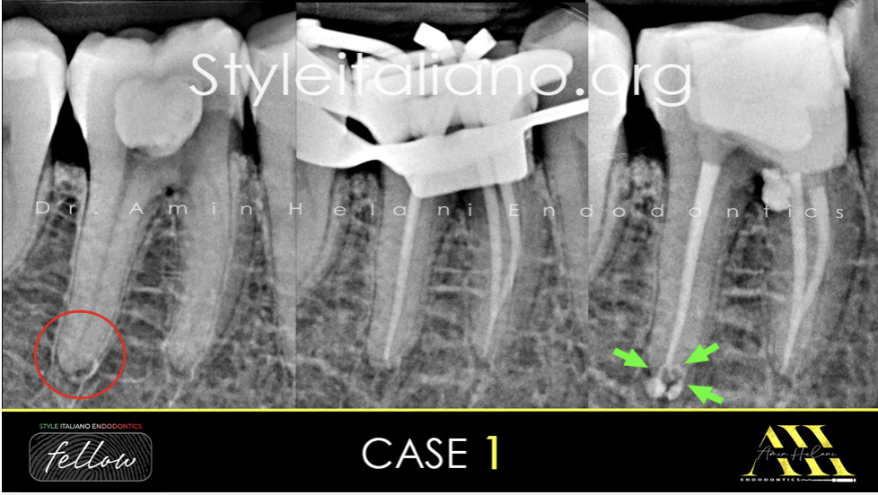
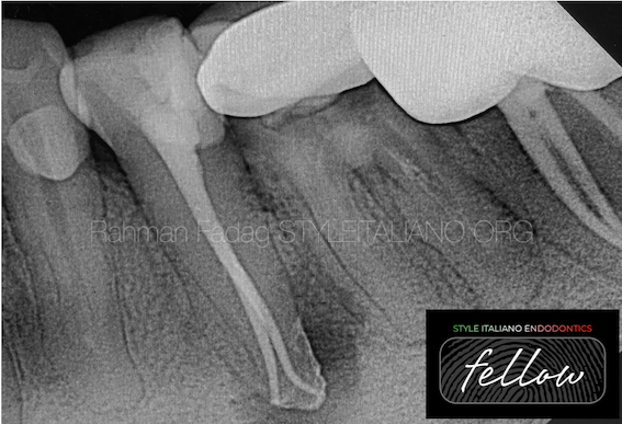
Fig. 1
Case 1:
A 55-year-old male patient presented to the clinic to repair a perforation caused by the previous clinician, who was unable to locate the mesial canals.
The radiograph shows the extent of caries and the perforation in the coronal portion, and a delta in the distal canal.
After access opening, the mesial canals were located, and following the shaping of all canals, the distal root was obturated using the squirt technique.
The post-operative radiograph revealed the presence of three portals of exit.
Shaping: VDW R25 Blue
Irrigation: NaOCL 5%, EDTA 17% with Ultrasonic Activation
Obturation: squirt technique with BC Sealer.
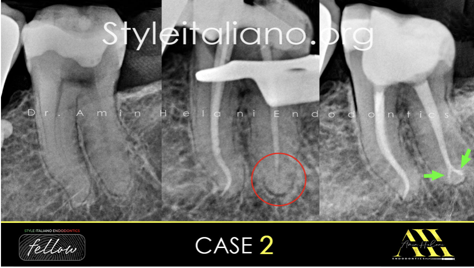
Fig. 2
Case 2:
A 40-year-old female patient presented to complete treatment after the previous clinician was unable to perform complete shaping of the canals.
The mesial root showed an S-shaped curvature, and in the distal root, I could not reach the apex due to a sharp curvature and a distal portal of exit, resulting in a sharp apical ledge.
Therefore, obturation was performed using the squirt technique. The post-operative radiograph revealed the presence of a delta and two portals of exit.
Shaping: VDW R25 Blue
Irrigation: NaOCL 5%, EDTA 17% with Ultrasonic Activation
Obturation: squirt technique with BC Sealer.
Video of the procedure.
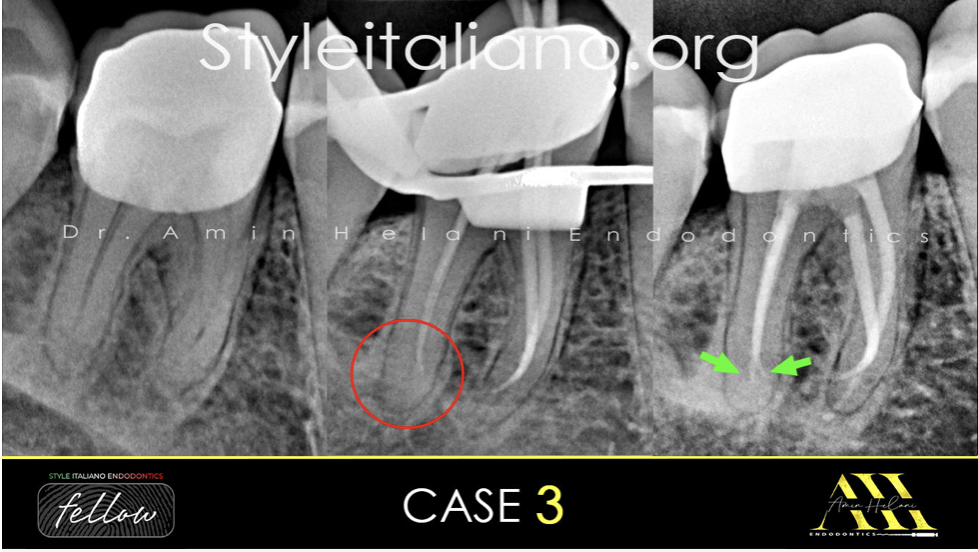
Fig. 3
Case 3:
A 37-year-old female patient presented with severe, sharp pain in the right mandibular first molar for the past 2 days and reported a history of pulp capping done 2 months ago. The pre-operative radiograph revealed a new, good-fitting crown and no apical findings.
The diagnosis was irreversible pulpitis.
Endodontic treatment was initiated. As seen on the master point radiograph, the gutta-percha tip is not centered in the canal and is directed towards the mesial, indicating the presence of a delta or another portal of exit.
Obturation was completed using the squirt technique, and the post-operative radiograph revealed that the second part of the delta was filled with the sealer.
Shaping: VDW R25 Blue
Irrigation: NaOCL 5%, EDTA 17% with Ultrasonic Activation
Obturation: squirt technique with BC Sealer.
Fig. 4
About the Author:
Amin Helani
• Dr. Amin Helani graduated in 2014 from the University of Aleppo in Syria.
• In 2021, he received a specialist degree in Endodontics from the German Society of Endodontics.
• He works as a Microscopic Endodontist for advanced root canal treatment in Berlin, Germany.
• He has been offering lectures, webinars, and hands-on courses to both general dentists and endodontists.
• He is a member of the German Society for Endodontology and Traumatology.
• He is a fellow member of Style Italiano Endodontics.
Conclusions
In conclusion, The Squirt Technique offers a viable obturation method in challenging cases, provided that canal shaping, adequate apical barriers, and thorough irrigation with activation are maintained, making it even suitable for beginners. The technique’s efficiency in obturation, particularly in hard-to-reach or unusually shaped canals, can reduce procedural times and improve workflow. While potential pitfalls such as overfilling or short obturations are real, these issues are mitigated with proper preparation and adherence to recommended protocols.
As with any endodontic technique, appropriate case selection and a thorough understanding of the method’s limitations are essential for success.
Bibliography
References:
- ⁃Yee FS, Marlin J, Krakow AA, Gron P. Three-dimensional obturation of the root canal using injection-molded, thermoplasticized dental gutta-percha. J Endod. 1977 May;3(5):168-74.
- ⁃ Marlin J, Krakow AA, Desilets RP Jr, Gron P. Clinical use of injection-molded thermoplasticized gutta-percha for obturation of the root canal system: a preliminary report. J Endod. 1981 Jun;7(6):277-81.
- ⁃ Stropko J. The System "S" technique: to seal the entire canal system for "success". Roots. 2008;4(3):6-18.
- ⁃ Kumar NS, Prabu PS, Prabu N, Rathinasamy S. Sealing ability of lateral condensation, thermoplasticized gutta-percha, and flowable gutta-percha obturation techniques: A comparative in vitro study. J Pharm Bioallied Sci. 2012 Aug;4(Suppl 2).
- ⁃Lottanti S, Tauböck TT, Zehnder M. Shrinkage of backfill gutta-percha upon cooling. J Endod. 2014 May;40(5):721-4.