Retreatment with pre-endodontic build up of a first upper premolar
23/04/2025
Fellow
Warning: Undefined variable $post in /home/styleendo/htdocs/styleitaliano-endodontics.org/wp-content/plugins/oxygen/component-framework/components/classes/code-block.class.php(133) : eval()'d code on line 2
Warning: Attempt to read property "ID" on null in /home/styleendo/htdocs/styleitaliano-endodontics.org/wp-content/plugins/oxygen/component-framework/components/classes/code-block.class.php(133) : eval()'d code on line 2
The success of endodontic treatment depends on removing microorganisms from root canal system, three dimensionally sealing and the placement of a good coronal seal to prevent communications between oral cavity and the periradicular tissues
The operator should be able to assess the endodontic case and know about tooth anatomy and the suspected anatomical variations one may face during treatment.
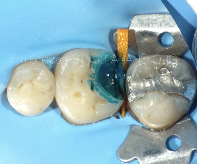
In most of the time we face severely destroyed teeth, which means a challenge for clinicians from the absolute isolation, the selection of the clamp, the isolation technique, etc. Therefore, we must know or resolve these situations with the help of restorative procedures, in this case with pre-endodontic build up.
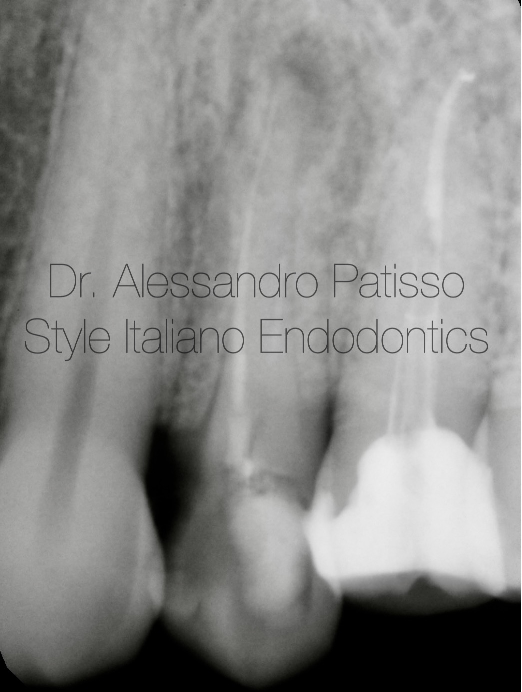
Fig. 1
A 48-year-old patient presented himself complaining of severe pain in the second quadrant. The radiological examination highlighted a periapical lesion affecting dental element 2.4 previously treated endodontically. The patient is explained that the old treatment was inappropriate and that the tooth needs to be retreated.
Pre-endodontic reconstruction allows you to work in a safer and more easily isolated field.
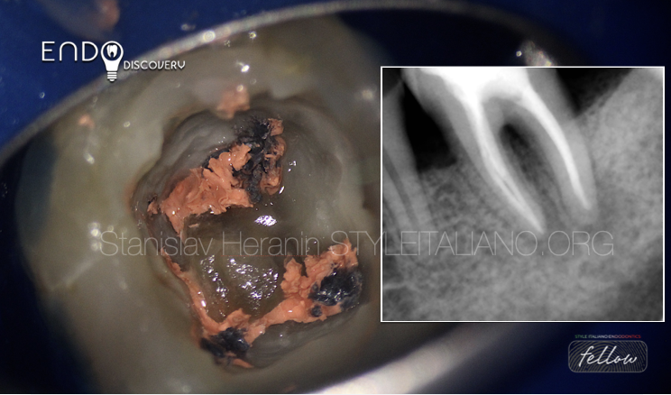
Thanks to the use of ultrasonic tips it was simple to remove the gutapercha and free the canal.
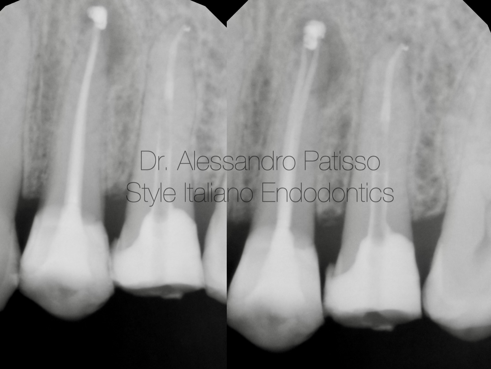
Fig. 2
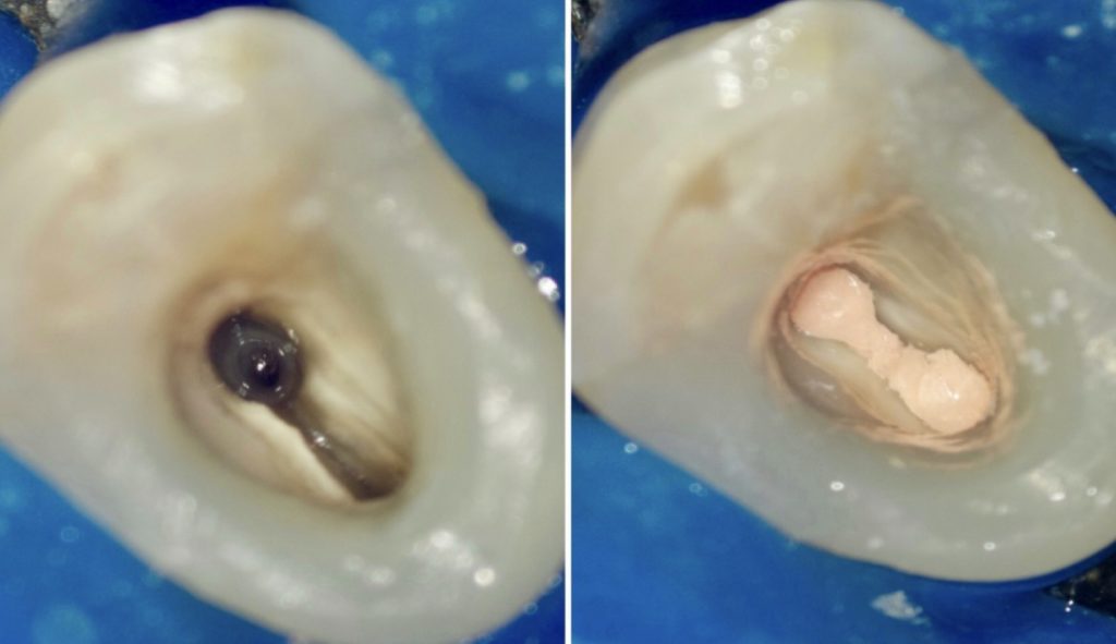
A three-dimensional hot obturation system allowed me to seal the entire root canal with the gutta percha.(BIOCONELESS)
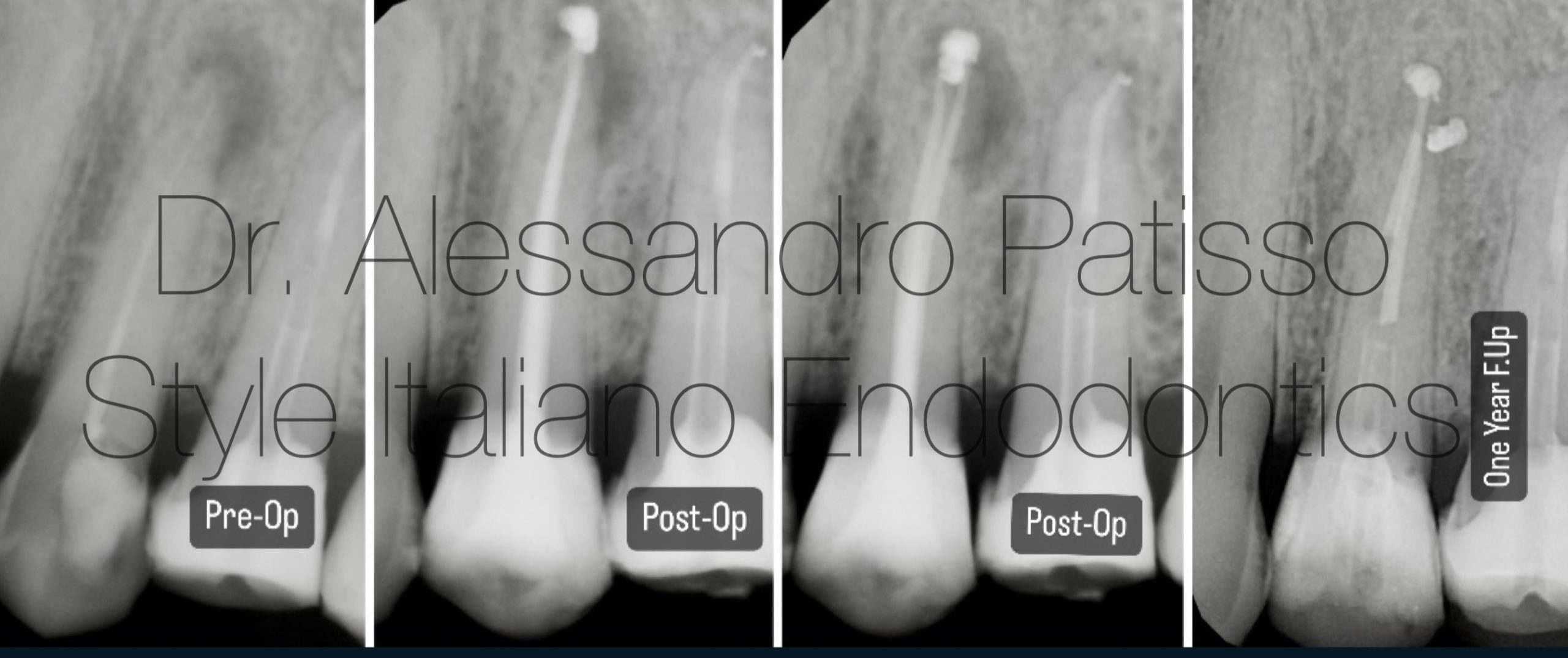
Fig. 3
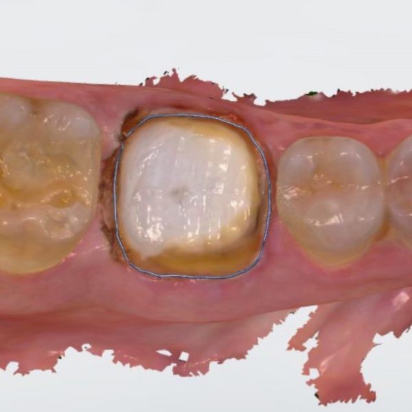
Follow-up One Year after reconstruction.

Fig. 4
Graduated in Dental Hygiene at the University of Bari in 2009 and Dentistry and Dental Prosthetics at the Universidad Europea de Madrid in 2015.
From the beginning he has practiced exclusively Clinical Edodontics, perfecting himself through numerous courses around Italy. Among the most recent he attended the courses of Dr Giuseppe Carrieri, finishing first in the course.
Today he works as a freelancer using only the microscope. Tutor in the courses of Dr Giuseppe Carrieri Fellows member Styleitaliano Endodontics.
Conclusions
In endodontic treatment, multiple variables can affect the outcome of a case. The use of pre-endodontic build-up will enhance by preventing marginal leakage before, during, and after treatment that is provided prior to placing a final restoration.
In cases where the root anatomy is complex, the use of the microscope associated with adequate instruments is essential to achieve a good result without running the risk of making serious errors, therefore we may have to have CBCT to estimate the situation .
Bibliography
-
- Ree M, Schwartz R. The Endo-Restorative Interface: Current Concepts, Dental Clinics of North America; 54: 345-374, 2010
- Vertucci FJ. Root canal Vertucci FJ. Root canal morphology and its relationship to endodontic procedures. Endod Topics. 2005;10:23–9.
- Association of Endodontists. Glossary of Endodontic Terms, 8th ed. Chicago: American Association of Endodontists; 2012.
- Siqueira JF, Barnett F. Interappointment pain: mechanisms, diagnosis, and treatment. Endodontic Topics 2004, 7, 93109
- Gavriil, D., Kakka, A., Myers, P., & O´Connor, C. J. (2021). Pre-endodontic restoration of structurally compromised teeth: current concepts. British Dental Journal, 231(6), 343–349. https://doi.org/10.1038/s41415-021-3467-0
- Iaculli, F., Rengo, C., Lodato, V., Patini, R., Spagnuolo, G., & Rengo, S. (2021). Fracture resistance of endodontically-treated maxillary premolars restored with different type of posts and direct composite reconstructions: A systematic review and meta-analysis of in vitro studies. In Dental Materials (Vol. 37, Issue 9, pp. e455–e484). Elsevier Inc. https://doi.org/10.1016/j.dental.2021.06.007